
Treatment sedule for tonsillitis
tonsillitis treatment management
tonsillitis acute inflamed tonsils pharynx
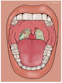
Summary : This illustration shows the inside of a human mouth with a focus on the throat, depicting inflamed tonsils with white-greenish patches, suggestive of tonsillitis or a similar infection. photo: Scene Overview : • Main subject is the open mouth and throat, viewed from the front. • The tonsils are visibly enlarged and covered with white-greenish exudate. • The uvula is centered and appears slightly swollen. • The tongue is prominent in the lower half of the image. • Teeth are visible along the top and bottom edges of the mouth. • The background is a solid, muted color, emphasizing the oral cavity. Technical Details : • No scale bar or magnification indicated. • No staining or medical imaging modality used; this is a digital illustration. • No on-image text except a faint watermark in the upper right corner. Spatial Relationships : • Tonsils are located on either side of the uvula, at the back of the throat. • White-greenish patches are localized to the tonsils, not extending to other oral structures. • The tongue occupies the foreground, with the throat and tonsils in the background. Analysis : • The key visual message is the presence of inflamed, exudate-covered tonsils, which is characteristic of acute tonsillitis or pharyngitis. • The illustration highlights the typical appearance of infected tonsils, useful for educational or diagnostic reference.
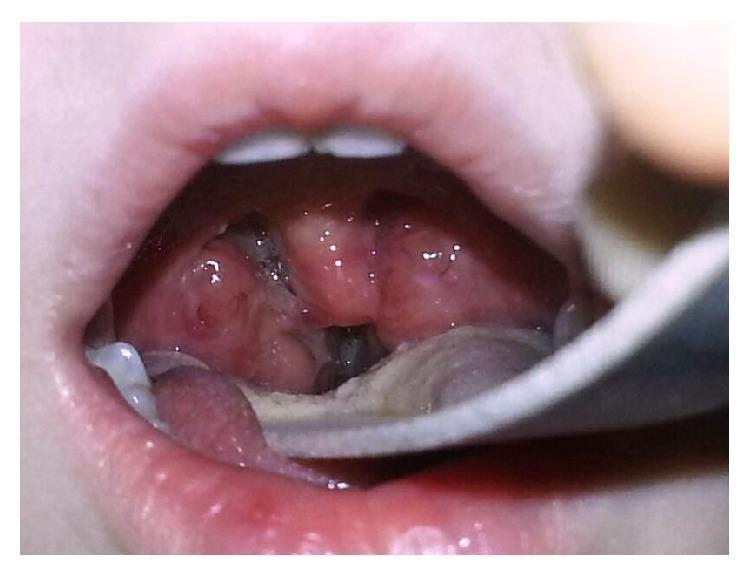
This clinical photograph displays an intraoral view of the oropharynx in a pediatric patient. The primary finding is significant bilateral palatine tonsillar hypertrophy. The tonsils appear erythematous and markedly enlarged, nearly meeting in the midline (kissing tonsils), which severely narrows the oropharyngeal airway. The surface of the tonsils is irregular and crypt-laden, with visible hyperemia and some localized white/grayish patches suggestive of either postoperative changes following radiofrequency ablation or exudative tonsillitis. A metal tongue depressor is visible in the lower right foreground, retracting the tongue to facilitate visualization of the posterior pharynx. The surrounding mucosa appears inflamed and erythematous. This image illustrates a common anatomical cause of obstructive sleep apnea (OSA) in children and serves as a teaching tool for grading tonsillar size and assessing oropharyngeal patency in an otolaryngology or primary care context.
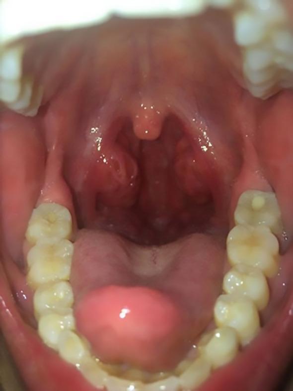
This clinical photograph displays an intraoral view of the oropharynx, highlighting a case of severe acute tonsillitis. The primary findings include significant bilateral tonsillar hypertrophy, with the palatine tonsils appearing markedly swollen and erythematous. Multiple small, white-to-yellowish purulent exudates are visible within the tonsillar crypts, a hallmark of bacterial infection. The surrounding oropharyngeal mucosa and soft palate exhibit generalized hyperemia. The uvula is midline and appears slightly edematous but not deviated. Other visible anatomical landmarks include the tongue and the maxillary and mandibular dentition. This visual representation is characteristic of infectious processes such as Lemierre syndrome precursors or streptococcal pharyngitis, demonstrating clinical signs that can lead to airway narrowing or systemic inflammatory response syndrome (SIRS).
Treatment Schedule for Tonsillitis
1. Initial Assessment - Centor Score
| Criterion | Points |
|---|---|
| Fever | +1 |
| Tonsillar exudate | +1 |
| Cervical lymphadenopathy | +1 |
| Absence of cough | +1 |
-
Score 0-1: No treatment needed (likely viral)
-
Score 2-3: GABHS rapid antigen test warranted; treat if positive
-
Score 4: Start antibiotics empirically
-
Schwartz's Principles of Surgery, p. 650
2. Determine Aetiology
- Viral (most common cause of acute tonsillitis): supportive care only; antibiotics not indicated
- GABHS (Group A beta-haemolytic Streptococcus): antibiotic therapy required
- Epstein-Barr Virus (EBV/Mononucleosis): mimics bacterial tonsillitis; look for lymphadenopathy, splenomegaly, hepatitis, atypical lymphocytes - avoid amoxicillin/ampicillin (causes maculopapular rash)
- Other bacteria: H. influenzae, M. catarrhalis, S. pneumoniae - managed similarly to GABHS
- Cummings Otolaryngology Head and Neck Surgery
3. Medical Treatment
A. First-Line Antibiotics (GABHS-confirmed or Centor score 4)
| Drug | Dose | Duration |
|---|---|---|
| Penicillin V (phenoxymethylpenicillin) | 500 mg PO twice daily (adults) | 10 days |
| Amoxicillin | 500 mg PO 3x daily OR 1g twice daily | 10 days |
B. Penicillin Allergy - Alternative Antibiotics
| Drug | Notes |
|---|---|
| First-generation cephalosporins (e.g. cefadroxil, cefalexin) | Preferred alternative; evidence suggests superior eradication rates |
| Clindamycin | Preferred for penicillin allergy or penicillin failure |
| Clarithromycin | Macrolide; note increasing macrolide resistance in GABHS |
| Azithromycin | Shorter course (5 days); macrolide resistance is a concern |
- Cummings Otolaryngology Head and Neck Surgery
C. Penicillin Failure (occurs in 7-37% of cases)
- Cephalosporins
- Clindamycin
- Amoxicillin/clavulanic acid (co-amoxiclav)
4. Symptomatic / Supportive Treatment
| Intervention | Details |
|---|---|
| Paracetamol (Acetaminophen) | Analgesia + antipyretic; first choice |
| NSAIDs (ibuprofen) | Effective analgesia and anti-inflammatory |
| Aspirin | Avoid - risk of Reye's syndrome in children |
| Corticosteroids | Not routinely indicated for uncomplicated tonsillitis |
| Adequate hydration | Oral fluids; IV fluids if dysphagia is severe |
| Salt water gargle | Soothing; no evidence-based benefit but commonly used |
| Rest | Until afebrile |
5. Treatment of Complications
| Complication | Management |
|---|---|
| Peritonsillar abscess (quinsy) | Aspiration or incision & drainage + IV antibiotics (amoxicillin-clavulanate, clindamycin); consider interval tonsillectomy |
| Cervical lymphadenitis | IV antibiotics; drainage if abscess forms |
| Parapharyngeal abscess | Surgical drainage + IV antibiotics |
| Retropharyngeal abscess | IV antibiotics; surgical drainage if needed; anti-TB regimen if tuberculosis |
| Rheumatic fever prevention | Complete 10-day penicillin course; steroids do NOT prevent RF |
| EBV/Mononucleosis | Avoid amoxicillin; supportive care; monitor for splenic rupture |
6. Surgical Treatment - Tonsillectomy
Indications (AAO-HNS Clinical Practice Guidelines / "Paradise Criteria")
- 7 or more episodes in the preceding year, OR
- 5 or more episodes per year in each of the preceding 2 years, OR
- 3 or more episodes per year in each of the preceding 3 years
- Fever >38.3°C
- Cervical lymphadenopathy
- Tonsillar exudate
- Positive GABHS test
- Poor response to antibiotic therapy
- Multiple antibiotic allergies/intolerances
- Significant quality-of-life impact (missed school/work)
- Periodic fever with aphthous stomatitis, pharyngitis, and adenitis (PFAPA)
- History of peritonsillar abscess
- Cummings Otolaryngology Head and Neck Surgery (Table 201.4)
Note: Children with less severe or less frequent disease are unlikely to have long-term benefits from tonsillectomy.
7. Summary Flow Chart
Sore throat + fever
       ↓
Centor Score
       ↓
0-1 → Supportive care only (viral most likely)
2-3 → Rapid strep test → Negative: supportive / Positive: antibiotics
 4  → Start penicillin/amoxicillin empirically
       ↓
Penicillin allergy? → Cephalosporin / Clindamycin / Macrolide
       ↓
Penicillin failure? → Clindamycin / Co-amoxiclav / Cephalosporin
       ↓
Recurrent (meets Paradise criteria)? → Consider tonsillectomy
- Cummings Otolaryngology Head and Neck Surgery (Chapter 201)
- Schwartz's Principles of Surgery, 11th Edition (Chapter 18, p. 650)
- Scott-Brown's Otorhinolaryngology Head & Neck Surgery