
orthopedic all sarcoma in details
Reading File
Finding Sources
Finding Sources
Finding Sources
Finding Sources
Now let me read the most comprehensive textbook sources in parallel:
Reading File
Reading File
Reading File
Reading File
Reading File
Reading File
Reading File
Reading File
Reading File
Reading File
Searching Images
osteosarcoma Codman triangle X-ray
Reading File
Searching Images
Ewing sarcoma onion skin periosteal reaction diaphysis
Searching Images
chondrosarcoma histology pelvis X-ray
Finding Sources
Reading File
Reading File
Orthopedic Sarcomas — Comprehensive Overview
Sarcomas are malignant tumors of mesenchymal origin (bone, cartilage, muscle, fat, vessels, nerves). They account for ~1% of all adult cancers but are disproportionately represented in children and young adults. This guide covers all major orthopedic sarcomas — both skeletal (bone) sarcomas and soft tissue sarcomas (STS).
EPIDEMIOLOGY AT A GLANCE
| Tumor | % of Primary Bone Cancers (Adults) | Peak Age |
|---|---|---|
| Chondrosarcoma | 40% | 40–60 yrs |
| Osteosarcoma | 28% | Bimodal: teens & elderly |
| Chordoma | 10% | 50–60 yrs |
| Ewing Sarcoma | 8% | <20 yrs |
| Others | 14% | — |
In children, osteosarcoma dominates (56%), followed by Ewing sarcoma (28%) and chondrosarcoma (6%). Approximately 3,970 new primary bone cancers are diagnosed annually in the US, and STS accounts for ~13,400 new cases/year.
PART I: SKELETAL (BONE) SARCOMAS
1. OSTEOSARCOMA (Osteogenic Sarcoma)
Definition
A malignant tumor that produces neoplastic osteoid (unmineralized bone matrix). It is the most common primary malignant tumor of bone (excluding hematopoietic tumors).
Epidemiology
- Bimodal age distribution: 75% occur in patients <20 years (adolescent growth spurt); a second, smaller peak occurs in older adults with predisposing conditions
- Male:Female ratio: 1.6:1
- Secondary osteosarcomas arise in the context of Paget disease, prior radiation, bone infarcts
Location
- Metaphysis of long bones (near the growth plate)
- ~50% involve the distal femur or proximal tibia (around the knee)
- Rarely involves hands/feet
Pathogenesis — Molecular
| Gene | Role |
|---|---|
| RB | Mutated in 70% of sporadic cases; germline mutation = 1000× ↑ risk |
| TP53 | Germline mutation in Li-Fraumeni syndrome; commonly mutated in sporadic tumors |
| MDM2 / CDK4 | Overexpressed in low-grade variants; inhibit p53 and RB function |
| CDKN2A (INK4a) | Inactivated in many cases (encodes p16 and p14) |
| MYC | Amplified in a subset |
No characteristic fusion gene (unlike Ewing sarcoma)
Radiology
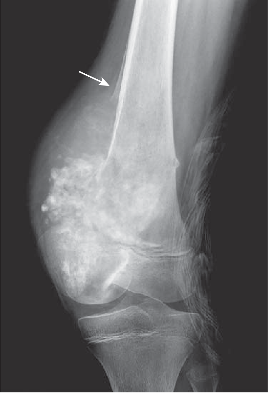
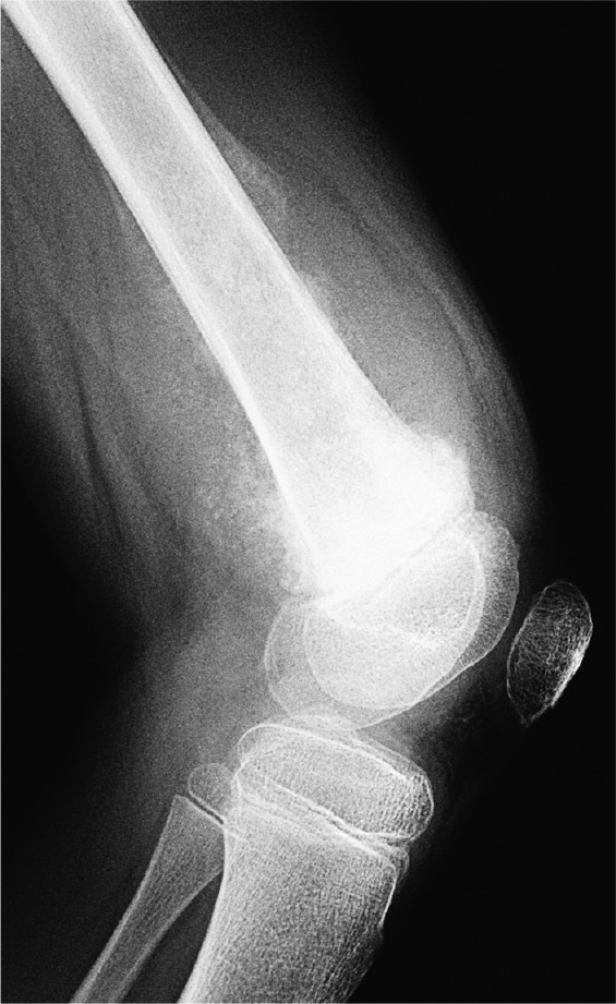
- Mixed lytic and sclerotic destructive mass with infiltrative margins
- Codman triangle: reactive periosteal bone when periosteum is lifted — indicates aggressive tumor (not pathognomonic)
- Sunburst pattern: perpendicular spicules of tumor bone extending into soft tissue
- Breaks through cortex into soft tissues
Pathology (Morphology)
- Gross: bulky, gritty, tan-white tumor; areas of hemorrhage; cortical destruction; soft tissue invasion
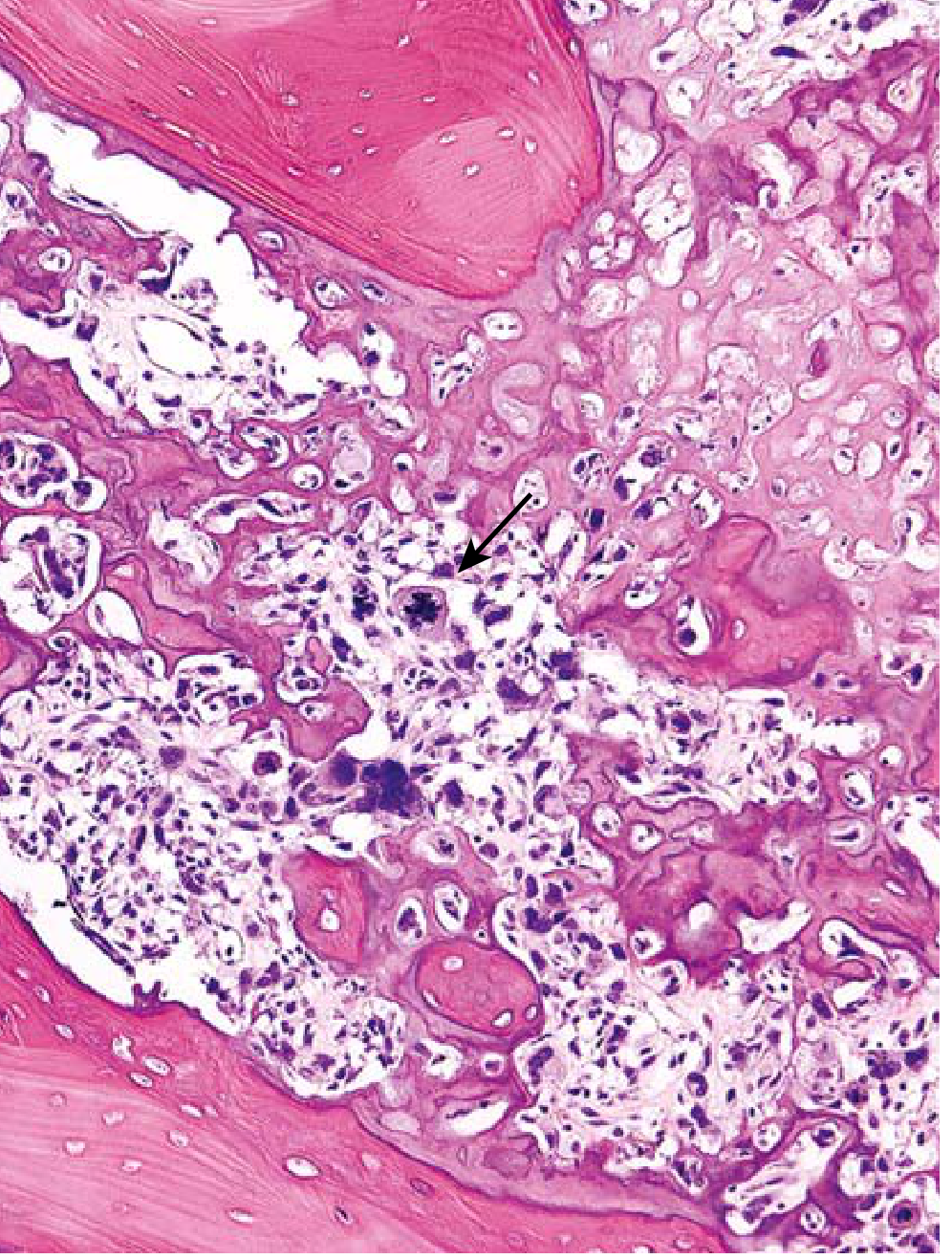
- Histology: pleomorphic cells, large hyperchromatic nuclei, bizarre giant cells, numerous mitoses; lacelike osteoid produced by malignant cells is diagnostic
- Extensive necrosis and intravascular invasion are common
Types of Osteosarcoma
| Type | Features |
|---|---|
| Conventional (intramedullary) | Most common; high grade; metaphyseal |
| Parosteal | Low grade; posterior distal femur; heavy ossification with "string sign" cleavage plane |
| Periosteal | Intermediate grade; cortical surface; chondroblastic |
| Telangiectatic | Lytic, blood-filled spaces; mimics ABC; high grade |
| Small cell | Resembles Ewing sarcoma; high grade |
| Secondary | Post-Paget disease, radiation, dedifferentiated |
| Extraskeletal | Arises in soft tissue; high grade |
Clinical Features
- Painful, progressively enlarging mass
- Pathologic fracture may be first sign
- Local warmth, tenderness, swelling
Staging — Enneking System (MSTS)
- Stage I (low grade): Ia — intracompartmental; Ib — extracompartmental
- Stage II (high grade): IIa — intracompartmental; IIb — extracompartmental
- Stage III — any grade with metastases
Treatment
- Neoadjuvant chemotherapy (MAP protocol: methotrexate, adriamycin/doxorubicin, cisplatin) — 3 cycles before surgery
- Wide surgical resection (limb salvage in ~90% of cases; local recurrence target <7%)
- Adjuvant chemotherapy post-surgery
- Chemotherapy response assessed histologically — ≥90% necrosis = good response (Huvos Grade III/IV)
Limb salvage vs. amputation: Early studies by Simon and Link showed equivalent local recurrence and survival for distal femoral osteosarcoma.
Reconstruction Options
- Osteoarticular allograft
- Intercalary allograft
- Megaprosthesis (endoprosthesis) — modular metal implant; 10-year survival 50–80%
- Rotational flaps (e.g., medial gastrocnemius flap for proximal tibial tumors)
Prognosis
- 5-year survival: ~70% without overt metastases at diagnosis
- With clinically evident metastases or secondary osteosarcoma: <20% 5-year survival
- Metastasizes hematogenously: lungs (most common) > bone > brain
2. EWING SARCOMA
Definition
A malignant small round blue cell tumor of bone (and occasionally soft tissue) characterized by a pathognomonic chromosomal translocation involving the EWSR1 gene.
Epidemiology
- Second most common primary bone sarcoma in children
- ~10% of primary malignant bone tumors
- 80% of patients are <20 years old; slight male predominance
- ~200 cases/year in the United States
- Rare in Black patients (genetic/ethnic predisposition)
Location
- Diaphysis of long bones (femur > tibia > fibula > humerus)
- Flat bones: pelvis, ribs, vertebrae
- 20% are extraskeletal (soft tissue Ewing sarcoma)
Pathogenesis — Molecular
- t(11;22)(q24;q12) in ~85% of cases → EWS–FLI1 fusion protein
- Remaining 15%: variant translocations (EWS-ERG, EWS-ETV1, EWS-FEV)
- EWS/FLI1 chimeric protein binds chromatin → dysregulates transcription → uncontrolled proliferation
- Cell of origin: likely mesenchymal stem cells or primitive neuroectodermal cells
All Ewing sarcoma family tumors (ESFT), including PNET and Askin tumor (chest wall), share the same translocation — they are considered the same entity.
Radiology
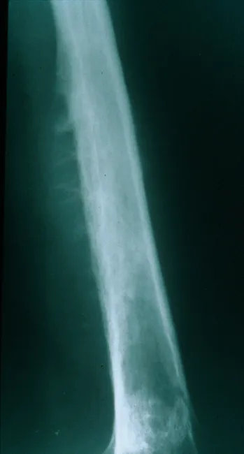
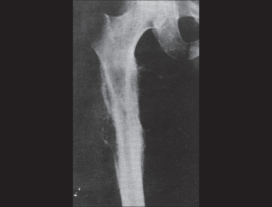
- Diaphyseal, permeative lytic lesion with ill-defined margins
- "Onion skin" periosteal reaction — concentric layers of reactive bone (classic)
- Large associated soft tissue mass
- May have aggressive sunburst pattern
Pathology (Morphology)
- Gross: soft, tan-white; hemorrhage and necrosis common; diaphyseal medullary origin with cortical and periosteal invasion
- Histology: sheets of uniform small round cells, slightly larger than lymphocytes, scant clear glycogen-rich cytoplasm
- Homer-Wright rosettes (cells arrayed around central fibrillary core) may be present
- No osteoid or cartilage production
- IHC: CD99 (MIC2) positive (diffuse membranous), FLI1 positive
Differential for small round blue cell tumors (SRBCT): Ewing sarcoma, neuroblastoma, rhabdomyosarcoma, lymphoma, small cell osteosarcoma — use IHC and molecular testing to distinguish.
Clinical Features
- Painful, enlarging mass at the diaphysis
- Affected site is tender, warm, and swollen (can mimic osteomyelitis — both may have fever, elevated ESR/CRP/WBC)
- Systemic symptoms (fever, weight loss) more common than in osteosarcoma
Treatment
- Systemic chemotherapy first (Ewing is considered a systemic disease from the outset)
- Standard regimen: VDC/IE (vincristine + doxorubicin + cyclophosphamide alternating with ifosfamide + etoposide) — typically 6 cycles before and after local treatment
- Local therapy: surgery, radiation, or both — choice depends on location and tumor factors
- Radiation is particularly useful for unresectable tumors (spine, sacrum, pelvis)
Prognosis
- With modern treatment: 75% 5-year survival; 50% long-term cure for non-metastatic disease
- Metastases at presentation → poor prognosis (<30% long-term survival)
- Chemotherapy response (% necrosis) is a key prognostic factor
3. CHONDROSARCOMA
Definition
A malignant tumor that produces hyaline cartilage. It is the second most common malignant matrix-producing bone tumor (after osteosarcoma).
Epidemiology
- Most common primary bone cancer in adults (40% of adult bone sarcomas)
- Peak age: 40–60 years; men affected 2× more than women
- ~15% are secondary (arising from enchondroma or osteochondroma)
Location
- Axial skeleton predilection: pelvis (most common), shoulder girdle, ribs
- Unlike osteosarcoma, which favors the metaphysis of long bones
- When peripheral: proximal long bones (femur, humerus)
Classification
| Type | Frequency | Features |
|---|---|---|
| Conventional (hyaline) | ~90% | Primary topic of discussion |
| Dedifferentiated | 10% | High-grade spindle cell component; very poor prognosis |
| Mesenchymal | Rare | Biphasic: small round cells + well-differentiated cartilage |
| Clear cell | Rare | Epiphyseal; low grade; recurs locally |
Pathogenesis — Molecular
- Arising in multiple hereditary exostosis: loss-of-function mutations in EXT1/EXT2 (regulate cartilaginous matrix protein synthesis)
- Sporadic and exostosis-related: IDH1 or IDH2 mutations (also seen in glioma, AML)
- CDKN2A silencing by DNA methylation common in sporadic tumors
Radiology
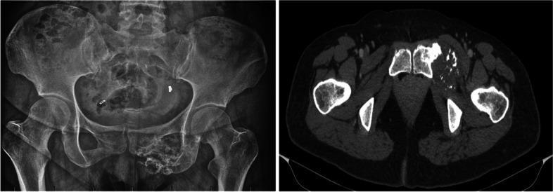
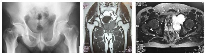
- Destroys cortex and may form a soft tissue mass
- "Popcorn" / "rings and arcs" calcifications — characteristic of chondroid matrix
- "Endosteal scalloping" (>2/3 width of cortex in enchondroma = worrisome for chondrosarcoma)
- On MRI: hyperintense on T2 (high water content of cartilaginous matrix); lobular architecture
Pathology (Morphology)
- Gross: bulky nodules of glistening gray-white translucent cartilage; gelatinous myxoid areas; focal calcifications; possible central necrosis
- Histology: cartilage infiltrates the marrow space, entrapping normal trabeculae
- Grading (correlates with prognosis):
- Grade 1: low cellularity, plump vesicular nuclei, small nucleoli
- Grade 2: increased cellularity, mild-moderate atypia
- Grade 3: high cellularity, extreme pleomorphism, bizarre giant cells, frequent mitoses
Clinical Features
- Painful, progressively enlarging mass
- Symptoms may be present for months to years before diagnosis (especially low-grade)
- Pathologic fracture may occur
Treatment
- Wide surgical excision is the definitive treatment
- Chemotherapy and radiation are NOT effective for conventional chondrosarcoma — this is a key distinguishing feature
- Pelvic chondrosarcomas often require hemipelvectomy or internal hemipelvectomy with complex reconstruction (megaprosthesis, 3D-printed implants)
Prognosis
| Grade | 5-Year Survival | Metastatic Rate |
|---|---|---|
| Grade 1 | 80–90% | Rarely metastasize |
| Grade 2 | ~60–70% | Moderate |
| Grade 3 | <50% | 70% spread hematogenously (to lungs) |
4. CHORDOMA
- Arises from notochordal remnants — skull base (clivus) and sacrococcygeal region
- Slow-growing, locally destructive; not truly a sarcoma but classified with bone tumors
- IHC: Brachyury positive (pathognomonic), S-100 positive, cytokeratin positive
- Treatment: wide resection; proton beam radiation for skull base lesions
- 10% of primary bone cancers in adults
PART II: SOFT TISSUE SARCOMAS (STS)
General Features of All Soft Tissue Sarcomas
Epidemiology
- ~70 distinct histologic subtypes arising from any anatomic site
- Tissues of origin: skeletal muscle, adipose, blood/lymphatic vessels, other mesoderm, peripheral nerves (neuroectoderm)
- ~13,400 new cases/year in the USA; ~5,140 deaths/year → 1% of cancer incidence
- Most common site: thigh (44% of extremity STSs); proximal limb > distal
- Majority arise sporadically
Genetic Syndromes Predisposing to STS
| Syndrome | Mutation | Associated STS |
|---|---|---|
| Li-Fraumeni | TP53 | Multiple sarcoma types |
| Neurofibromatosis type 1 | NF1 (neurofibromin) | MPNST (10% lifetime risk) |
| Hereditary retinoblastoma | RB1 | Osteosarcoma, fibrosarcoma |
| Gardner syndrome | APC | Desmoid tumor |
| Werner syndrome | WRN | Various sarcomas |
General Principles — Diagnosis
- STS present as painless enlarging masses >5 cm (pain is possible)
- Any deep mass >5 cm warrants biopsy
- MRI is the gold standard for local staging
- CT chest for pulmonary metastasis staging
- Biopsy should be performed at a sarcoma center — biopsy-related complications and changes in outcome are significantly lower when done at specialized centers
General Principles — Staging (AJCC/Enneking)
Enneking system:
- Stage I — low grade (Ia: intracompartmental; Ib: extracompartmental)
- Stage II — high grade (IIa: intracompartmental; IIb: extracompartmental)
- Stage III — any grade with regional/distant metastases
Metastatic Pattern
- Lung = most common site of STS metastasis (hematogenous)
- Lymph node metastasis is rare (~5%) — except in specific subtypes: E-S-A-R-C mnemonic:
- Epithelioid sarcoma (most common STS of the hand, young adults)
- Synovial sarcoma
- Angiosarcoma
- Rhabdomyosarcoma
- Clear cell sarcoma
General Treatment Principles
- Wide surgical resection (goal: negative margins)
- Radiation — adjuvant for high-grade, large tumors (reduces local recurrence)
- Chemotherapy — doxorubicin ± ifosfamide as first-line for advanced/metastatic disease
Major STS Subtypes
1. Undifferentiated Pleomorphic Sarcoma (UPS)
(Formerly: Malignant Fibrous Histiocytoma — MFH)
- Most common STS in adults >40 years
- Deep soft tissues of extremities
- Histology: spindle and histiocytic cells arranged in storiform (cartwheel) pattern; slit-like vessels; chronic inflammatory cells
- High grade; frequent recurrence and metastasis
2. Liposarcoma
- Most common STS in adults overall (or UPS — varies by series)
- Rarely in subcutaneous tissue; typically deep retroperitoneum or thigh
- IHC: MDM2 positive (especially well-differentiated/dedifferentiated)
- Histology: lipoblasts (signet ring-shaped cells) are hallmark
- Subtypes:
Subtype Grade Features Well-differentiated (WDLPS) Low MDM2/CDK4 amplification Dedifferentiated (DDLPS) High WDLPS + high-grade spindle cell component Myxoid Low–intermediate t(12;16) → FUS-DDIT3 fusion Round cell High High-grade variant of myxoid Pleomorphic High Rare; worst prognosis
3. Leiomyosarcoma
- Arises from smooth muscle (uterus, retroperitoneum, blood vessel walls)
- Histology: fascicular growth with spindle cells intersecting at right angles ("herringbone" variant)
- IHC: smooth muscle actin (SMA) and desmin positive
- Treatment: surgical resection; gemcitabine + docetaxel for metastatic disease
4. Rhabdomyosarcoma (RMS)
- Most common STS in children
- Occurs in young adults as well
- Subtypes: embryonal (most common, best prognosis), alveolar (t(2;13) → PAX3-FOXO1 or t(1;13) → PAX7-FOXO1), pleomorphic (rare, adults)
- IHC: myogenin, MyoD1, desmin positive
- Treatment: multimodality (chemotherapy + surgery ± radiation); VAC regimen
- Lymph node metastasis common (ESARC)
5. Synovial Sarcoma
- Misnomer — does not arise from synovium; likely from pluripotent mesenchymal cells
- Common in young adults; para-articular location (near joints, especially knee)
- t(X;18) → SYT-SSX1 or SYT-SSX2 fusion products (2 most common)
- Histology: biphasic (epithelial + spindle cell) or monophasic
- IHC: TLE1 positive, cytokeratin positive, EMA positive
- Lymph node metastasis possible (ESARC)
- Treatment: surgery + radiation ± chemotherapy (ifosfamide-sensitive)
6. Angiosarcoma
- Resembles endothelium of blood vessels
- Highly malignant — worst prognosis among STS
- Stewart-Treves syndrome: angiosarcoma arising in chronic lymphedematous arm (post-mastectomy)
- Cutaneous and lymph node metastases common
- Infiltrative with poor margins — amputation may be necessary
- IHC: CD31, CD34, ERG positive
7. Malignant Peripheral Nerve Sheath Tumor (MPNST)
- Arises de novo or in neurofibromatosis type 1 (NF1)
- NF1 patients have 10% lifetime risk of MPNST
- Histology: spindle cells in sweeping fascicles with nodular/whorled areas
- IHC: S-100 positive (often focal)
- High grade; poor prognosis
8. Epithelioid Sarcoma
- Young adults; most common STS of the hand/wrist
- Two types: classical (distal) and proximal (more aggressive)
- IHC: cytokeratin positive, EMA positive, INI1 (SMARCB1) lost
- Lymph node metastasis common (ESARC)
9. Clear Cell Sarcoma
(Melanoma of soft parts)
- Young adults; common in lower extremity/foot (attached to tendons and aponeuroses)
- t(12;22) → EWS-ATF1 fusion
- IHC: S-100 and HMB-45 positive (melanocytic markers)
- Lymph node metastasis (ESARC)
10. Fibrosarcoma
- Histology: fasciculated spindle cells with scanty cytoplasm separated by interwoven collagen — "herringbone" pattern (intersecting fascicles with transverse vs. longitudinal nuclei in adjacent fascicles)
- Can arise de novo or from radiation
PART III: BONE MICROENVIRONMENT & SARCOMA BIOLOGY
The bone microenvironment is rich in growth factors, signaling molecules, and mesenchymal stem cells — ideal "soil" for tumor cell seeding:
- RANKL / RANK axis: RANKL (produced by osteoblasts) binds RANK on osteoclast precursors → stimulates osteoclastogenesis. Tumor cells exploit this "vicious cycle" to promote bone destruction.
- CXCL12 / CXCR4 axis: mesenchymal stem cells secrete CXCL12; circulating tumor cells (e.g., breast cancer) express CXCR4 → homing to marrow
- Denosumab (anti-RANKL monoclonal antibody): used in giant cell tumor of bone and bone metastases
DIAGNOSTIC APPROACH SUMMARY
Step 1: History & Exam
  Age → helps narrow differential (age-specific tumor chart)
  Pain characteristics (night pain, at rest = more aggressive)
  Personal history: cancer, radiation, genetic syndromes
  Physical: mass size, depth, lymph nodes, café-au-lait spots (NF1)

Step 2: Labs
  CBC, ESR, CRP, LDH (elevated in Ewing), ALP (elevated in osteosarcoma)
  PSA (if metastatic disease suspected in older male)

Step 3: Imaging
  Plain X-ray FIRST (location, matrix, periosteal reaction, zone of transition)
  MRI (gold standard for local staging and soft tissue sarcomas)
  CT chest (lung metastases)
  Bone scan or PET (skip lesions, multifocal disease)

Step 4: Biopsy
  MUST be performed at sarcoma center (specialist placement critical)
  Core needle biopsy preferred
  Biopsy tract must be excised en bloc with specimen at definitive surgery
  Gene fusion testing (molecular) where applicable

Step 5: Staging (Enneking/AJCC)
  Grade + compartment + metastases
QUICK COMPARISON TABLE
| Feature | Osteosarcoma | Ewing Sarcoma | Chondrosarcoma |
|---|---|---|---|
| Age | Bimodal (teens, elderly) | <20 years | 40–60 years |
| Location | Metaphysis, long bones | Diaphysis, flat bones | Axial skeleton, pelvis |
| X-ray | Sunburst, Codman triangle | Onion-skin, permeative lysis | Rings/arcs calcification |
| Genetics | RB, TP53 (no fusion) | t(11;22) EWS-FLI1 | IDH1/2, EXT1/EXT2 |
| Histology | Malignant osteoid | Small round blue cells | Malignant cartilage |
| Chemo | Sensitive (MAP) | Sensitive (VDC/IE) | Resistant |
| Radiation | Resistant | Sensitive | Resistant |
| 5-yr survival | ~70% | ~75% (non-met) | 50–90% (grade-dependent) |
| Metastasis site | Lungs | Lungs, bone | Lungs |
STAGING: ENNEKING / MSTS SYSTEM (Applicable to All Bone & STS)
| Stage | Grade | Compartment | Metastasis |
|---|---|---|---|
| Ia | Low (G1) | Intracompartmental (T1) | None (M0) |
| Ib | Low (G1) | Extracompartmental (T2) | None (M0) |
| IIa | High (G2) | Intracompartmental (T1) | None (M0) |
| IIb | High (G2) | Extracompartmental (T2) | None (M0) |
| III | Any | Any | Regional or distant (M1) |
This system, described by Enneking, Spanier, and Goodman (1980), is the most correlative system for predicting sarcoma outcomes and remains standard in orthopedic oncology.
Sources: Robbins & Kumar Basic Pathology; Sabiston Textbook of Surgery; Miller's Review of Orthopaedics 9th Ed; Grainger & Allison's Diagnostic RadiologyThis is a comprehensive reference covering all orthopedic sarcomas. Here's a summary of what was covered:
What Was Covered
Skeletal (Bone) Sarcomas:
- Osteosarcoma — most common in adolescents; metaphyseal; Codman triangle / sunburst on X-ray; RB/TP53 mutations; treated with MAP chemotherapy + wide resection; 70% 5-year survival
- Ewing Sarcoma — diaphyseal; <20 yrs; t(11;22) EWS-FLI1; "onion-skin" periosteal reaction; small round blue cells; treated with VDC/IE chemotherapy ± surgery/radiation; 75% survival
- Chondrosarcoma — most common adult bone sarcoma; axial/pelvis; "popcorn" calcification; IDH1/2 mutations; chemo/radiation-resistant — surgery only; grade-dependent prognosis
- Chordoma — notochordal origin; Brachyury+; sacrum/clivus
Soft Tissue Sarcomas (10 subtypes):
UPS, Liposarcoma, Leiomyosarcoma, Rhabdomyosarcoma, Synovial Sarcoma, Angiosarcoma, MPNST, Epithelioid Sarcoma, Clear Cell Sarcoma, Fibrosarcoma — each with characteristic histology, molecular markers, and key clinical features
Key Frameworks:
- Enneking staging system (Stage I–III)
- Diagnostic approach (imaging → biopsy protocol)
- ESARC mnemonic for STSs with lymph node metastases
- Bone microenvironment and RANKL biology
now make summarize comparision
Orthopedic Sarcomas — Summary Comparison
🦴 BONE SARCOMAS — Master Comparison Table
| Feature | Osteosarcoma | Ewing Sarcoma | Chondrosarcoma | Chordoma |
|---|---|---|---|---|
| Incidence | Most common primary bone sarcoma (children) | 2nd most common (children) | Most common (adults, 40%) | 10% of adult bone tumors |
| Age | Bimodal: teens + elderly | <20 yrs (80%) | 40–60 yrs | 50–60 yrs |
| Sex | M > F (1.6:1) | M > F | M > F (2:1) | M > F |
| Location | Metaphysis, long bones (distal femur, proximal tibia) | Diaphysis, flat bones (femur, pelvis, ribs) | Axial (pelvis, shoulder, ribs) | Sacrum / clivus |
| Origin | Osteoblastic mesenchyme | Mesenchymal stem cell / primitive neuroectodermal | Chondroblastic mesenchyme | Notochordal remnant |
| Key Genetics | RB (70%), TP53, MDM2, CDK4 — no fusion gene | t(11;22) → EWS-FLI1 (85%) | IDH1/IDH2, EXT1/EXT2, CDKN2A | Brachyury amplification |
| X-ray appearance | Sunburst pattern + Codman triangle (lifted periosteum); mixed lytic-sclerotic | Onion-skin periosteal reaction; permeative lysis + soft tissue mass | Rings and arcs / popcorn calcification; endosteal scalloping | Midline lytic sacral/clival lesion |
| Histology | Pleomorphic cells producing lacelike osteoid | Sheets of uniform small round blue cells; Homer-Wright rosettes; glycogen-rich clear cytoplasm | Malignant chondrocytes in lobules; graded 1–3 by cellularity/atypia | Physaliphorous cells in myxoid stroma |
| IHC | No specific marker | CD99 (MIC2)+, FLI1+ | S-100+ | Brachyury+, S-100+, CK+ |
| Chemo sensitive? | ✅ Yes (MAP protocol) | ✅ Yes (VDC/IE) | ❌ No | ❌ No |
| Radiation sensitive? | ❌ Resistant | ✅ Yes | ❌ Resistant | Partial (proton beam) |
| Treatment | Neoadjuvant chemo → Wide resection → Adjuvant chemo | Systemic chemo → Surgery ± Radiation | Wide surgery alone | Wide resection + proton XRT |
| Chemo response marker | Huvos grade (≥90% necrosis = good) | % necrosis post-chemo | N/A | N/A |
| Metastasis route | Hematogenous → Lungs (most common) > bone | Hematogenous → Lungs, bone | Hematogenous → Lungs | Local invasion; late distant mets |
| 5-year survival | ~70% (non-met); <20% (metastatic) | ~75% (non-met); <30% (metastatic) | 80–90% (Gr1); <50% (Gr3) | ~50–60% |
| Mimic / DDx | Ewing, osteomyelitis, metastasis | Osteomyelitis (both can have fever!), Ewing family | Enchondroma (benign) | Giant notochordal cell tumor |
🧬 GENETIC / MOLECULAR SNAPSHOT
| Sarcoma | Key Alteration | Clinical Significance |
|---|---|---|
| Osteosarcoma | RB + TP53 mutations | Li-Fraumeni → ↑ risk; no targeted therapy yet |
| Ewing Sarcoma | t(11;22) EWS-FLI1 | Diagnostic; 15% have variant translocations |
| Chondrosarcoma | IDH1/IDH2 mutations | Shared with glioma/AML; ivosidenib under study |
| Synovial Sarcoma | t(X;18) SYT-SSX1/2 | Diagnostic; ifosfamide-sensitive |
| Liposarcoma | MDM2/CDK4 amplification | Diagnostic for WDLPS/DDLPS |
| Rhabdomyosarcoma | PAX3-FOXO1 t(2;13) | Alveolar type; worse prognosis |
| Clear Cell Sarcoma | EWS-ATF1 t(12;22) | Melanocytic markers (HMB-45+) |
🔬 SOFT TISSUE SARCOMAS — Subtype Snapshot
| Subtype | Key Feature | Age | IHC | Unique Point |
|---|---|---|---|---|
| UPS (Undifferentiated Pleomorphic Sarcoma) | Storiform "cartwheel" pattern | Adults >40 | — | Formerly MFH; most common adult STS |
| Liposarcoma | Lipoblasts (signet ring cells) | Adults | MDM2+ | Most common deep STS; rarely subcutaneous |
| Leiomyosarcoma | Spindle cells at right angles | Adults | SMA+, Desmin+ | Retroperitoneum, uterus, vessels |
| Rhabdomyosarcoma | Embryonal / alveolar subtypes | Children | Myogenin+, MyoD1+ | Most common pediatric STS |
| Synovial Sarcoma | Biphasic; near joints | Young adults | TLE1+, CK+, EMA+ | Misnomer — not from synovium; t(X;18) |
| Angiosarcoma | Endothelial-like cells | Any | CD31+, CD34+, ERG+ | Stewart-Treves (lymphedema); worst prognosis |
| MPNST | Sweeping fascicles, whorled | NF1 patients | S-100+ (focal) | 10% lifetime risk in NF1 |
| Epithelioid Sarcoma | Granuloma-like nodules | Young adults | CK+, EMA+, INI1 lost | Most common STS of the hand |
| Clear Cell Sarcoma | Melanocytic markers | Young adults | S-100+, HMB-45+ | "Melanoma of soft parts"; lower extremity |
| Fibrosarcoma | "Herringbone" spindle cells | Adults | — | Intersecting fascicles |
🏥 LYMPH NODE METASTASIS — ESARC Mnemonic
Most STS spread to lungs (not lymph nodes). Only 5% spread to lymph nodes — but these 5 types do:
| Letter | Sarcoma |
|---|---|
| E | Epithelioid sarcoma |
| S | Synovial sarcoma |
| A | Angiosarcoma |
| R | Rhabdomyosarcoma |
| C | Clear cell sarcoma |
📊 ENNEKING STAGING — All Bone & STS
| Stage | Grade | Compartment | Metastasis |
|---|---|---|---|
| Ia | Low | Intracompartmental | None |
| Ib | Low | Extracompartmental | None |
| IIa | High | Intracompartmental | None |
| IIb | High | Extracompartmental | None |
| III | Any | Any | Present |
⚡ ONE-LINE MEMORY HOOKS
| Sarcoma | Memory Hook |
|---|---|
| Osteosarcoma | "Sunburst at the knee in a teen → RB/p53 gone" |
| Ewing Sarcoma | "Onion skin at the diaphysis → EWS-FLI1 fusion, mimics infection" |
| Chondrosarcoma | "Popcorn in the pelvis in a 50-year-old → surgery only, chemo useless" |
| Synovial Sarcoma | "Young adult, near a joint, t(X;18) → not actually synovial" |
| Liposarcoma | "Deep thigh mass, lipoblasts, MDM2+" |
| RMS | "Rhabdo = kids, myogenin+, treat with chemo" |
| Angiosarcoma | "Post-mastectomy arm edema + mass = Stewart-Treves" |
| MPNST | "NF1 patient + new deep mass = until proven otherwise" |
| Epithelioid | "Hand mass in young adult, INI1 lost" |
| Clear cell | "Foot/ankle tumor with melanin markers but no sun exposure" |
Sources: Robbins & Kumar Basic Pathology; Sabiston Textbook of Surgery; Miller's Review of Orthopaedics 9th Ed
This is a shared conversation. Sign in to Orris to start your own chat.