I now have solid content from both the medical textbook and clinical nursing resources. Let me compile a thorough pediatric pneumonia nursing care plan.
Nursing Care Plan: Pediatric Pneumonia
Overview
Pneumonia is one of the most common serious infections in children, with an incidence of 34-40 cases per 1,000 children in the US and Europe. It is characterized by inflammation of the lung parenchyma and can be caused by bacteria, viruses, fungi, or aspiration. The causative organism often differs by age group. Younger children typically present with tachypnea, nasal flaring, and fever; older children more often show rales, tactile fremitus, pleural rub, and bronchial breath sounds. - Swanson's Family Medicine Review, p. 584
Nursing Assessment
History:
- Onset and duration of fever, cough, respiratory distress
- Immunization status (pneumococcal, Hib, influenza vaccines)
- Exposure history, daycare attendance, sick contacts
- Underlying conditions (congenital heart disease, immunodeficiency, asthma)
Physical Assessment:
| Parameter | Findings in Pediatric Pneumonia |
|---|
| Respiratory rate | Tachypnea (RR >60 in infants, >50 in 1-5 yrs, >40 in >5 yrs) |
| Breath sounds | Decreased, bronchial, crackles/rales, rhonchi |
| Work of breathing | Nasal flaring, intercostal/subcostal retractions, grunting |
| Auscultation | Dullness to percussion (consolidation), pleural friction rub |
| Temperature | Fever (often >38.5°C) |
| Oxygen saturation | SpO2 may be <92-94% |
| Color | Pallor, cyanosis (severe cases) |
| Feeding/hydration | Reduced intake, signs of dehydration |
Diagnostics:
- Pulse oximetry (recommended in all children with respiratory distress)
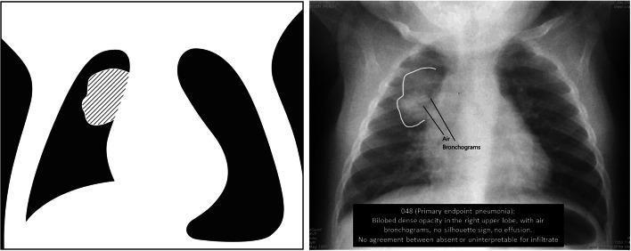
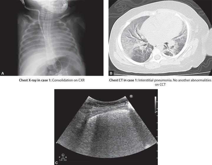
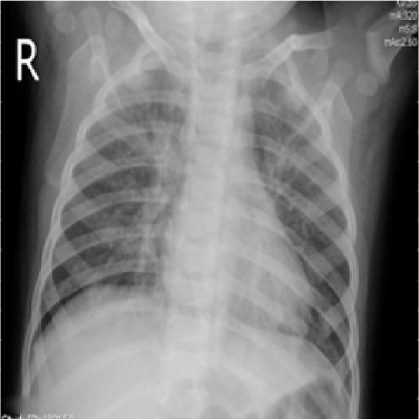
- Chest X-ray - shows consolidation or interstitial infiltrates
- CBC (WBC elevation suggests bacterial cause)
- Blood culture (severe or hospitalized cases)
- Sputum culture if obtainable (>25 leukocytes, <10 squamous epithelial cells per LPF)
- ABG in severe cases
Nursing Problem Priorities
- Impaired gas exchange
- Ineffective airway clearance
- Ineffective breathing pattern
- Hyperthermia
- Risk for fluid volume deficit
- Activity intolerance
- Imbalanced nutrition (less than body requirements)
- Anxiety/fear (child and parent)
- Risk for infection spread / deficient knowledge
Nursing Care Plans (NANDA-I Aligned)
Care Plan 1 - Impaired Gas Exchange
NANDA-I Diagnosis:
Impaired Gas Exchange related to alveolar-capillary membrane changes secondary to inflammatory exudate and consolidation, as evidenced by SpO2 <94%, tachypnea, nasal flaring, and cyanosis.
Expected Outcomes:
- Child maintains SpO2 ≥94% (or per physician target) on room air or supplemental oxygen
- Respiratory rate returns to age-appropriate normal range within 24-48 hours
- Absence of cyanosis and decreased work of breathing
Nursing Interventions and Rationales:
| Intervention | Rationale |
|---|
| Monitor SpO2 continuously or per policy; apply pulse oximetry | Provides early detection of hypoxia before clinical cyanosis appears |
| Administer supplemental oxygen via nasal cannula, face mask, or high-flow nasal cannula as ordered | Corrects hypoxemia; method chosen based on severity and child's tolerance |
| Position child with head of bed elevated 30-45° (or in parent's lap for infants if tolerated) | Promotes optimal chest expansion and diaphragmatic descent; reduces aspiration risk |
| Assess ABGs or capillary blood gas as ordered | Detects hypoxemia, hypercapnia, and acid-base disturbances |
| Monitor for signs of respiratory failure (increasing RR, apnea, severe retractions, altered mental status) | Rapid deterioration can occur in infants; early recognition allows timely escalation |
| Cluster nursing activities to minimize oxygen demand | Reduces metabolic demand and prevents desaturation episodes |
Evaluation:
SpO2 ≥94% maintained, RR within normal range, no cyanosis or increased work of breathing.
Care Plan 2 - Ineffective Airway Clearance
NANDA-I Diagnosis:
Ineffective Airway Clearance related to increased secretions, pain with coughing, and fatigue, as evidenced by abnormal breath sounds (crackles/rhonchi), productive cough, tachypnea, and retractions.
Expected Outcomes:
- Child demonstrates clear or improved breath sounds on auscultation
- Child/caregiver performs effective airway clearance techniques
- No signs of secretion-related airway obstruction
Nursing Interventions and Rationales:
| Intervention | Rationale |
|---|
| Encourage adequate oral/IV fluid intake appropriate for age and weight | Adequate hydration thins secretions and facilitates expectoration |
| Use humidified oxygen when supplemental O2 is indicated | Prevents drying of mucosa and thickening of secretions |
| Perform nasopharyngeal suctioning in infants and young children unable to cough effectively | Young children cannot cough on command; suctioning clears the upper airway |
| Encourage older children to cough deeply every 2-4 hours | Voluntary cough mobilizes secretions toward the upper airway for expectoration |
| Perform chest physiotherapy (percussion and postural drainage) as ordered | Mechanical loosening and gravity-assisted drainage of mucus from affected lung segments |
| Auscultate lung fields before and after airway clearance interventions | Tracks effectiveness of intervention; identifies areas of persistent consolidation |
| Administer mucolytics or bronchodilators as prescribed | May reduce viscosity of secretions and relieve bronchospasm in reactive airways disease |
Evaluation:
Improved or clear breath sounds; effective cough; reduced work of breathing.
Care Plan 3 - Ineffective Breathing Pattern
NANDA-I Diagnosis:
Ineffective Breathing Pattern related to inflammatory process, pain, and fatigue, as evidenced by tachypnea, nasal flaring, intercostal retractions, and grunting.
Expected Outcomes:
- Respiratory rate within age-appropriate normal limits
- Absence of grunting, nasal flaring, and retractions
- Child verbalizes or demonstrates reduced breathing difficulty
Nursing Interventions and Rationales:
| Intervention | Rationale |
|---|
| Assess respiratory rate, rhythm, depth, and effort every 1-4 hours | Frequent monitoring detects early deterioration or improvement |
| Place child in semi-Fowler's or high Fowler's position | Reduces pressure of abdominal contents on diaphragm; promotes lung expansion |
| Encourage slow, deep breathing and breathing exercises (age-appropriate) | Improves lung expansion and prevents atelectasis in cooperative older children |
| Administer analgesics as prescribed if pleuritic pain limits breathing | Pain relief encourages deeper breathing efforts; splinting due to pain worsens hypoventilation |
| Administer antibiotics or antivirals as ordered; monitor response | Treating the underlying infection reduces the inflammatory burden on the airways |
Evaluation:
RR within normal range; absence of retractions, nasal flaring, and grunting; no accessory muscle use.
Care Plan 4 - Hyperthermia
NANDA-I Diagnosis:
Hyperthermia related to infectious process (bacterial/viral), as evidenced by elevated temperature (>38.5°C), tachycardia, and warm, flushed skin.
Expected Outcomes:
- Temperature maintained within normal range (36.5-37.5°C) within 24-48 hours
- No febrile seizures
- Child remains hydrated and comfortable
Nursing Interventions and Rationales:
| Intervention | Rationale |
|---|
| Monitor temperature every 2-4 hours or per protocol | Tracks fever trend; guides antipyretic therapy |
| Administer antipyretics (paracetamol/acetaminophen or ibuprofen) per weight-based dosing as ordered | Reduces fever and associated discomfort; prevents febrile seizure risk in susceptible children |
| Apply cooling measures as needed (tepid sponging, remove excess clothing/blankets) | Facilitates heat dissipation through evaporation and convection |
| Encourage oral fluids or administer IV fluids as ordered | Fever increases insensible water loss; hydration prevents dehydration |
| Monitor for febrile seizures, especially in children 6 months to 5 years | This age group is at highest risk; prompt recognition enables immediate management |
| Administer antibiotics as prescribed and on schedule | Treating the infection is the definitive management of fever; timely dosing maintains therapeutic drug levels |
Evaluation:
Temperature returns to normal; child afebrile or controlled with medication; no seizure activity.
Care Plan 5 - Risk for Deficient Fluid Volume
NANDA-I Diagnosis:
Risk for Deficient Fluid Volume related to increased insensible losses (fever, tachypnea), decreased oral intake (fatigue, respiratory distress, anorexia).
Expected Outcomes:
- Child maintains adequate hydration (moist mucous membranes, normal skin turgor, urine output ≥1 mL/kg/hr)
- No clinical signs of dehydration
Nursing Interventions and Rationales:
| Intervention | Rationale |
|---|
| Monitor fluid intake and urine output strictly; weigh daily | Provides objective measure of fluid balance |
| Assess for dehydration signs: sunken fontanelle (infants), dry mucous membranes, decreased skin turgor, delayed capillary refill | Early identification prompts IV fluid therapy before severe dehydration |
| Encourage age-appropriate oral fluids (breastmilk, formula for infants; water, oral rehydration solutions, clear liquids) | Maintains hydration while maintaining the child's preferred feeding method |
| Administer IV fluids as ordered if oral intake is inadequate | Bypasses the GI route when the child cannot maintain adequate intake |
| Monitor electrolytes as ordered | Fever, vomiting, poor intake, and increased respiratory losses can cause electrolyte imbalances |
Evaluation:
Child remains hydrated; urine output adequate; no signs of dehydration.
Care Plan 6 - Activity Intolerance
NANDA-I Diagnosis:
Activity Intolerance related to imbalance between oxygen supply and demand secondary to impaired gas exchange, as evidenced by fatigue, SpO2 drop with minimal activity, tachycardia on exertion.
Expected Outcomes:
- Child tolerates age-appropriate activities without significant SpO2 desaturation or tachycardia
- Child demonstrates energy conservation techniques
Nursing Interventions and Rationales:
| Intervention | Rationale |
|---|
| Cluster nursing care activities and provide rest periods between | Reduces overall oxygen demand and prevents fatigue |
| Allow child to assume a position of comfort; encourage rest | Children naturally self-limit activity when unwell; forced rest reduces O2 demand |
| Monitor SpO2 during and after activity | Detects desaturation during exertion |
| Gradually increase activity as tolerated as child improves | Progressive activity rebuilds tolerance without stressing the recovering respiratory system |
| Involve parents in comforting activities (holding, reading) that minimize distress | Reduces crying and agitation, which significantly increase O2 consumption in infants |
Evaluation:
Child tolerates increasing activity levels; SpO2 stable during activity; fatigue decreasing over hospital course.
Care Plan 7 - Imbalanced Nutrition (Less Than Body Requirements)
NANDA-I Diagnosis:
Imbalanced Nutrition: Less Than Body Requirements related to anorexia, fatigue, and increased metabolic demands of illness, as evidenced by decreased food intake and weight loss.
Expected Outcomes:
- Child maintains stable weight during hospitalization
- Child resumes age-appropriate dietary intake as illness resolves
Nursing Interventions and Rationales:
| Intervention | Rationale |
|---|
| Offer small, frequent meals; provide preferred foods within dietary restrictions | Small meals reduce fatigue during eating; preferred foods improve acceptance |
| Encourage breastfeeding in infants; it provides antibodies and optimal nutrition | Breast milk provides immunological support in addition to nutrition |
| Position upright during and after feeding to reduce aspiration risk | Gravity aids in gastric emptying; reduces aspiration risk, especially in ill children |
| Monitor daily weight and document intake | Tracks nutritional adequacy and response to dietary interventions |
| Consult dietitian if intake remains poor beyond 48-72 hours | Specialist guidance helps optimize caloric and nutritional targets during illness |
Evaluation:
Weight maintained or appropriate gain; adequate oral intake; no new aspiration events.
Care Plan 8 - Anxiety and Fear (Child and Parents)
NANDA-I Diagnosis:
Anxiety/Fear related to hospitalization, dyspnea, painful procedures, and uncertainty, as evidenced by crying, clinging to caregiver, parental verbalization of worry.
Expected Outcomes:
- Child appears calmer and less distressed with nursing interventions
- Parents verbalize understanding of child's condition, treatment plan, and expected outcomes
Nursing Interventions and Rationales:
| Intervention | Rationale |
|---|
| Encourage parental presence at bedside (family-centered care) | Parental presence reduces child anxiety, lowers oxygen demand from crying, and improves cooperation |
| Use age-appropriate language and play therapy when explaining procedures | Reduces fear of the unknown; therapeutic play helps children process medical experiences |
| Provide honest, clear explanations to parents about diagnosis, treatment, and expected progress | Informed parents are better partners in care; reduces anxiety from uncertainty |
| Allow child to bring a comfort object (stuffed animal, toy) | Familiar objects reduce separation anxiety and provide emotional security |
| Administer procedural pain management (sucrose for infants, topical anesthetics, distraction) | Reduces pain and fear associated with procedures; builds trust |
Evaluation:
Reduced crying and distress; parents verbalize understanding of illness and treatment; cooperative child.
Care Plan 9 - Deficient Knowledge (Parents/Caregiver)
NANDA-I Diagnosis:
Deficient Knowledge related to unfamiliarity with disease process, home management, and prevention, as evidenced by parental questions and incorrect home management practices.
Expected Outcomes:
- Parents verbalize understanding of pneumonia causes, signs, and symptoms
- Parents demonstrate correct medication administration technique
- Parents identify warning signs requiring immediate return to care
Discharge Teaching Topics:
| Teaching Topic | Key Points |
|---|
| Antibiotic adherence | Complete the full 7-10 day course even if child looks better; stopping early risks relapse and resistance |
| Warning signs | Return immediately for: increasing respiratory distress, SpO2 <92%, inability to drink, seizures, or worsening fever |
| Follow-up | Schedule follow-up visit; repeat CXR only if symptoms do not resolve (not routine) |
| Hydration | Continue encouraging fluids at home; watch for decreased wet diapers |
| Fever management | Weight-based acetaminophen or ibuprofen dosing; avoid aspirin in children |
| Vaccination | Ensure pneumococcal conjugate vaccine (PCV13), Hib, and influenza vaccines are up to date; these reduce CAP incidence |
| Hand hygiene | Teach family proper handwashing to reduce household spread |
| Avoid antibiotic misuse | Do not use leftover antibiotics for future illnesses; avoids antibiotic resistance |
Evaluation:
Parents demonstrate correct administration of medications; correctly identify warning signs; verbalize follow-up plan.
Pediatric-Specific Considerations
- Hospitalization criteria: Infants <3 months with fever, toxic-appearing children, SpO2 <92%, inability to maintain hydration, or failure to improve outpatient therapy require admission. - Swanson's Family Medicine Review, p. 584
- Antibiotic selection: Based on the child's age and clinical picture. Amoxicillin is typically first-line for community-acquired bacterial pneumonia in children. Macrolides cover atypical organisms (Mycoplasma, Chlamydia).
- Viral vs. bacterial: Radiographic infiltrates and CBC do not reliably distinguish viral from bacterial CAP; clinical judgement guides antibiotic use.
- Prevention: PCV13, Hib, and influenza vaccines significantly reduce CAP incidence and severity.
Quick Reference Summary Table
| NANDA Diagnosis | Related to | As Evidenced by | Key Interventions |
|---|
| Impaired Gas Exchange | Alveolar consolidation | SpO2 <94%, cyanosis, tachypnea | O2 therapy, position, monitor ABG |
| Ineffective Airway Clearance | Increased secretions | Rhonchi, productive cough | Suctioning, hydration, CPT, coughing |
| Ineffective Breathing Pattern | Inflammation, pain, fatigue | Tachypnea, retractions, grunting | Positioning, analgesia, antibiotics |
| Hyperthermia | Infectious process | Temp >38.5°C, tachycardia | Antipyretics, fluids, cooling |
| Risk for Fluid Deficit | Fever, decreased intake | Dehydration signs | I&O monitoring, IV/oral fluids |
| Activity Intolerance | O2 supply/demand imbalance | Fatigue, SpO2 drop on exertion | Rest, cluster care, gradual mobilization |
| Imbalanced Nutrition | Anorexia, increased demand | Decreased intake, weight loss | Small frequent meals, breastfeeding |
| Anxiety/Fear | Hospitalization, dyspnea | Crying, clinging | Family-centered care, education |
| Deficient Knowledge | Unfamiliarity with disease | Parental questions | Discharge teaching, medication education |