
Recent updates in diagnosis of endometrial intraepithelial neoplasia and who classification of endometrial hyperplasia for md pathology 10 marks answer in corporate Pathogenesis morphology
endometrial intraepithelial neoplasia EIN classification diagnosis
endometrial hyperplasia with atypia EIN histology
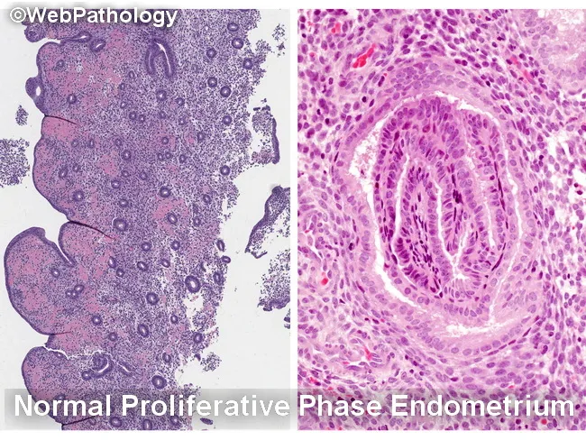
Histology image illustrating the endometrium in two complementary views. The left panel depicts normal proliferative phase endometrium with relatively sparse glands embedded in a cellular stroma, producing a low gland-to-stroma ratio and open gland spacing. The right panel shows characteristic epithelial morphology from proliferative endometrium with elongated, back-to-back glands and prominent, basally oriented nuclei at high power. The specimen emphasizes normal architectural patterns and avoids overcalling hyperplasia when glands are well separated or when fragmentation and telescoping create artifactual crowding. Key differential considerations include endometrial hyperplasia (EH) with or without atypia, disordered proliferative endometrium, reparative or regenerative changes, and endocervical/metaplastic proliferations. Within EH, nonatypical EH must be distinguished from atypical endometrial hyperplasia/endometrioid intraepithelial neoplasia (AEH/EIN), and AEH/EIN must be differentiated from endometrioid carcinoma and from endocervical lesions. Normal proliferative glands show minimal cytologic atypia, whereas AEH/EIN demonstrates architectural complexity (crumpled, crowded glands) and cytologic atypia. Artifacts such as glandular fragmentation, fragmentation- or telescoping-related crowding, and sample artifact can mimic AEH/EIN if the specimen is not intact. This image supports educational differentiation for gynecologic pathology, endometrial biopsy interpretation, and clinical correlation with bleeding patterns and imaging findings. Keywords: endometrium, proliferative phase, hyperplasia, AEH, EIN, histology, biopsy, gland-stroma ratio, artifacts, differential diagnosis teaching.
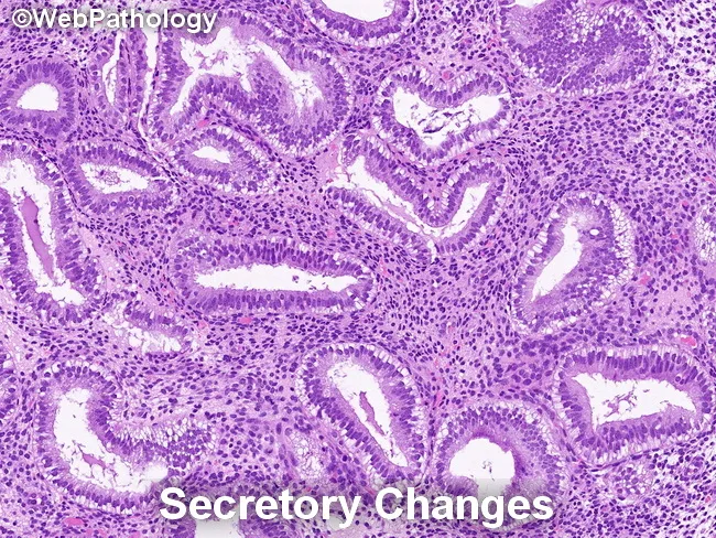
Endometrial histology in secretory phase demonstrated by light microscopy on a hematoxylin and eosin stained endometrial biopsy specimen. Glandular architecture shows irregular serration with gland crowding in focal areas, while glands are lined by tall, columnar secretory epithelium. Nuclei are enlarged and may display mild pleomorphism and loss of polarity, consistent with secretory transformation. The background stroma is edematous and commonly predecidualized, with cytoplasmic vacuolization of stromal cells. Together, these findings reflect physiologic secretory changes rather than overt endometrial hyperplasia. Notably, secretory endometrium can mimic endometrial hyperplasia/endometrioid intraepithelial neoplasia (EIN) in some cases, but the absence of marked architectural crowding, complex glandular patterns, or cytologic atypia argues against a neoplastic process. Differential considerations include secretory change, simple endometrial hyperplasia without atypia, and EIN in a background of secretory endometrium; careful architectural assessment and clinical correlation are essential. Diagnostic significance lies in distinguishing benign secretory transformation from premalignant or malignant entities to avoid overtreatment. This image is educational for pathology training, differential diagnosis exercises, and research on endometrial cycle pathology. Correlate with patient menstrual history, pregnancy status, and cycle timing; repeat sampling or adjunct studies may be warranted when discordance is suspected. Interpret interpretation should integrate histology with clinical data and follow-up.
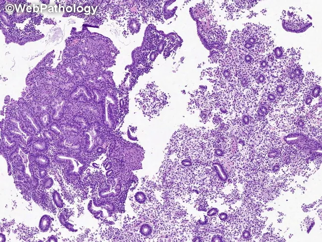
Imaging modality: Light microscopy of a hematoxylin and eosin stained endometrial tissue section, showing features used to diagnose atypical endometrial hyperplasia/endometrioid intraepithelial neoplasia (AEH/EIN). The specimen is endometrium within the uterine cavity; glandular epithelium with stromal backdrop. The histology demonstrates dense glands with back-to-back arrangement, yielding a gland:stroma ratio greater than 1, consistent with AEH/EIN criteria. Glands are cytologically and architecturally atypical relative to adjacent non-atypical glands, with nuclear enlargement, hyperchromasia, and irregular contours. The focus typically exceeds 1 mm in a single linear dimension and commonly comprises more than five to ten glands; multiple foci should not be pooled to satisfy the size criterion. Squamous morular areas are not part of the AEH/EIN assessment and are excluded from this ratio. Artifacts such as gland telescoping or artifactual displacement may create transient density increases but lack true cytologic atypia and architectural disorder. Benign mimics and invasion must be excluded; no myometrial invasion is visible at this plane. This image serves as a reference for diagnosing premalignant endometrial lesions, guiding differential diagnosis (benign hyperplasia without atypia, endometrial carcinoma, adenocarcinoma in situ), and informing clinical management decisions, including surveillance, risk stratification, and potential treatment strategies in gynecologic oncology.
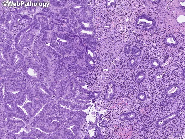
Histology, bright-field microscopy of an H&E stained endometrial tissue section. The specimen represents endometrium with atypical hyperplasia/endometrioid intraepithelial neoplasia (AEH/EIN). In this field, glands exhibit a gland-to-stroma ratio greater than 1, with crowding and architectural complexity consistent with preinvasive endometrial neoplasia. The left half demonstrates crowded, irregular glands that are cytologically and architecturally distinct from the adjacent normal background endometrium on the right. The proliferating glands show mild-to-moderate cytologic atypia, including increased nuclear size, hyperchromasia, and occasional nuclear stratification, while maintaining overall glandular architecture without overt invasion. Glands that are cystically dilated, atrophic glands, or glands with squamous morular metaplasia are absent, helping to differentiate AEH/EIN from benign mimics. The lesion focus exceeds 1 mm in greatest dimension, satisfying the size criterion for AEH/EIN. Benign mimics such as polyps, metaplasias, disordered proliferative endometrium, fragmentation, telescoping artifact, repair, and noninvasive patterns are excluded in favor of a precursor lesion. This histologic pattern denotes a premalignant endometrial condition with significant risk of progression to endometrioid adenocarcinoma if untreated. Clinical risk factors include prolonged estrogen exposure, obesity, and nulliparity; management typically involves careful surveillance or surgical intervention depending on clinical context. This finding aids risk stratification and informs targeted management decisions for patients.
Endometrial Intraepithelial Neoplasia (EIN) and WHO Classification of Endometrial Hyperplasia
Introduction
Classification Systems: Historical to Current
Old WHO 1994 Classification (Four-Tier System)
| Category | Risk of Progression to Carcinoma |
|---|---|
| Simple hyperplasia without atypia | ~1% |
| Complex hyperplasia without atypia | ~3% |
| Simple atypical hyperplasia | ~8% |
| Complex atypical hyperplasia | ~29% |
New WHO 2014/Current Two-Tier Classification
| WHO Category | Former Equivalent | Risk of Carcinoma |
|---|---|---|
| Hyperplasia without atypia | Simple + Complex hyperplasia without atypia | ~1-3% |
| Atypical hyperplasia / EIN | Simple + Complex atypical hyperplasia | ~25-45% (up to 40-50% concurrent carcinoma at hysterectomy) |
The EIN Concept: Background and Rationale
- Endometrial hyperplasia with atypia (atypical hyperplasia) represents a clonal neoplastic proliferation, not merely reactive hyperplasia.
- These lesions share specific molecular driver mutations with endometrioid endometrial carcinoma - particularly PTEN mutations.
- Risk stratification for progression is better predicted by the presence/absence of atypia and clonality than by architectural complexity alone.
- Benign endometrial hyperplasia = nonatypical hyperplasia (polyclonal, estrogen-driven, low malignant potential)
- EIN = premalignant monoclonal neoplasia, replaces "atypical hyperplasia"
- Well-differentiated endometrioid adenocarcinoma = the malignant end of the spectrum
Pathogenesis
Hormonal/Estrogenic Pathway
- Obesity - peripheral aromatization of androgens to estrogens in adipose tissue is the most common cause
- Anovulatory cycles - failure of corpus luteum formation (perimenopause, PCOS)
- Polycystic ovarian syndrome (PCOS)
- Functioning granulosa-theca cell tumors of the ovary (estrogen-secreting)
- Exogenous estrogen - hormone replacement therapy without progestin, tamoxifen (partial agonist effect on endometrium)
- Excessive ovarian cortical function (cortical stromal hyperplasia)
- Cowden syndrome (germline PTEN mutations) - markedly elevated risk
Molecular Pathogenesis of EIN
- PTEN encodes a lipid phosphatase that negatively regulates the PI3K/AKT signaling pathway
- Loss of PTEN function leads to overactivation of PI3K/AKT, promoting cell proliferation and survival
- PTEN mutations are found in >20% of hyperplasias (both with and without atypia) and in 30-80% of endometrial carcinomas
- This shared mutation landscape is the molecular basis for the precursor relationship between EIN and endometrioid carcinoma
- Of note: PI3K/AKT signaling enhances estrogen receptor-mediated transcription, creating a synergistic pro-proliferative loop when PTEN is lost
- Microsatellite instability (MSI) - mismatch repair gene alterations (seen in Lynch syndrome-associated cases)
- KRAS mutations - found in a subset
- PAX2 loss - PAX2 immunohistochemistry can be used diagnostically (see below)
- p53 mutations - rare in EIN (more characteristic of Type II serous carcinoma pathway)
- MLH1 promoter methylation - epigenetic silencing of mismatch repair genes
Morphology
Hyperplasia Without Atypia
- The cardinal feature is an increased gland-to-stroma ratio compared with normal proliferative endometrium
- Glands show variation in size and shape, may be cystically dilated
- Glandular outlines range from smooth (simple pattern) to irregular and branching (complex pattern)
- Some intervening stroma is retained even with back-to-back glands focally
- No nuclear atypia - cells are tall columnar with pseudostratified, basally oriented nuclei; mitoses may be present but are not atypical
- May undergo cystic atrophy when estrogen is withdrawn
Atypical Hyperplasia / EIN
- Gland-to-stroma ratio >1 (glands occupy >50% of the lesion volume) - this is a key EIN criterion
- Glands are back-to-back with minimal intervening stroma
- Complex, irregular outlines due to branching, budding, and intraluminal tufting
- Represent clonal outgrowths - cytologically distinct from the background endometrium
- Lesion size criterion: >1 mm in greatest linear dimension (to separate from tangential sectioning artifacts)
- Nuclear rounding and enlargement - cells lose their normal elongated shape and perpendicular orientation to the basement membrane
- Open (vesicular) chromatin with prominent nucleoli
- Nuclear crowding and loss of polarity
- Cytoplasmic eosinophilia with increased cytoplasm-to-nucleus ratio
- Cells are cytologically different from the background non-atypical glands - this contrast is essential for diagnosis
| Criterion | Description |
|---|---|
| Demographics | Exclude mimics (benign, carcinoma) |
| Excluded benign conditions | Polyp, secretory changes, repair, lower uterine segment |
| Minimal size | >1 mm in greatest dimension |
| Outgrown stroma | Area of glands >stroma (gland:stroma ratio >1) |
| Nuclear cytology altered** | Cytologically distinct from background endometrium |
Distinction from Well-Differentiated Endometrioid Carcinoma
- Atypical hyperplasia/EIN and well-differentiated endometrioid adenocarcinoma share considerable morphologic and genetic overlap
- Features favoring invasion/carcinoma: desmoplastic stromal reaction, irregular infiltration, stromal foam cells, papillary patterns without fibrovascular cores
- Up to 40% of patients with a biopsy diagnosis of atypical hyperplasia/EIN are found to have concurrent carcinoma at hysterectomy - this is a critical clinical point
- Definitive distinction often requires hysterectomy specimen evaluation
Recent Updates in Diagnosis of EIN
1. WHO 2020 Classification Update
2. Immunohistochemistry in EIN Diagnosis
- PTEN IHC: Loss of PTEN expression (absent brown nuclear staining) marks clonal EIN foci against PTEN-positive background - useful when morphologic distinction is difficult. A PTEN-null gland cluster is strong evidence of EIN even with borderline morphology.
- PAX2 IHC: PAX2 (a transcription factor) is expressed in normal endometrial glands but is lost in EIN - aberrant loss serves as a marker of clonal neoplasia. Used alongside PTEN.
- MLH1 IHC: Loss indicates mismatch repair deficiency - important for Lynch syndrome screening.
- Ki-67: Elevated proliferative index in EIN compared to nonatypical hyperplasia.
- p53: Diffuse strong positivity would raise concern for serous EIC (a different entity, not the EIN pathway).
3. Molecular Classification (TCGA-based)
- POLE-ultramutated subset: favorable prognosis even when progressing to carcinoma
- MMR-deficient (MSI-high): Lynch syndrome-associated; ~10-15% of EIN cases
- Copy number-low (NSMP): Most common, corresponds to classic estrogen-driven pathway
- TP53-mutant/copy number-high: Rare in true EIN; suggests possible reclassification as serous precursor
4. EIN vs. Serous Endometrial Intraepithelial Carcinoma (SEIC)
| Feature | EIN (Endometrioid) | SEIC (Serous) |
|---|---|---|
| Synonyms | Atypical hyperplasia/EIN | Endometrial intraepithelial carcinoma |
| Pathway | Type I / estrogen-driven | Type II / estrogen-independent |
| Background | Hyperplastic endometrium | Atrophic endometrium |
| Nuclear atypia | Mild-moderate | Severe, pleomorphic |
| p53 IHC | Wild-type | Aberrant (diffuse or null) |
| TP53 mutation | Absent | Present (>90%) |
| Malignant potential | Precursor (years) | Already behaves as carcinoma (spreads without myometrial invasion) |
| Treatment | Hysterectomy | Hysterectomy with full staging |
5. Diagnostic Challenges and Updates
- Interobserver reproducibility: Even with the two-tier system, distinction of EIN from nonatypical hyperplasia has moderate interobserver agreement (kappa ~0.4-0.6). Panel IHC (PTEN + PAX2) improves reproducibility.
- Fragmented biopsy specimens: A common pitfall - glandular crowding due to tissue fragmentation must not be mistaken for EIN. The size criterion (>1 mm) and cytologic contrast from background are essential safeguards.
- Lower uterine segment (LUS) sampling: LUS glands are naturally more compact and can mimic EIN - anatomic site awareness is critical.
- Lynch syndrome screening: Per current guidelines, all EIN/atypical hyperplasia specimens should undergo MMR IHC (MLH1, MSH2, MSH6, PMS2) as a screening step for Lynch syndrome.
Clinical Implications and Management
| Diagnosis | Risk | Management |
|---|---|---|
| Hyperplasia without atypia | 1-3% | Progestin therapy (oral/LNG-IUD), observation, repeat biopsy |
| EIN / Atypical hyperplasia | ~25-45% + ~40-50% concurrent carcinoma | Hysterectomy (definitive); progestin + LNG-IUD if fertility preservation desired |
- LNG-IUD (levonorgestrel intrauterine device) is the preferred medical treatment for EIN when fertility preservation is needed, with regression rates superior to oral progestins (supported by 2024 meta-analysis, Suzuki et al., JNCI 2024, PMID 38305500).
- Repeat biopsy every 3 months is mandatory during conservative management.
- Recurrence risk after progestin therapy approaches 50%.
Histologic Images
Summary / Key Points for Exam
- WHO 2014/2020 uses a two-tier system: hyperplasia without atypia vs. atypical hyperplasia/EIN - replacing the older four-tier 1994 system.
- EIN = clonal premalignant lesion driven by PTEN loss and unopposed estrogen; morphologically defined by gland:stroma >1 + cytologic atypia + cytologic contrast from background + size >1 mm.
- Pathogenesis: prolonged estrogen excess → PTEN mutation (PI3K/AKT activation) → clonal EIN → endometrioid carcinoma.
- 40-50% concurrent carcinoma risk at hysterectomy in EIN/atypical hyperplasia.
- IHC panel (PTEN loss, PAX2 loss, MMR proteins, p53) now integral to diagnosis and molecular triage.
- SEIC (serous intraepithelial carcinoma) is a separate entity: p53-aberrant, arises in atrophic endometrium, spreads without myometrial invasion.
- Management: hysterectomy for EIN (definitive); LNG-IUD preferred for fertility preservation.