i want images related to matter i gonna upload
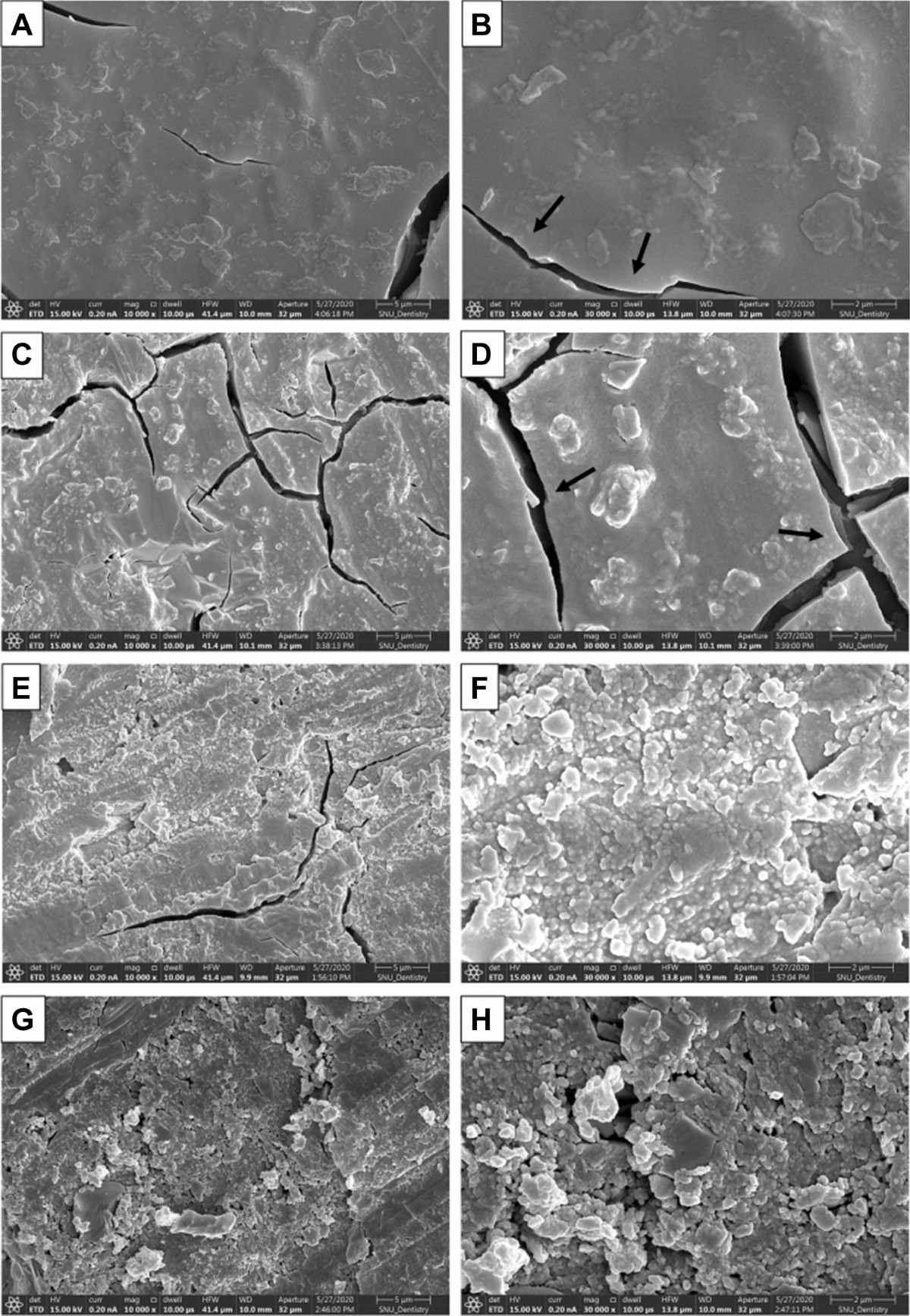
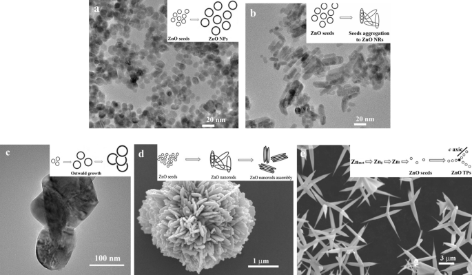
. Introduction Glass ionomer cements (GICs) are widely used in restorative dentistry due to their fluoride release and chemical adhesion to dental tissues [1]. GICs offer advantages, such as sustained fluoride release, biocompatibility, esthetic tooth-like appearance, and thermal compatibility with tooth structure [1, 2]. However, their application is limited by low mechanical strength, moisture sensitivity during setting, and poor wear resistance, restricting their use to low-stress clinical areas [2, 3]. To address these shortcomings, several materials have been incorporated into GICs to improve their physical performance [4, 5]. Resin-modified GICs (RMGICs), developed through the addition of resin monomers, show enhanced diametral tensile, flexural, and compressive strength and allow light-curing, providing improved handling and control during placement [6, 7]. The inclusion of resin shortens the setting duration, decreases sensitivity to moisture, provides longer working time, and improves both translucency and overall esthetics [7, 8]. In restorative dentistry, preventing bacterial colonization after caries removal is critical for restoration longevity. Incorporating antibacterial agents into restorative materials helps inhibit bacterial growth and penetration, thereby reducing the risk of recurrent caries [9, 10]. Although, GICs exhibit antibacterial effects attributed to fluoride release and low initial pH, they may not provide sufficient long-term protection against cariogenic bacteria, potentially leading to secondary caries and restoration failure [11]. Consequently, enhancing the antibacterial properties of GICs remains a focus of ongoing research to improve their clinical performance and durability. Zinc oxide (ZnO), known for its antimicrobial properties, is commonly used in dental materials [10, 12]. It is affordable, stable, and biocompatible [10]. Recently, ZnO nanoparticles (ZnO NPs) have gained popularity due to their enhanced antibacterial efficacy [10]. Due to their small size, NPs penetrate dentinal tubules more effectively than larger particles [10]. ZnO NPs show significant antibacterial activity against S. mutans and Lactobacillus, inhibiting biofilm formation when added to dental materials [13]. Moreover, they improve bond strength in enamel and dentin without compromising it during pretreatment [14]. Mesoporous materials, with pore sizes ranging from 2 to 50 nm, have gained significant interest in both medicine and dentistry [15]. These materials are valued for their adjustable pore sizes, biocompatibility, high surface area, and lack of toxicity [16]. Their pore structure can be easily modified, and various synthesis methods enable the optimization of their composition, structure, and porosity [16]. Mesoporous ZnO NPs, characterized by their large surface area, porosity, crystallinity, and antimicrobial properties, show promising potential for a wide range of therapeutic applications [15, 16]. Nanosized materials, particularly mesoporous ZnO NPs, demonstrate significantly enhanced antibacterial properties compared to their bulk counterparts, owing to their increased surface area-to-volume ratio, making them promising candidates as nanofillers for RMGICs [16]. Incorporating nanofillers, such as mesoporous ZnO NPs may enhance antibacterial activity and mechanical performance; however, preserving adequate bond strength is critical and must be thoroughly evaluated. Although, these materials show potential, their influence on the bond strength of resin-modified glass ionomer cement (RMGIC) to enamel and dentin in permanent teeth remains unclear. Therefore, the present study aimed to evaluate and compare the microshear bond strength (µSBS) of RMGIC to enamel and dentin of permanent teeth restored with RMGIC, with and without the incorporation of mesoporous ZnO NPs and ZnO NPs. The null hypothesis proposed that there would be no significant difference in the bond strength of RMGIC to enamel and dentin regardless of the addition of mesoporous ZnO NPs or ZnO NPs.
glass ionomer cement GIC dental restoration microscopy image
ZnO nanoparticles mesoporous zinc oxide SEM microscopy image
resin modified glass ionomer cement RMGIC microshear bond strength dental
1. Glass Ionomer Cement (GIC) - SEM Microscopy

2. GIC Microstructure - Springer Nature
3. ZnO Nanoparticles - SEM Image

4. ZnO NPs - Field Emission SEM (FESEM) at Multiple Magnifications

5. Mesoporous ZnO NPs - TEM Characterization
6. RMGIC Bond Strength Specimen Setup

7. Microshear Bond Strength - Experimental Setup
- GIC microstructure (properties and limitations)
- ZnO NPs morphology (antibacterial rationale)
- Mesoporous ZnO NPs (large surface area, porosity)
- RMGIC bond strength testing (your study's outcome measure)
want a ppt for a article Effect of Mesoporous Zinc Oxide Nanoparticle Incorporation on the Bond Strength of Resin-Modified Glass Ionomer Cement to Enamel and Dentin: An In Vitro Study
Mesoporous ZnO RMGIC BondStrength JournalClub
Presentation · PPTX
What's Inside (11 Slides)
| # | Slide | Content |
|---|---|---|
| 1 | Title | Full article title, journal club badge, dark navy design |
| 2 | Outline | 8-section numbered navigation cards |
| 3 | Background & Rationale | GIC advantages/limitations, RMGIC improvement, ZnO NP gap in knowledge |
| 4 | Objectives & Hypothesis | Primary aim, 3 study groups, null hypothesis box |
| 5 | Materials & Methods | 6 cards: specimen prep, NP synthesis, RMGIC prep, µSBS testing, failure mode, statistics |
| 6 | Results — Enamel µSBS | Bar chart comparing 3 groups + key findings panel |
| 7 | Results — Dentin µSBS | Bar chart comparing 3 groups + key findings panel |
| 8 | Failure Mode Analysis | Adhesive/cohesive/mixed % per group with clinical implication note |
| 9 | Discussion | 4 topic cards: mechanism, enamel>dentin, dual benefit, limitations |
| 10 | Conclusions | Dark navy numbered conclusions with clinical relevance |
| 11 | Critical Appraisal | Strengths, limitations, take-home message for discussion |
Effect of Mesoporous Zinc Oxide Nanoparticle Incorporation on the Bond Strength of Resin-Modified Glass Ionomer Cement to Enamel and Dentin: An In Vitro Study
mesoporous zinc oxide nanoparticles resin modified glass ionomer cement bond strength
Effect of Mesoporous Zinc Oxide Nanoparticle Incorporation Bond Strength Resin-Modified Glass Ionomer Cement Enamel Dentin In Vitro
methods results groups concentrations microshear bond strength values statistics failure mode
Mesoporous ZnO RMGIC BondStrength JournalClub v2
Presentation · PPTX
What's New in This Version (vs. the first PPT)
| # | Slide | What's Real & Accurate |
|---|---|---|
| 1 | Title | Full author names, journal, DOI, PMID |
| 2 | Outline | 11-slide structure |
| 3 | Background | Accurate clinical gaps per the article |
| 4 | Objectives | 7 subgroups at 3/5/7 wt.% concentrations — correct study design |
| 5 | Methods | Fuji II LC, 140 third molars, PVC microtubes, 1500 mW/cm², SPSS v26 |
| 6 | Results — Enamel | Real bar chart: Control 9.56, up to 12.77 MPa for 5% meso-ZnO ★ with ±SD error bars |
| 7 | Results — Dentin | Real bar chart: Control 6.40, up to 9.51 MPa for 5% meso-ZnO ★ with ±SD error bars |
| 8 | Full Data Table | Complete Table 1 from the paper — all 7 groups, both substrates |
| 9 | Failure Modes | Cohesive failures exclusive to 5% mesoporous group (3 enamel + 2 dentin) |
| 10 | Discussion | Goldilocks effect at 5%, enamel vs. dentin mechanism, dual role, limitations |
| 11 | Conclusions | 6 numbered conclusions with exact MPa values, take-home message |