
A 42 years old obese lady presented with local upper abdominal pain and vague fever for last seven days. Pain worse with meals. She also complains of nausea and anorexia. Her look is non-toxic and she is comfortable during exam. On examination, epigastrium is moderately tender. So what should be the diagnosis?
cholecystitis gallstones ultrasound upper abdominal pain
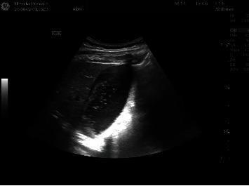
This abdominal diagnostic ultrasound image focuses on the right upper quadrant, specifically visualizing the gallbladder and liver parenchyma. The gallbladder is prominently distended with anechoic contents. Notably, the ultrasound lacks classic diagnostic signs of acute cholecystitis, such as gallbladder wall thickening (greater than 3mm), pericholecystic fluid, or the presence of intraluminal gallstones. In the perihepatic space, there is a small, localized collection of hypoechoic fluid, described as a minimal perihepatic fluid pocket. The adjacent hepatic tissue appears relatively homogenous. This image is clinically relevant for teaching the differential diagnosis of abdominal pain in obstetric or general surgical patients, where distention may occur in the absence of primary inflammatory cholecystitis. It highlights the importance of distinguishing between simple gallbladder distension and acute cholecystitis during sonographic evaluation.
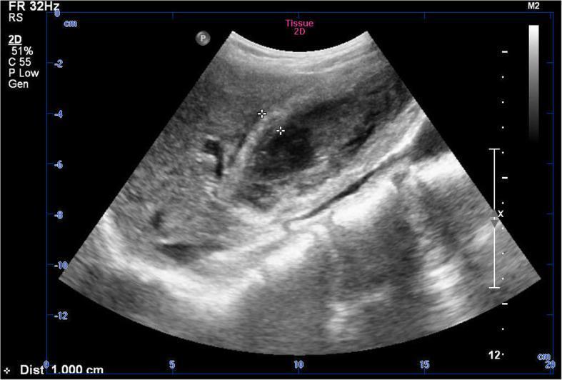
This diagnostic image is a 2D grayscale abdominal ultrasound focusing on the gallbladder. The scan reveals a significantly thickened gallbladder wall, highlighted by calipers measuring a distance of approximately 1.000 cm. Within the gallbladder lumen, there is a prominent, irregular hyperechoic structure that lacks the acoustic shadowing typically associated with gallstones, suggesting intraluminal material such as gallbladder sludge, hemorrhage, or organized clots. The surrounding hepatic parenchyma appears largely homogeneous. This imaging is clinically significant for evaluating cholecystitis, specifically hemorrhagic cholecystitis, as it demonstrates inflammatory wall changes and atypical intraluminal echoes. The visual evidence supports the differential diagnosis between inflammatory processes and potential biliary malignancy in a patient presenting with right upper quadrant pain.
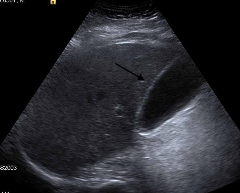
This abdominal ultrasound image shows a longitudinal view of the gallbladder and adjacent liver parenchyma. The gallbladder appears as a clear, ovoid, anechoic (black) structure, indicating it is filled with fluid (bile) without internal echoes, which is consistent with the absence of cholelithiasis (gallstones) or sludge. The gallbladder wall is thin and hyperechoic (bright), showing no signs of thickening or pericholecystic fluid, which helps rule out acute cholecystitis in a clinical setting of abdominal pain. The liver parenchyma exhibits a normal, homogeneous intermediate echogenicity. Posterior acoustic enhancement is visible behind the anechoic gallbladder, a common artifact of fluid-filled structures. This diagnostic image is used to evaluate the biliary system in patients presenting with symptoms such as epigastric or right upper quadrant pain, particularly to differentiate between biliary etiologies and other conditions like acute pancreatitis.
Diagnosis: Chronic Cholecystitis with Cholelithiasis (Gallstones)
Why This Patient Fits the Classic Profile
Symptom-by-Symptom Analysis
| Feature | Significance |
|---|---|
| Obese female, 42 yrs | Classic gallstone demographic |
| Upper abdominal / epigastric pain x 7 days | Typical location for biliary pain (RUQ or epigastric) |
| Pain worse with meals (especially fatty foods) | Postprandial pain = hallmark of biliary disease ("qualitative dyspepsia") |
| Nausea, anorexia | Common in cholecystitis; vomiting is less frequent than in peptic ulcer |
| Vague low-grade fever | Reflects mild gallbladder wall inflammation |
| Non-toxic appearance, comfortable at rest | Suggests chronic/subacute process, not acute emergency |
| Epigastric tenderness on exam | Consistent with inflamed gallbladder |
Key Clinical Sign to Look For
- Murphy's sign - inspiratory arrest on deep palpation of the RUQ while the patient inhales. Sensitivity ~65%, specificity ~87%, LR+ 5.0 (Symptom to Diagnosis, 4th Ed.). Its absence does not exclude cholecystitis.
Working Diagnosis Hierarchy
- Most likely: Chronic cholecystitis with cholelithiasis - prolonged history (7 days of dull pain), non-toxic appearance, postprandial worsening, low-grade fever
- Alternative: Acute cholecystitis - if fever/WBC elevated and Murphy's sign positive; develops from prolonged cystic duct obstruction (>4-6 hours)
- Rule out: Acute pancreatitis - epigastric pain radiating to the back, markedly elevated lipase; more severe presentation
- Rule out: Peptic ulcer disease - pain often relieved (not worsened) by food, different character
- Rule out: Acute hepatitis - elevated liver enzymes, jaundice, systemic symptoms
Investigations Needed
- Abdominal ultrasound (first-line) - gold standard for gallstones; also looks for gallbladder wall thickening (>3mm), pericholecystic fluid, acoustic shadowing
- CBC - leukocytosis present in ~52-63% of acute cholecystitis
- LFTs, lipase, amylase - to rule out CBD stone, cholangitis, pancreatitis
- CRP - elevated in inflammation
Ultrasound Example