
Give me easy diagram for parotid gland
Reading File
Finding Sources
Reading File
Reading File
I found two great diagrams in Gray's Anatomy for Students. Let me fetch both images.
Loading Image
Loading Image
Here are two clear diagrams of the parotid gland from Gray's Anatomy for Students:
Diagram 1 - Position & Relations (Simple Overview)
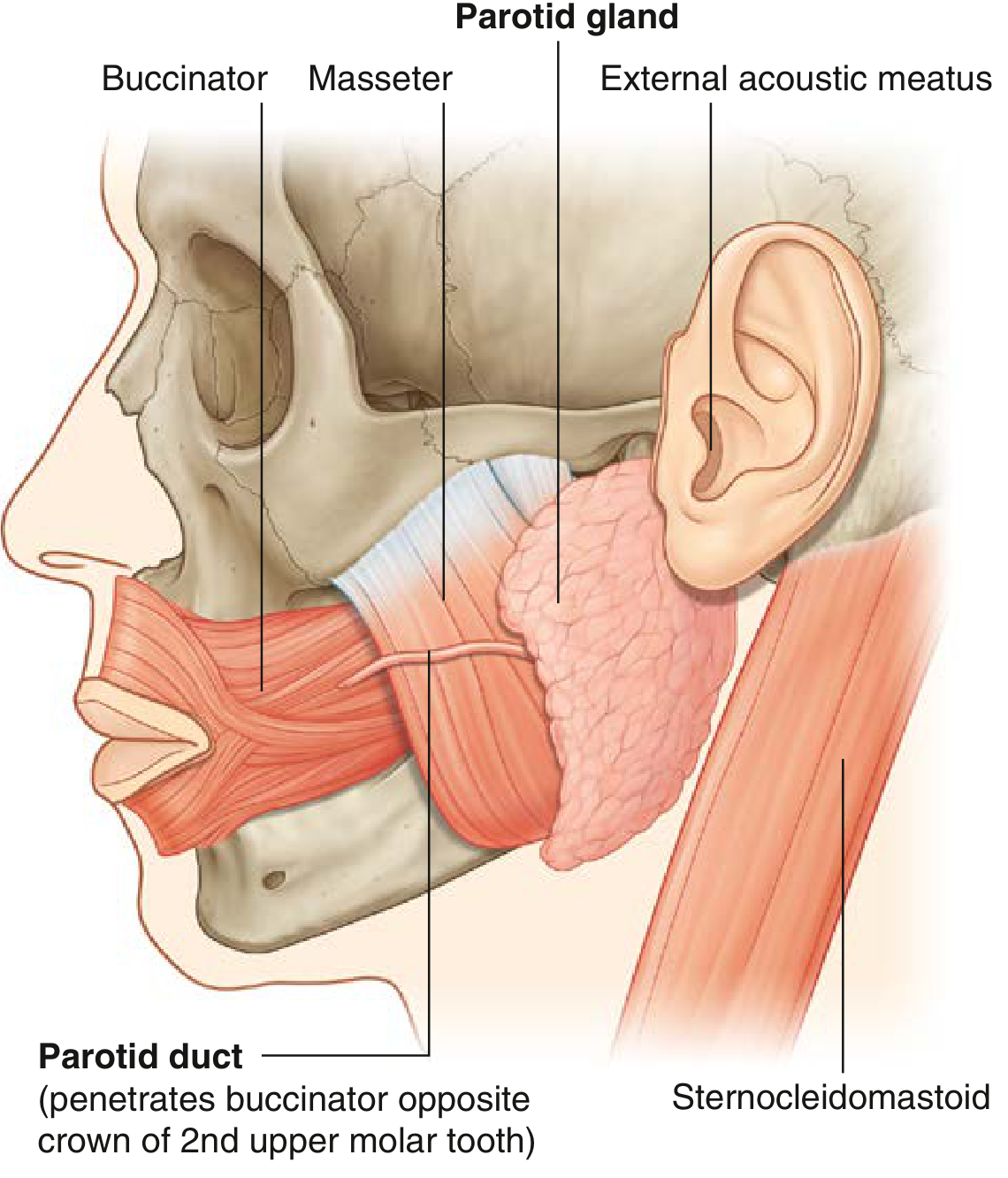
This shows the position of the parotid gland and its key neighbors:
- Sits anterior and inferior to the ear, overlying the masseter muscle
- Bounded by the sternocleidomastoid (behind) and ramus of mandible (front)
- The parotid duct crosses the masseter and pierces the buccinator to open opposite the crown of the 2nd upper molar tooth
Diagram 2 - Detailed Relations (Lateral + Cross-Section)
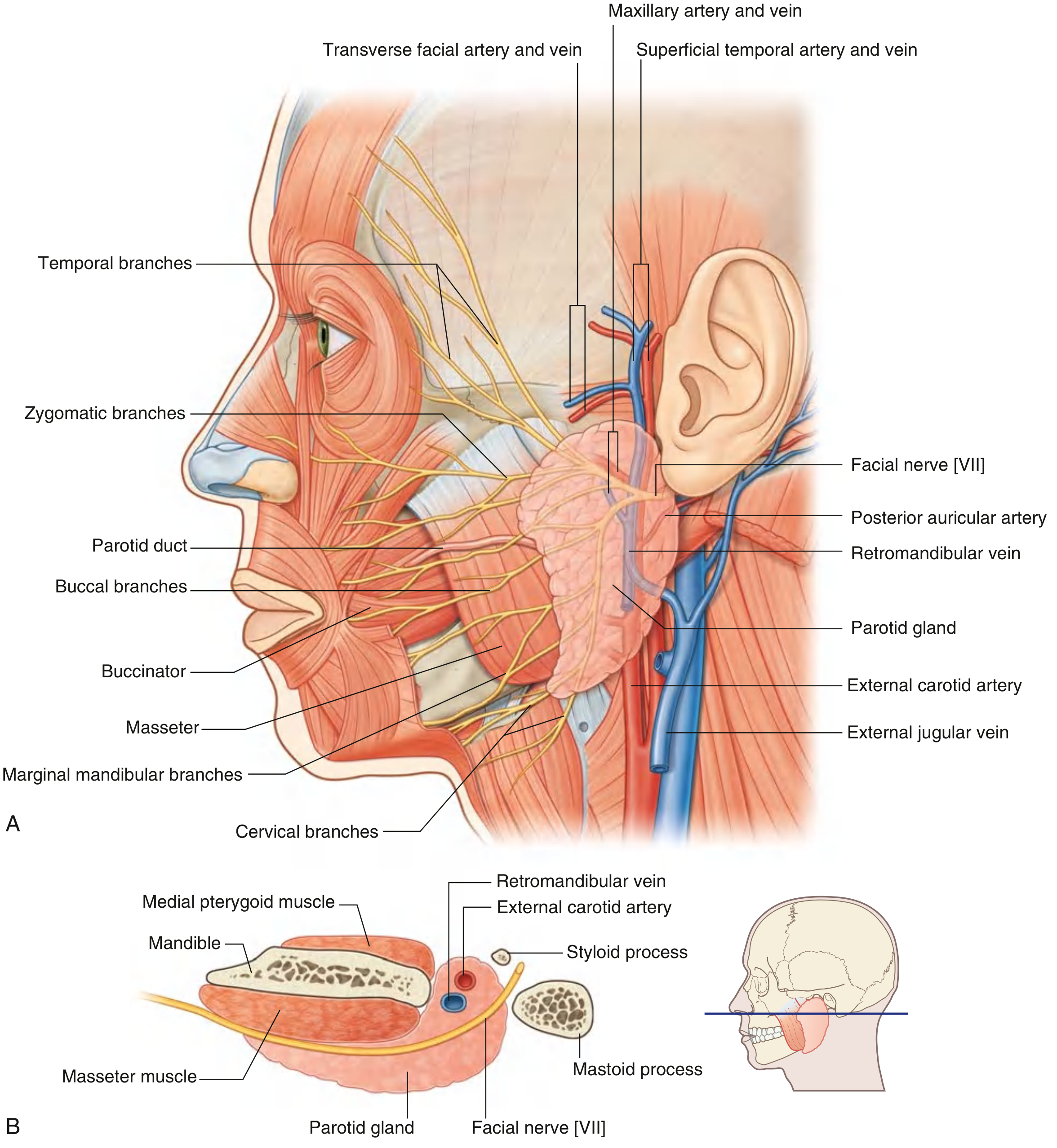
(A) Lateral view shows the facial nerve [VII] branches fanning out from the gland:
- Temporal, Zygomatic, Buccal, Marginal mandibular, Cervical branches
(B) Cross-section shows structures passing through the gland (from superficial to deep):
| Structure | Notes |
|---|---|
| Facial nerve [VII] | Most superficial - exits stylomastoid foramen, divides inside gland |
| Retromandibular vein | Formed by maxillary + superficial temporal veins |
| External carotid artery | Divides into maxillary + superficial temporal arteries inside gland |
Quick Summary
| Feature | Detail |
|---|---|
| Type | Largest salivary gland (serous) |
| Location | Pre-auricular - anterior/inferior to ear |
| Duct | Stensen's duct - opens at 2nd upper molar |
| Key nerve | Facial nerve [VII] - divides within it |
| Key vessels | External carotid artery, retromandibular vein |
| Innervation (secretomotor) | Glossopharyngeal nerve [IX] via lesser petrosal nerve |
Source: Gray's Anatomy for Students, Figs. 8.65 & 8.267
This is a shared conversation. Sign in to Orris to start your own chat.