
Tinea sorosis
- Tinea capitis - scalp ringworm
- Tinea corporis - ringworm of the body
- Tinea unguium (onychomycosis) - nail fungal infection
- Tinea versicolor (Pityriasis versicolor) - a common skin fungal condition
Tinea sorosis dermatology fungal infection
"Tinea Sorosis" - Not a Recognized Medical Term
- Standard dermatology references (Fitzpatrick's, Andrews' Diseases of the Skin)
- Medical microbiology textbooks (Jawetz)
- Clinical databases or peer-reviewed literature
Recognized Tinea Infections (by body site)
| Name | Site |
|---|---|
| Tinea capitis | Scalp and hair |
| Tinea corporis | Body/trunk (ringworm) |
| Tinea cruris | Groin ("jock itch") |
| Tinea pedis | Feet ("athlete's foot") |
| Tinea manuum | Hands |
| Tinea unguium | Nails (onychomycosis) |
| Tinea barbae | Beard area |
| Tinea faciei | Face |
| Tinea favosa | Scalp (favus type) |
| Tinea nigra | Palms/soles (dark pigmentation) |
| Tinea versicolor | Trunk (actually caused by Malassezia, not a dermatophyte) |
Could You Mean Something Else?
- Tinea circinata - another name for tinea corporis (circular lesions)
- Tinea incognito - tinea whose appearance is altered by incorrect steroid treatment
- Sycosis (not "sorosis") - tinea barbae is sometimes called tinea sycosis, referring to follicular involvement of the beard
Could this be a misspelling or regional term?
Most Probable Matches
1. Tinea Sycosis (closest phonetic match)
- "Sorosis" → "Sycosis" is very plausible (regional accent or phonetic mishearing)
- Tinea sycosis = tinea barbae with deep follicular involvement of the beard/mustache area
- Caused by Trichophyton species
- Presents as pustular, boggy, inflammatory plaques in the beard region
2. Tinea Circinata (spelling confusion)
- "Sorosis" could be a corruption of "circinata" in non-English speaking regions
- Another name for tinea corporis - the classic ring-shaped body ringworm
3. Tinea Versicolor (color-related confusion)
- "Sorosis" is actually the name of a type of fruit (a fleshy multiple fruit like pineapple/mulberry)
- Some regional languages use descriptive fruit/color-based nicknames for skin conditions
- Tinea versicolor causes multicolored/varied patches, which might evoke such a name colloquially
4. Tinea Scyosis / Scosis (orthographic variant)
- Some older or non-English dermatology texts spell sycosis differently
Most Likely Answer
Corporis
tinea corporis ringworm skin lesion
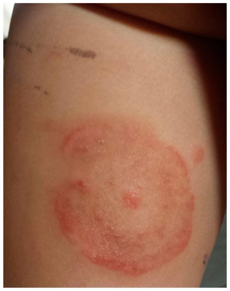
This clinical photograph displays a dermatological presentation of tinea corporis (ringworm) in a pediatric patient. The primary feature is an annular, erythematous plaque located on the trunk or proximal limb. The lesion is characterized by a well-demarcated, raised red border with evidence of mild scaling or vesiculation, indicating active peripheral growth. The center of the plaque shows partial clearing with a lighter pink hue, though it remains occupied by a few scattered inflammatory papules. Above the primary lesion, incidental linear hyperpigmented marks are visible, likely representing excoriations or minor superficial trauma. The image serves as a classic educational example of a fungal skin infection, specifically demonstrating the centrifugal spread and central clearing characteristic of Microsporum canis dermatophytosis.
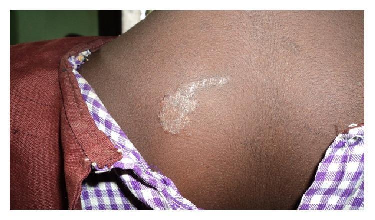
Clinical photograph of a skin lesion on the upper back of a pediatric patient with a dark skin tone, consistent with Tinea corporis (ringworm). The image demonstrates a hypopigmented, well-circumscribed patch with an irregular, slightly raised, and scaly border. Centrally, the lesion shows characteristic clearing but maintains a whitish, fine-textured scaling. A linear extension of the scaling is visible, trailing away from the primary circular focus. This presentation illustrates a common superficial fungal infection (dermatophytosis) of the glabrous skin. The anatomical location is the posterior trunk near the right scapular region. The visual features are diagnostic of a fungal etiology, highlighting the annular morphology and peripheral scale distribution typically seen in localized tinea infections.
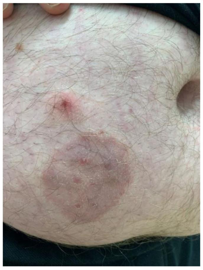
This clinical photograph displays a dermatological presentation of tinea corporis (zoonotic dermatophytosis) on the abdomen of an adult male. The primary feature is a well-circumscribed, annular, erythematous plaque approximately 4-5 cm in diameter. The lesion exhibits a classic 'ringworm' morphology with a slightly raised, scaly inflammatory border and a partially cleared central area containing subtle papular elements. Superior to the primary plaque is a secondary, smaller, raised erythematous papule with a central punctum or crust. The surrounding skin shows mild diffuse erythema and scattered smaller inflammatory macules. Hair follicles are visible throughout the affected and unaffected regions. This image serves as an educational example of fungal infection (likely Trichophyton mentagrophytes) transmitted from a pet (guinea pig) to a human host, emphasizing the visual diagnostic features of annular inflammatory skin lesions.
Tinea Corporis (Tinea Circinata / Ringworm of the Body)
Definition
- Dermatology 2-Volume Set 5e, p.1604
Clinical Features
- Classic lesion: Annular (ring-shaped), sharply circumscribed, slightly erythematous plaque with a raised, scaly, active advancing border and central clearing - this centrifugal spread is the hallmark
- Lesions may widen to many centimeters in diameter
- Concentric circles or polycyclic lesions can form intricate patterns
- Scale is usually present but may be absent if topical corticosteroids have been used (see tinea incognito)
- Pruritus and burning are common associated symptoms
- Incubation period: 1 to 3 weeks
- Pustules within the active border are suggestive of tinea
Causative Organisms
| Type | Key Species | Notes |
|---|---|---|
| Anthropophilic | T. rubrum | Most common overall; can produce concentric rings; causes Majocchi granuloma |
| T. tonsurans | Seen in adults caring for children with tinea capitis | |
| T. indotineae | Terbinafine-resistant; causes large widespread lesions | |
| Zoophilic | M. canis | Pet exposure (cats/dogs); multiple small lesions |
| T. verrucosum | Cattle exposure; mimics bacterial furunculosis | |
| T. behnamiae | Guinea pig exposure | |
| Geophilic | M. gypseum | Outdoor/soil exposure; may be bullous |
- Andrews' Diseases of the Skin, p.347
Clinical Variants
- Tinea incognito - atypical appearance due to topical steroid use; lacks raised scaly border; may have multiple edges with no central clearing
- Majocchi granuloma (nodular perifolliculitis) - deep dermatophyte folliculitis with follicular papulopustules/granulomatous nodules, usually from T. rubrum; common in women who shave their legs with tinea pedis/onychomycosis
- Tinea profunda - excessive inflammatory response, granulomatous/verrucous; may mimic cutaneous TB
- Tinea imbricata - caused by T. concentricum; concentric rings resembling erythema gyratum repens; chronic infections in South Pacific, Asia, Central/South America
- Tinea gladiatorum - in wrestlers, spread via opponents, equipment, and mats
- Bullous tinea - vesicular/bullous lesions, more common on the feet
Diagnosis
- KOH preparation of skin scrapings - look for branching septate hyphae (beware "mosaic false hyphae" artifact at keratinocyte junctions)
- Fungal culture - growth apparent within 1-2 weeks; identifies genus by colony morphology
- Biopsy - for chronic refractory cases (tinea incognita)
- Note: Recent topical antifungal use can cause false-negative KOH/culture
Differential Diagnosis
- Nummular eczema
- Pityriasis rosea
- Tinea versicolor
- Erythema annulare centrifugum
- Granuloma annulare
- Subacute lupus erythematosus
- Impetigo
- Secondary/tertiary syphilis
- Psoriasis, seborrheic dermatitis
Treatment
- Topical antifungals for 2-4 weeks (twice daily): clotrimazole, miconazole, econazole, naftifine, ketoconazole, ciclopirox, butenafine
- Terbinafine (once daily) - course can be shortened to 1 week
- Avoid combination products with potent corticosteroids (e.g., clotrimazole/betamethasone) - these cause widespread tinea and fungal folliculitis
-
Systemic therapy required: griseofulvin, terbinafine, itraconazole, or fluconazole
-
Trichophyton spp. respond better to terbinafine (shorter courses)
-
Microsporum spp. respond better to griseofulvin
-
Andrews' Diseases of the Skin, p.347
Special Considerations
- Immunocompromised patients (e.g., AIDS): widespread tinea corporis may be a presenting sign; lesions can be deep and nodular
- Topical steroid use leads to tinea incognito and deeper follicular infections
- T. indotineae is an emerging concern due to terbinafine resistance, causing large, widespread, difficult-to-treat lesions
Versicolour
tinea versicolor pityriasis versicolor skin discoloration patches
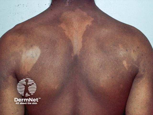
Clinical photography of the back showing large hypopigmented patches consistent with pityriasis versicolor (tinea versicolor). The dorsal trunk demonstrates irregular, well-demarcated macules with reduced pigment relative to surrounding skin and slight surface scaling. Lesions are primarily on the upper back and shoulders, sometimes extending centrally, with a predominantly hypo pigmented appearance in darker skin tones and mixed hypo/hyperpigmentation in lighter tones. The patches may appear less conspicuous in sun-exposed areas and more noticeable in cooler seasons. On close inspection, there is minimal erythema or itching; scale, when present, is fine and compelling under dermoscopy or magnification. The imaging modality is clinical photography, without contrast, intended for documentation and educational reference. The pattern is truncal, symmetric or patchy, often affecting the upper back, shoulders and central chest in typical presentations. In practice, differential diagnoses include pityriasis alba, post-inflammatory hypopigmentation, chronic eczema variants, and hypopigmented tinea corporis. Clinically, pityriasis versicolor is caused by Malassezia species; diagnosis is supported by KOH showing spaghetti-and-meatballs and Wood's lamp fluorescence. Management involves topical azoles or oral antifungals in extensive disease. This image supports dermatology training, telemedicine consultations, and research into pigmentary disorders, fungal colonization, and treatment response. It serves as a reference for pattern recognition globally.
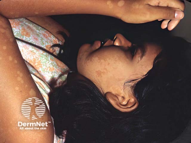
This dermatology clinical photograph depicts hypopigmented patches consistent with pityriasis versicolor (tinea versicolor) involving sun-exposed skin on the face and forearm of a patient labeled PV-patient1. The lesions appear as flat, well-demarcated, hypoesthetic macules with subtle surface scale. Facial patches are lighter than surrounding skin, while forearm lesions show a similar pigmentary disturbance. The appearance is characteristic for PV, caused by superficial Malassezia overgrowth that metabolizes lipids, producing pigmentary changes in the stratum corneum. Sun exposure can accentuate hypopigmentation, and Wood’s lamp examination may reveal fluorescence supportive of diagnosis. Clinically, recognize it as a benign, treatable dermatosis that should be differentiated from vitiligo, pityriasis alba, and post-inflammatory hypopigmentation. Management typically includes topical antifungal therapy (ketoconazole, itraconazole, terbinafine) or selenium sulfide, with potential recurrence. This image serves educational purposes in dermatology training, remote assessment, and AI-driven lesion recognition, illustrating characteristic distribution on the face and arms, the macular morphology, and the essential differential diagnoses in pigmentary disorders.
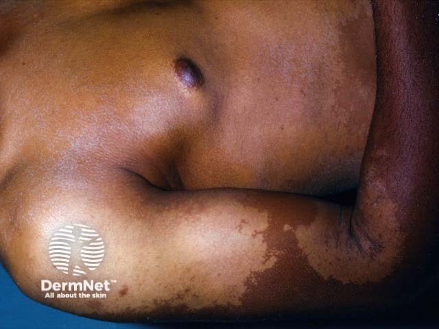
Clinical photograph of in vivo human forearm skin showing hypopigmented, confluent patches compatible with pityriasis versicolor (tinea versicolor). The image demonstrates localized involvement of the volar forearm with pale, irregularly shaped macules that coalesce into larger hypopigmented patches. The lesions display subtle surface scaling and well-defined but variable borders; surrounding skin is darker, giving a contrast typical for this condition. The distribution is on sun-exposed or humid skin regions and may appear more prominent after tanning. Pathophysiology involves superficial Malassezia yeast colonization of the stratum corneum with production of acids that disrupt melanin synthesis, leading to pigmentary heterogeneity. Clinically, pityriasis versicolor presents as nonpruritic hypo- or hyperpigmented patches that fluoresce under Wood's lamp. Differential diagnoses include vitiligo, postinflammatory hypopigmentation, and pityriasis alba; however, tinea versicolor generally preserves skin texture and shows minimal scale on closer inspection. Diagnostic confirmation is often via KOH preparation revealing the spaghetti-and-meatballs pattern of yeast and hyphae. Management is antifungal (topical azoles, selenium sulfide, or oral therapy for extensive disease). This image is valuable for dermatology education, pigmentary disorder assessment, and search-indexing for clinical skin disease repositories, aiding learners in recognition and differential diagnosis of superficial fungal hypopigmentation.
Tinea Versicolor (Pityriasis Versicolor)
Key Concept First
- Dermatology 2-Volume Set 5e
Causative Organism
- Primary species: Malassezia globosa
- Also: M. restricta, M. sympodialis, M. furfur, M. obtusa, M. slooffiae
- Malassezia is a lipophilic yeast - part of normal follicular flora; overgrowth leads to disease
- Andrews' Diseases of the Skin, p.358
Predisposing Factors
- Hot, humid weather - more common in summer
- Oily skin - favors seborrheic areas
- Immunosuppression (face involvement in immunocompromised)
- Antibiotic therapy (disrupts normal flora)
- Organ/marrow transplant recipients
- Lipid-rich hyperalimentation (can cause Malassezia sepsis)
Clinical Features
- Multiple oval-to-round macules, patches, or thin plaques with fine, mild scale
- Scale is subtle - best revealed by scratching or stretching the skin surface
- Lesions are often confluent centrally
- "Versicolor" = variable colors in the same patient:
- Hypopigmented (whitish-tan) - most conspicuous in dark-skinned people
- Hyperpigmented (brown)
- Pink/erythematous - mild inflammation variant
- Sternal region, sides of chest, abdomen, upper back, shoulders
- Neck, pubis, intertriginous areas (antecubital/popliteal fossae)
- Scalp, palms, soles (less common)
- Face - in infants and immunocompromised patients
- "Inverse" tinea versicolor = flexural area involvement
Clinical Images

Pathophysiology of Hypopigmentation
- Malassezia metabolizes skin surface lipids, producing dicarboxylic acids (e.g., azelaic acid)
- These acids inhibit melanin production and reduce melanocyte activity
- Abnormally small, poorly melanized melanosomes are not properly transferred to keratinocytes
- Hypopigmentation may persist for weeks to months after the infection is cured - this is NOT treatment failure
- UV exposure can speed re-pigmentation
- Andrews' Diseases of the Skin, p.358
Diagnosis
| Test | Finding |
|---|---|
| KOH preparation | Classic "spaghetti and meatballs" pattern - short thick hyphae (spaghetti) + large round spores (meatballs) |
| Wood's lamp | Yellow-green fluorescence of lesions and adjacent follicles; accentuates pigment changes |
| Tape stripping | Can be used instead of skin scraping |
| Biopsy | Thick basket-weave stratum corneum with hyphae and spores; atrophic variant shows effacement of rete ridges |
| Culture | Rarely done; requires lipid-enriched media (organism is lipophilic) |
Differential Diagnosis
| Condition | Distinguishing Feature |
|---|---|
| Vitiligo | Complete depigmentation, no scale |
| Seborrheic dermatitis | Erythematous yellowish tint, greasy scale, glabella/nasolabial/scalp distribution |
| Pityriasis rosea | Herald patch, Christmas tree distribution |
| Pityriasis alba | Usually in children/atopics, face |
| Confluent & reticulated papillomatosis | Verrucous papules, reticular pattern |
| Macular syphilis | <1 cm pink lesions, positive serology |
| Hansen disease (leprosy) | Hypoesthetic patches |
- Andrews' Diseases of the Skin, p.358
Treatment
Topical (first-line):
- Selenium sulfide lotion - very cost-effective; apply daily for 1 week, wash off after 10 min; or single overnight application repeated monthly for prophylaxis
- Zinc pyrithione soap - cost-effective, well tolerated for treatment and prophylaxis
- Imidazoles (ketoconazole, clotrimazole, econazole, bifonazole), ciclopirox olamine
- Salicylic acid, propylene glycol, benzoyl peroxide preparations
Systemic (extensive disease):
- Itraconazole 200 mg once daily x 7 days, OR 400 mg single dose
- Fluconazole 400 mg single dose (can be repeated monthly as prophylaxis)
- Oral terbinafine is NOT effective (despite topical terbinafine working well)
Prophylaxis (to prevent relapse - very important):
- Weekly zinc pyrithione bar wash
- Monthly overnight selenium sulfide or ketoconazole/econazole shampoo
- Monthly oral fluconazole or itraconazole
Key point to tell patients: Pigmentation changes (hypo- or hyperpigmentation) will persist for weeks to months after cure. This is not treatment failure - the skin simply needs time to re-pigment.
Related Condition: Malassezia (Pityrosporum) Folliculitis
- Chronic, moderately itchy, monomorphic dome-shaped follicular papules and tiny pustules
- Upper back and adjacent areas; can involve face and scalp
- Often associated with tinea versicolor or seborrheic dermatitis
- More common after antibiotic therapy (especially acne treatment) and in transplant recipients
- Diagnosed by Wood's lamp fluorescence of papules or biopsy
- Responds promptly to antifungal therapy