
Splenomegaly with portal vein dilatation etiology, evaluation, and management
Reading File
Finding Sources
Finding Sources
Searching PubMed
splenomegaly portal hypertension evaluation management
Finding Sources
Finding Sources
Reading File
Reading File
Reading File
Reading File
Reading File
Reading File
Finding Sources
Reading File
Reading File
Loading Image
Reading File
Reading File
I now have all the key content I need. Let me compile a comprehensive answer.
Splenomegaly with Portal Vein Dilatation: Etiology, Evaluation, and Management
Overview
Splenomegaly with portal vein dilatation is almost always a sign of portal hypertension - the final common pathway of multiple hepatic and extrahepatic diseases. Portal hypertension is defined as a portal venous pressure gradient >5-7 mmHg. When the portal vein pressure rises, splenic venous outflow is impaired, causing passive congestion, blood pooling, and progressive splenic enlargement. The portal vein normally measures <13 mm in diameter; dilatation (>13 mm) is a reliable ultrasonographic marker of portal hypertension.
- Goldman-Cecil Medicine, p. 3883
- Current Surgical Therapy 14e, p. 455
CT Appearance
The image below shows the classic CT findings of compensated cirrhosis with portal hypertension: heterogeneous liver parenchyma, splenomegaly, and portosystemic collateral vessels.
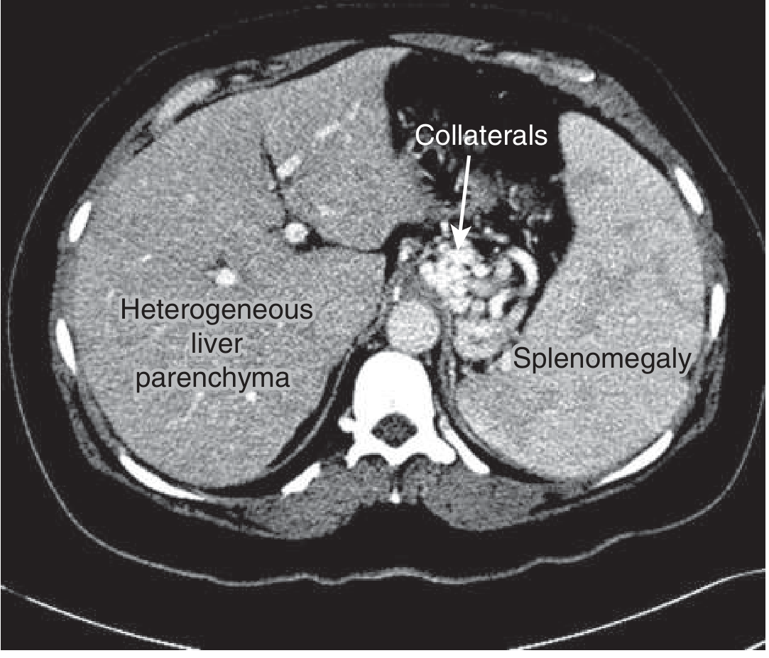
CT in compensated cirrhosis: heterogeneous liver parenchyma, splenomegaly, and portosystemic collaterals. Goldman-Cecil Medicine.
Part 1: Etiology
Portal hypertension (and thus congestive splenomegaly) is classified anatomically as prehepatic, intrahepatic (presinusoidal, sinusoidal, or postsinusoidal), or posthepatic. In North America, cirrhosis is the leading cause (~90% of cases). Worldwide, schistosomiasis, Budd-Chiari, and portal vein thrombosis are more common non-cirrhotic causes.
Classification by Site of Obstruction
| Level | Conditions |
|---|---|
| Prehepatic | Portal vein thrombosis, splenic vein thrombosis, arteriovenous fistula, increased splenic flow (massive splenomegaly from other causes) |
| Intrahepatic - Presinusoidal | Schistosomiasis (most common cause worldwide), primary biliary cholangitis (early), nodular regenerative hyperplasia, hepatic sarcoidosis, congenital hepatic fibrosis |
| Intrahepatic - Sinusoidal | Cirrhosis (viral, alcoholic, NAFLD/NASH, autoimmune, metabolic), acute alcoholic hepatitis |
| Intrahepatic - Postsinusoidal | Veno-occlusive disease (sinusoidal obstruction syndrome), hepatic vein thrombosis (Budd-Chiari) |
| Posthepatic | Budd-Chiari syndrome, constrictive pericarditis, right heart failure, inferior vena cava obstruction, tricuspid regurgitation |
- Current Surgical Therapy 14e, p. 455
- Goldman-Cecil Medicine, p. 4023
Additional Causes of Splenomegaly (Grouped by Mechanism)
Harrison's (22e) groups the broader differential of splenomegaly by pathogenic mechanism:
1. Increased demand for splenic function (reticuloendothelial hyperplasia)
- Malaria, leishmaniasis (kala-azar - produces the largest spleens globally)
- Hereditary spherocytosis, early sickle cell anemia, thalassemia
- Autoimmune hemolytic anemia, autoimmune neutropenia, ITP
2. Abnormal splenic or portal blood flow (congestive splenomegaly)
- Cirrhosis (all etiologies)
- Portal vein thrombosis, splenic vein thrombosis
- Cardiac failure (right-sided), constrictive pericarditis
- Budd-Chiari syndrome
3. Infiltration of the spleen
- Malignant: leukemias (CML produces the largest spleens in the malignant category), lymphomas, hairy cell leukemia, myelofibrosis, metastatic tumors
- Benign: Gaucher's disease, Niemann-Pick disease, amyloidosis, sarcoidosis
4. Unknown mechanism
-
Felty's syndrome (RA + splenomegaly + neutropenia)
-
SLE, systemic mastocytosis
-
Harrison's Principles of Internal Medicine 22e, Table 70-2
The Hepatic Venous Pressure Gradient (HVPG) Localizes the Level
| HVPG | Interpretation |
|---|---|
| Normal (3-5 mmHg) | Prehepatic cause (e.g., portal vein thrombosis) |
| Elevated (≥6 mmHg) | Sinusoidal or postsinusoidal (cirrhosis, veno-occlusive disease) |
| ≥10 mmHg | "Clinically significant" portal hypertension - predicts decompensation |
| ≥12 mmHg | Varices develop; risk of bleeding escalates |
| >20 mmHg | Predicts recurrent variceal hemorrhage and death |
- Goldman-Cecil Medicine, p. 4023
Part 2: Evaluation
History and Physical Examination
Key features to elicit:
- Symptoms: Left upper quadrant heaviness or pain, early satiety (stomach compression), symptoms of hypersplenism (fatigue from anemia, bleeding from thrombocytopenia)
- Risk factors: Alcohol use, viral hepatitis exposure (HBV/HCV), travel to malaria/schistosomiasis-endemic areas, hypercoagulable state (for portal vein thrombosis), cardiac disease
- Physical exam: Spleen is palpable only when enlarged ~3x; record centimeters below the costal margin. Look for stigmata of liver disease (jaundice, palmar erythema, spider angiomata, caput medusae, ascites), elevated JVP (cardiac cause)
A normal-sized spleen on ultrasound has a craniocaudal length ≤13 cm. Splenomegaly is present in ~2% of U.S. adults but is far more prevalent in endemic regions (up to 60% in parts of New Guinea).
- Harrison's Principles of Internal Medicine 22e, p. 522-524
Laboratory Evaluation
First-line blood tests:
- CBC with differential: cytopenias suggest hypersplenism (↓Hgb, ↓WBC, ↓platelets)
- LFTs, albumin, bilirubin, INR: assess hepatic synthetic function and degree of liver disease
- Peripheral blood smear: spherocytes (hereditary spherocytosis, hemolysis), teardrop cells (myelofibrosis), blasts (leukemia)
- Reticulocyte count
Directed serology based on clinical suspicion:
- Viral hepatitis panel (HBsAg, anti-HCV)
- ANA, anti-smooth muscle Ab, AMA (autoimmune liver disease)
- Iron studies, ferritin (hemochromatosis)
- Ceruloplasmin, slit-lamp exam (Wilson's disease in young patients)
- Serum protein electrophoresis (amyloid, myeloma)
- JAK2 mutation (myeloproliferative neoplasm, Budd-Chiari)
- Hypercoagulability workup (Factor V Leiden, prothrombin mutation, antiphospholipid Ab) if portal vein thrombosis suspected
- Thick blood film for malaria (if travel history)
- Serology for leishmaniasis in endemic areas
Imaging
Ultrasound with Doppler (first-line):
- Measures spleen size, portal vein diameter (>13 mm = dilated), flow velocity and direction (hepatofugal flow = severe portal hypertension), detects ascites, liver echogenicity
- Assess for portal vein thrombosis, cavernous transformation, collaterals
CT abdomen with contrast:
- Best overall assessment: liver morphology (cirrhotic vs. normal), spleen size, portal vein patency, collateral vessels (varices, recanalized umbilical vein), splenic or hepatic lesions
- Contrast-enhanced CT can also show arteriovenous fistulae
MRI/MRCP:
- Superior soft-tissue characterization; useful for infiltrative diseases (Gaucher's, amyloid), biliary pathology, hepatic venous anatomy (Budd-Chiari)
Liver stiffness measurement (transient elastography / FibroScan):
-
Non-invasive assessment of liver fibrosis; correlates with HVPG
-
A liver stiffness >20-25 kPa is highly predictive of clinically significant portal hypertension
-
Note: overestimates stiffness in postprandial state, hepatic inflammation, cholestasis, and right heart failure
-
Sleisenger & Fordtran's Gastrointestinal and Liver Disease, p. 3338
-
Goldman-Cecil Medicine, p. 4023
Hepatic Venous Pressure Gradient (HVPG):
- Gold standard for quantifying portal pressure; performed by hepatic vein catheterization
- Direct portal vein catheterization is rarely needed and carries risks
- Guides prognosis and monitors treatment response
Endoscopy (EGD):
- Detects and grades esophageal/gastric varices
- Varices graded: small (straight, minimally elevated), medium (<1/3 lumen), large (>1/3 lumen)
- White nipple sign, adherent clot, or active bleeding from a varix confirms variceal hemorrhage
Bone marrow biopsy: If myeloproliferative neoplasm or infiltrative disorder is suspected
Liver biopsy: Remains the gold standard for diagnosing the cause and stage of liver disease; used when non-invasive workup is inconclusive
Part 3: Management
Management targets three goals: (1) treat the underlying cause, (2) reduce portal pressure, and (3) prevent/manage complications.
Treat the Underlying Cause
| Etiology | Specific Treatment |
|---|---|
| Viral hepatitis B | Antiviral therapy (tenofovir, entecavir) - can reverse cirrhosis |
| Viral hepatitis C | Direct-acting antivirals (DAAs) - SVR reduces portal pressure |
| Alcoholic cirrhosis | Abstinence from alcohol |
| NASH | Weight loss, metabolic control |
| Autoimmune hepatitis | Corticosteroids + azathioprine |
| Wilson's disease | D-penicillamine, trientine, zinc |
| Hemochromatosis | Phlebotomy |
| Schistosomiasis | Praziquantel |
| Malaria | Antimalarials (artemisinin-based) |
| Portal vein thrombosis | Anticoagulation (LMWH or direct anticoagulants) |
| Budd-Chiari | Anticoagulation; TIPS or liver transplantation if refractory |
| Right heart failure | Treat cardiac cause |
Reduce Portal Pressure - Medical Therapy
Non-selective beta-blockers (NSBBs):
- Propranolol or nadolol: reduce portal pressure by ~20%; first-line for primary and secondary prophylaxis of variceal bleeding
- Carvedilol (alpha1 + non-selective beta): superior portal pressure reduction; increasingly preferred
- Target: reduce HVPG to <12 mmHg or by >20% from baseline; or heart rate to 55-60 bpm
Vasopressin analogues / somatostatin analogues (acute bleeding):
- Terlipressin or octreotide/somatostatin: acute variceal hemorrhage; reduce splanchnic blood flow
Endoscopic Management
- Endoscopic variceal ligation (EVL): primary prophylaxis (large varices with high-risk features), acute bleeding, secondary prophylaxis
- Endoscopic sclerotherapy: less favored due to higher complication rate vs. EVL
- Combined EVL + NSBBs: standard for secondary prophylaxis
Interventional Radiology - TIPS
Transjugular Intrahepatic Portosystemic Shunt (TIPS):
-
Creates a shunt between portal and hepatic veins through liver parenchyma
-
Rapidly reduces portal pressure
-
Indications: refractory ascites, refractory variceal bleeding, Budd-Chiari, hepatic hydrothorax
-
Limitations: risk of hepatic encephalopathy (reduced hepatic perfusion); contraindicated in severe hepatic encephalopathy, right heart failure, polycystic liver disease
-
TIPS has largely replaced surgical shunts in current practice
-
Current Surgical Therapy 14e, p. 455-456
Surgical Shunts (Selective Indication)
Used when TIPS is not feasible or fails; also for non-cirrhotic portal hypertension:
- Distal splenorenal shunt (Warren shunt): selective decompression while preserving hepatic portal perfusion; lower encephalopathy rate
- Portocaval shunt: non-selective; high risk of encephalopathy
- Mesocaval shunt: bridges portal to IVC via graft
Management of Hypersplenism
-
Splenomegaly with cytopenia (hypersplenism): usually managed conservatively
-
Splenectomy indications: symptomatic massive splenomegaly, refractory cytopenias, splenic rupture, staging (Hodgkin's lymphoma in select cases), hairy cell leukemia, hereditary spherocytosis
-
Pre-splenectomy vaccines (mandatory): pneumococcal, meningococcal, Hib - given at least 2 weeks before elective splenectomy
-
Splenic artery embolization: partial embolization as alternative to splenectomy in high-surgical-risk patients
-
Harrison's Principles of Internal Medicine 22e, p. 524
-
Bailey and Love's Short Practice of Surgery 28e, p. 3857-3860
Liver Transplantation
- Definitive treatment for end-stage liver disease with portal hypertension
- Cures both the liver dysfunction and portal hypertension
- Gold standard for cirrhotic portal hypertension when patient is a suitable candidate
- Non-cirrhotic etiologies (e.g., portal vein thrombosis, schistosomiasis) are generally not indications for transplant
Management of Ascites
- Sodium restriction (<2g/day)
- Spironolactone ± furosemide (aldosterone antagonist is primary)
- Large-volume paracentesis + albumin infusion for tense ascites
- TIPS for refractory ascites
Acute Variceal Hemorrhage (Emergency Management)
- Airway protection, IV access, transfuse to Hgb ~7-8 g/dL
- Vasoactive agents: octreotide or terlipressin (started immediately, before endoscopy)
- Antibiotics: short-course norfloxacin or ceftriaxone (reduces bacterial translocation, improves survival)
- Urgent EGD within 12 hours: EVL is treatment of choice
- TIPS if bleeding uncontrolled or early rebleeding
- Balloon tamponade (Sengstaken-Blakemore tube): bridge to definitive therapy
Summary Algorithm
Splenomegaly + Portal Vein Dilatation on Imaging
           ↓
  Assess for portal hypertension
           ↓
  Doppler US: portal vein >13mm, hepatofugal flow?
           ↓
  Classify: Prehepatic / Intrahepatic / Posthepatic
           ↓
  LFTs, CBC, viral hepatitis, autoimmune, metabolic workup
           ↓
  Liver stiffness measurement (FibroScan/elastography)
           ↓
  EGD: screen for varices
           ↓
  Treat underlying cause + Portal pressure reduction (NSBBs)
           ↓
  Endoscopic prophylaxis (EVL) for high-risk varices
           ↓
  TIPS or surgical shunts for refractory cases
           ↓
  Liver transplant evaluation for end-stage disease
Recent Evidence (2024-2026)
- Splenic transient elastography for portal hypertension (PMID: 40900859) - A 2025 review supports splenic stiffness measurement (via SWE or ARFI) as a promising non-invasive marker for clinically significant portal hypertension, complementing liver stiffness
- Multiparametric ultrasound for splenic pathology (PMID: 39417855) - A 2025 review highlights contrast-enhanced US and elastography as key additions to B-mode US for characterizing splenomegaly
- TIPS for ectopic variceal bleeding (PMID: 38935315) - 2024 systematic review confirms TIPS as an effective rescue therapy for ectopic varices refractory to endoscopy
Key sources:
- Harrison's Principles of Internal Medicine 22e (2025), Ch. 70
- Goldman-Cecil Medicine, Ch. 154 (Splenomegaly), Ch. 144 (Portal Hypertension)
- Current Surgical Therapy 14e, "Portal Hypertension: Role of Shunting Procedures"
- Sleisenger & Fordtran's Gastrointestinal and Liver Disease, Ch. 93
- Bailey & Love's Short Practice of Surgery 28e, Ch. 70
This is a shared conversation. Sign in to Orris to start your own chat.