
Gastric reflux physiology
"gastroesophageal reflux" AND "pathophysiology"
gastroesophageal reflux GERD pathophysiology lower esophageal sphincter diagram
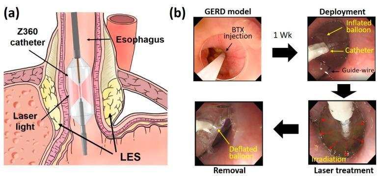
Educational figure illustrating Endoscopic Balloon-assisted Laser Treatment (EBLT) for Gastroesophageal Reflux Disease (GERD). (a) Anatomical diagram showing a Z360 balloon catheter positioned within the distal esophagus at the lower esophageal sphincter (LES). The catheter features a transparent inflated balloon and a central diffusing applicator that emits circumferential laser light to target the LES muscle layers. (b) A series of four endoscopic frames detailing the procedural stages in a porcine model: 1. GERD model induction via botulinum toxin (BTX) injection to weaken the LES; 2. Deployment stage showing the catheter and inflated balloon guided by a guide-wire; 3. Laser treatment stage showing active irradiation (indicated by red arrows); 4. Removal stage following balloon deflation. The educational focus is on the minimally invasive delivery of laser energy to the esophagogastric junction to induce tissue remodeling and increase LES pressure without mucosal injury.
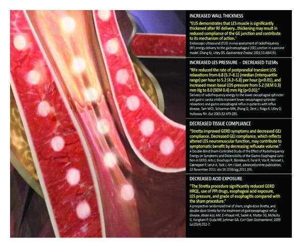
This medical illustration demonstrates the mechanism of action of the Stretta procedure, a minimally invasive treatment for gastroesophageal reflux disease (GERD). The visual depicts a cross-section of the esophageal wall near the gastroesophageal junction. Key components include multiple nickel-titanium needle electrodes delivering radiofrequency (RF) energy into the muscularis propria of the esophagus, represented by bright yellow circular glows within the red-textured tissue layers. The anatomical focus is on the lower esophageal sphincter (LES) region. Accompanying text panels summarize clinical outcomes and pathophysiology: increased wall thickness, increased LES basal pressure with decreased transient LES relaxations (TLESRs), decreased tissue compliance at the gastroesophageal junction, and significantly decreased esophageal acid exposure. The image highlights the device's ability to deliver thermal energy to induce tissue remodeling while protecting the overlying mucosa via irrigation. This illustration serves as an educational tool for gastroenterologists and surgical trainees to understand non-surgical antireflux interventions and their physiological impact on LES neuromuscular function.
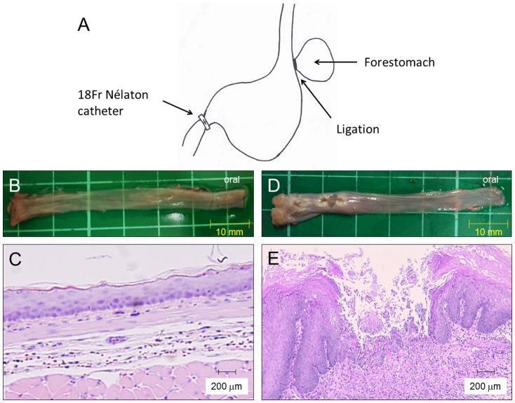
This composite image illustrates a rat model of acid reflux esophagitis, comparing normal and pathological findings across macroscopic and microscopic scales. Panel A presents a surgical diagram depicting the induction of reflux via forestomach ligation and pyloric ring constriction using an 18Fr Nélaton catheter. Panel B shows the macroscopic appearance of a normal esophagus, characterized by a smooth, uniform mucosal surface. In contrast, Panel D displays a diseased esophagus with visible erosions and ulcers in the middle and lower regions. Panel C provides a histological view (H&E stain, 200µm scale) of normal esophageal tissue, showing a relatively thin squamous epithelium and minimal inflammatory infiltrate. Panel E demonstrates the pathophysiology of reflux esophagitis, featuring significant mucosal thickening, basal cell hyperplasia, and dense inflammatory cell infiltration within the lamina propria and submucosa. This figure serves as an educational tool for identifying structural and cellular changes associated with gastroesophageal reflux disease (GERD).
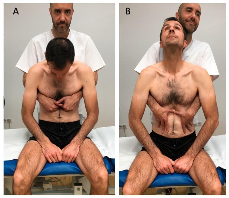
Two-panel clinical photograph demonstrating a visceral osteopathic manual technique targeting the lower esophageal sphincter, typically used for gastroesophageal reflux disease (GERD). Panel A (Initial Position) shows a male patient seated upright on a treatment table in spinal flexion with his neck flexed and head looking down. A practitioner stands behind him with hands placed horizontally in the epigastric region, specifically contacting the area just inferior to the costal margins. Panel B (Final Position) illustrates the completion of the maneuver: the patient has moved into spinal extension with his neck extended and gaze upward. Simultaneously, the practitioner has shifted hand placement to apply caudal (downward) pressure within the epigastrium. The visual demonstrates the coordination of patient respiratory/postural movement with manual tissue mobilization by the therapist. This procedural image is intended for medical students and physical therapy or osteopathic practitioners to understand the manual application and patient positioning for visceral abdominal therapy.
Gastric Reflux (GERD) Physiology
Definition
1. The Anti-Reflux Barrier
| Component | Role |
|---|---|
| Lower esophageal sphincter (LES) | Primary pressure barrier; resting tone 10-45 mmHg |
| Crural diaphragm | External pinch-cock mechanism during inspiration and Valsalva |
| Phrenoesophageal ligament | Anchors LES to crural diaphragm |
| Angle of His | Oblique entry of esophagus into stomach creates a flap-valve effect |
2. Mechanisms of Reflux
a) Transient LES Relaxations (TLESRs) - the most common mechanism
- These are spontaneous relaxations not triggered by swallowing
- Mediated by a vagal reflex stimulated by gastric distension
- In healthy people, TLESRs cause brief physiologic reflux episodes
- In GERD patients, TLESRs are more frequent and prolonged, and are the dominant mechanism (responsible for ~70% of reflux episodes)
b) Low resting LES pressure
- A chronically hypotensive LES (<6 mmHg) allows passive reflux, especially when intra-abdominal pressure rises
- Factors that reduce LES tone: progesterone, smoking, fatty foods, caffeine, theophylline, nitrates, calcium channel blockers
c) Increased intra-abdominal pressure overcoming normal LES pressure
-
Seen in obesity, pregnancy, and straining
-
Associated with rumination syndrome and supragastric belching variants of GERD
-
Goldman-Cecil Medicine, p. 1429
3. Role of Hiatal Hernia
- Widens the GEJ opening
- Decreases effective LES pressure
- Increases reflux during TLESRs and with elevated gastric pressure
- Creates a hernia sac that acts as a reservoir - when the LES relaxes during swallowing, trapped fluid in the hernia refluxes back up into the esophagus ("re-reflux")
4. The Gastric Acid Pocket
- The acid pocket is longer and more proximate to the GEJ
- Displacement of this pocket into a hiatal hernia dramatically increases acid exposure
- This is why postprandial symptoms are so common in GERD
5. Obesity and GERD
- Raises intragastric pressure, increasing the gastroesophageal pressure gradient
- Increases frequency of TLESRs
- Enhances the spatial separation of crural diaphragm from LES, predisposing to hiatal hernia
- The associated metabolic syndrome may independently promote esophageal injury
6. Esophageal Clearance and Mucosal Defense
- Pre-epithelial barrier - an unstirred water layer + bicarbonate from saliva and submucosal glands
- Epithelial barrier - tight intercellular junctions, cell membrane ion transporters, and intracellular buffers
- Postepithelial barrier - the esophageal blood supply
7. Clinical Syndromes (Montreal Classification)
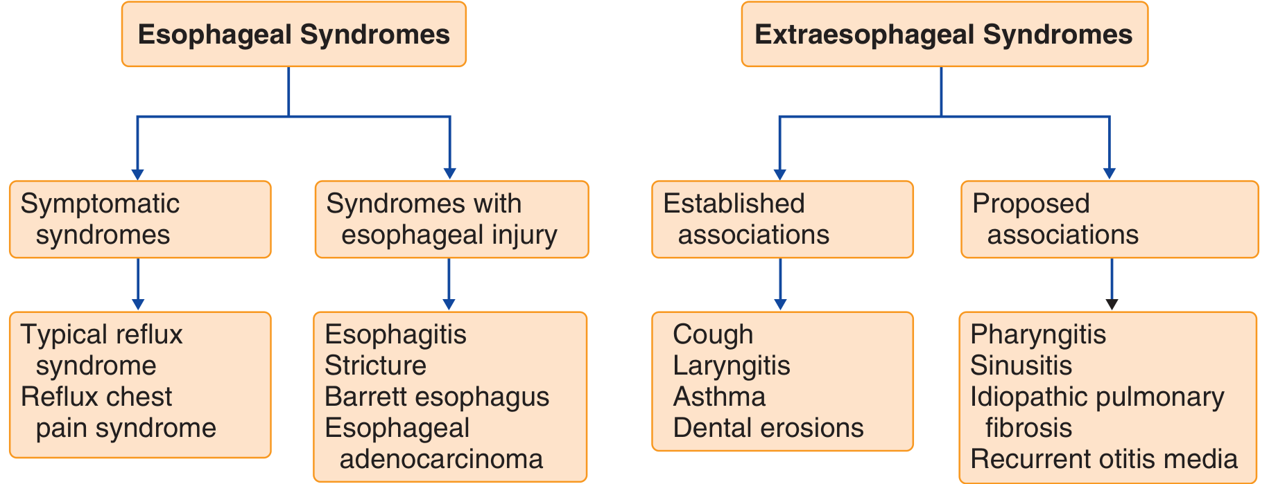
- Symptomatic: typical reflux syndrome (heartburn, regurgitation), reflux chest pain syndrome
- With esophageal injury: esophagitis, stricture, Barrett's esophagus, adenocarcinoma
- Established: chronic cough, laryngitis, asthma, dental erosions
- Proposed: pharyngitis, sinusitis, idiopathic pulmonary fibrosis, recurrent otitis media
8. Pathologic Refluxate Components
- Hydrochloric acid - primary mucosal injuriant; pH < 4 correlates with symptom generation
- Pepsin - activated at low pH, directly damages epithelial protein; works synergistically with acid
- Bile acids - particularly damaging in alkaline (duodenogastric) reflux; implicated in Barrett's metaplasia
- Trypsin - contributes to mucosal injury in mixed acid-bile reflux
Summary: Pathophysiology at a Glance
Increased TLESRs
     +
Low resting LES pressure     →   Reflux of acid/pepsin/bile
     +                                      ↓
Hiatal hernia              Overcomes esophageal clearance
     +                                      ↓
Impaired peristalsis         Mucosal barrier breakdown
     +                                      ↓
Impaired salivation           Chemokine release → inflammation
     +                                      ↓
Acid pocket displacement     Symptoms + complications
- Goldman-Cecil Medicine International Edition, p. 1429
- Sleisenger and Fordtran's Gastrointestinal and Liver Disease
- Goodman & Gilman's Pharmacological Basis of Therapeutics