
Psoriasis ayurveda treatment prescription
psoriasis Ayurveda treatment prescription Kushtha herbs panchakarma
https://pmc.ncbi.nlm.nih.gov/articles/PMC11994298
psoriasis ayurveda herbal treatment
psoriasis plaque skin lesion clinical photo
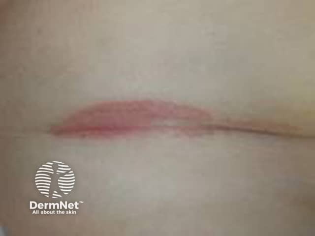
Clinical photography of the antecubital fossa region shows a single erythematous plaque within a flexural skin fold. The lesion is well-demarcated, with a smooth pink-to-erythematous surface and minimal scales evident, reflecting inverse/ flexural psoriasis features in moist environments where scaling is less conspicuous. The plaque lies centrally within the fold, with intact surrounding skin and no obvious crusting or excoriation. Lighting emphasizes a uniform coloration without effusion or ulceration. The regional skin may appear slightly edematous or glistening from moisture, a common appearance in intertriginous psoriasis. In clinical practice, such lesions are often symmetric or multipart and may co-occur with plaques on extensor surfaces; history of chronic plaque psoriasis or family history supports the diagnosis. Differential considerations include irritant dermatitis, candidal intertrigo, or seborrheic dermatitis; biopsy would reveal epidermal hyperplasia with parakeratosis and neutrophilic microabscesses typical of plaque psoriasis, though histology is not demonstrated here. The image is intended for dermatology education, aiding recognition of flexural psoriasis, differentiation from dermatitis, and guiding topical therapy, including high-potency corticosteroids or vitamin D analogs with occlusion, while avoiding excessive moisture. This photo supports image-based teaching, clinical pattern recognition, and correlation with patient history in practice and research.
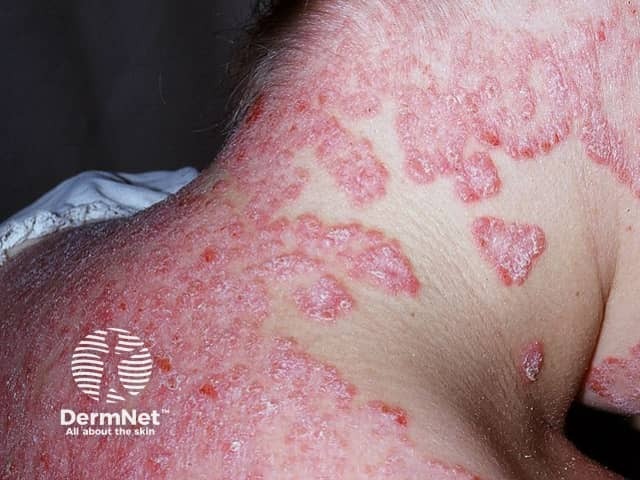
Clinical photography of facial skin demonstrates classic psoriatic plaques involving the malar region and surrounding cheeks. The lesions are erythematous to violaceous, well-demarcated, and markedly scaly with silvery-white scale, consistent with plaque-type psoriasis (psoriasis vulgaris). Involved areas show surface coarseness, thickening (acanthosis) and mild induration, while adjacent skin remains relatively spared. Perilesional inflammation creates a ring of erythema, and some plaques exhibit superficial fissuring. The distribution is facial rather than scalp, trunk, or limbs, suggesting involvement limited to the head-and-face region in this image. No vesiculation or purulence is evident. The appearance may mimic seborrheic dermatitis, eczema, or contact dermatitis; however, the well-circumscribed plaques with thick scale and the typical red-silver presentation strongly favor psoriasis. Skin photo shows high-contrast textural differences between lesional and non-lesional skin. Clinically, facial psoriasis can be challenging due to cosmetic and functional concerns, including itching and social impact. This image supports differential diagnosis discussions in dermatology education and serves as a reference for recognizing facial plaque psoriasis, assessing severity, and guiding treatment planning (topical steroids, vitamin D analogs, or phototherapy). It is useful for teaching photodocumentation, lesion morphology, and pattern recognition in psoriasis spectrum. This image thus enhances reasoning for diagnosis, prognosis, and management planning.
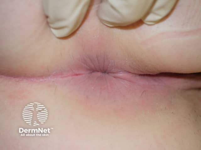
Clinical photograph of external genital skin showing an erythematous, well-demarcated plaque in an intertriginous fold. The lesion is pink to red, with a smooth surface and minimal peripheral scaling within moist skin areas; a slight fissure is seen along the crease. The surrounding skin is mildly inflamed but otherwise unremarkable. This appearance is characteristic of genital psoriasis, most compatible with inverse psoriasis in moist genital folds where scaling is less prominent than on extensor surfaces. Differential diagnoses include candidal balanitis or vulvitis, irritant contact dermatitis, lichen sclerosus, eczema, and less commonly erosive lesions. Clinical context such as past or concurrent psoriasis elsewhere, absence of itch or burning, and response to topical steroids can help confirmation. Histopathology would typically show epidermal hyperplasia with parakeratosis, regular elongation of rete ridges, and a dermal perivascular lymphocytic infiltrate if biopsy were performed; in practice, biopsy is not required for typical genital psoriasis. The diagnostic significance lies in recognizing genito-pubic psoriasis to guide therapy and avoid misdiagnosis as infection. This image is useful for education on genital psoriasis recognition, differential diagnosis, and management considerations including topical corticosteroids, vitamin D analogs, and calcineurin inhibitors, with referrals for systemic therapy if extensive disease. Clinical photo for reference.
Psoriasis - Ayurveda Treatment Prescription
Ayurvedic Correlation
- Ekakushtha - silvery, fish-scale-like patches; non-sweating skin (closest to plaque psoriasis)
- Kitibha - dry, rough, blackish plaques (resembles psoriasis with lichenification)
- Vata: Dryness, scaling, itching, cracking
- Kapha: Thickening, induration, chronicity
- Pitta (secondary): Inflammation, erythema, burning sensation
- Rakta dushti (blood vitiation): Central to pathogenesis - blood-purifying therapies are key
Treatment Framework
- Shodhana Chikitsa (Purification/Detoxification)
- Shamana Chikitsa (Palliation with internal + external medicines)
Phase 1 - Poorvakarma (Preparatory Procedures)
| Procedure | Method | Duration |
|---|---|---|
| Deepana-Pachana | Trikatu churna or Chitrakadi vati to digest Ama (toxins) | 7-10 days |
| Snehana (internal oleation) | Mahatikta ghrita or Tikta ghrita: 30-60 mL on empty stomach, increasing daily | 5-7 days |
| Snehana (external) | Abhyanga (whole-body oil massage) with Ksheerabala taila or Dashamula taila | Daily |
| Swedana (sudation) | Steam bath / Bashpa sweda | Daily after Abhyanga |
Mahatikta Ghrita is the classical choice for Kushtha with Rakta dushti - it is both Shodhana (purifying) and Rasayana (rejuvenating).
Phase 2 - Panchakarma (Shodhana Chikitsa)
A. Vamana (Therapeutic Emesis) - For Kapha-dominant psoriasis
- Indication: Thick, indurated plaques; Kapha symptoms
- Drug: Madanaphala + Vacha + Rock salt in warm water
- Pre-procedure: Internal oleation for 5-7 days, then Vamana
- Frequency: 1 session (3 to 7 Vegas/emetic episodes)
B. Virechana (Therapeutic Purgation) - Primary and most recommended
- Indication: Psoriasis with inflammation, Pitta-Rakta involvement; most cases
- Drug: Trivrut lehya (Operculina turpethum) or Haritaki churna with warm water
- Dose: Trivrut lehya 30-60 g in warm water (given post Poorvakarma)
- Vegas: 20-30 purgative episodes expected
C. Basti (Medicated Enema) - For Vata-dominant / chronic psoriasis
- Niruha Basti (decoction): Dashamula kashaya + honey + salt + oil
- Anuvasana Basti: Mahatikta ghrita or Tiktaka ghrita
- Schedule: Kala Basti (15 days) or Yoga Basti (8 days)
D. Raktamokshana (Bloodletting) - For severe Rakta dushti
- Leech therapy (Jalaukavacharana): Most suitable; applied at lesional sites
- Indication: Localized, inflammatory plaques with burning
- Siravyadha (venesection) for large-area involvement
Phase 3 - Shamana Chikitsa (Internal Medicines)
Classical Oral Formulations
| Medicine | Dose | Timing | Action |
|---|---|---|---|
| Arogyavardhini Vati | 500 mg - 1 g twice daily | After food with warm water | Liver detox, skin purifier, Kapha-Pitta pacifier |
| Khadirarishta | 20 mL twice daily | After food (mixed with 40 mL water) | Blood purifier, antipsoriatic - Khadir (Acacia catechu) as base |
| Mahamanjishthadi Kashaya | 20 mL twice daily | Before food (empty stomach) | Rakta shodhaka (blood purifier), Pitta-shamaka |
| Patolkaturohinyadi Kashaya | 20 mL twice daily | 1 hour before food | Anti-inflammatory, Pitta-Kapha reducer |
| Punarnavadi Kashaya | 20 mL twice daily | Mixed with 50 mL warm water before food | Shothahara (anti-inflammatory), Rakta prasadaka |
| Gandhaka Rasayana | 500 mg - 1 g twice daily | After food | Rasayana (rejuvenation), antimicrobial, skin-healing |
| Kaisora Guggulu | 500 mg - 1 g twice daily | After food | Vatarakta, inflammation, joint involvement (psoriatic arthritis) |
| Tiktaka Ghrita | 10-20 mL daily | On empty stomach in morning | Pitta-Rakta dushti; systemic absorption of tikta (bitter) dravyas |
Khadirarishta is the most well-documented Ayurvedic formulation for psoriasis. It has been used consistently across classical texts and is supported by this PMC case report (2025).
Phase 4 - Bahya Chikitsa (External / Topical Applications)
| Preparation | Application | Action |
|---|---|---|
| Winsoria oil (or Karanja taila) | Apply twice daily over lesions | Vranaropana (wound healing), anti-inflammatory, skin complexion |
| Mahamarichyadi Taila | Apply twice daily | Kills Krimis (microbes), drying, anti-scaling |
| Neem (Azadirachta indica) paste or oil | Apply over plaques, leave 30 min | Antibacterial, anti-inflammatory, antipruritic |
| Turmeric (Haridra) + Neem paste | Apply twice daily | Curcumin - down-regulates inflammatory kinases in psoriasis (source: Dermatology 5e) |
| Coconut oil + Karpura (camphor) | Apply overnight for scaling | Moisturizing, anti-itch, keratolytic |
| Takradhara (medicated buttermilk pour) | 45-60 min daily for 7-14 days | Cooling, Pitta-shamaka, reduces erythema |
Per the Dermatology 2-Volume Set 5e in the textbook library: "topical and oral curcumin are efficacious in psoriasis. Curcumin is thought to down-regulate kinases in patients with psoriasis."
Sample Full Prescription (Moderate Plaque Psoriasis)
| # | Medicine | Dose | Timing |
|---|---|---|---|
| 1 | Arogyavardhini Vati | 500 mg x 2 | After meals with warm water |
| 2 | Khadirarishta | 20 mL x 2 | After meals diluted with 40 mL water |
| 3 | Mahamanjishthadi Kashaya | 20 mL x 2 | Before meals |
| 4 | Kaisora Guggulu (if arthritis) | 500 mg x 2 | After meals |
| 5 | Gandhaka Rasayana | 500 mg x 2 | After meals |
| 6 | Tiktaka Ghrita | 10 mL x 1 (morning) | On empty stomach |
| 7 | Winsoria oil / Karanja taila | Topical, twice daily | Apply on lesions |
| 8 | Neem + Haridra paste | Topical, once daily | Apply and wash after 30 min |
Pathyapathya (Diet & Lifestyle)
Pathya - Recommended (Do)
- Old rice, barley, moong dal (green gram), bitter gourd, drumstick
- Garlic, turmeric, neem leaves in diet
- Warm water for drinking
- Sunlight exposure (moderate - natural phototherapy)
- Yoga, Pranayama (stress reduction is key - stress triggers psoriasis flare)
- Meditation, regular sleep
Apathya - Avoid (Don't)
- Salt (restrict severely), sour foods, fermented food, curd (yogurt)
- Fish, shellfish, red meat
- Alcohol, smoking
- Milk + fish (Viruddha ahara / incompatible food combination)
- Spicy, oily, heavy food
- Late nights, day-sleep, emotional stress
- Excessive sweating or exposure to extreme cold
Duration of Treatment
| Phase | Duration |
|---|---|
| Poorvakarma | 7-14 days |
| Panchakarma (Virechana / Vamana) | 1-3 sessions over 1-3 months |
| Internal medicines (Shamana) | Minimum 1 year for chronic psoriasis |
| Dietary restrictions | 3 years recommended |
| Follow-up Panchakarma | Every 6-12 months |
Important Safety Notes
- Arogyavardhini Vati contains Parada (Mercury) in some formulations - only use GMP-certified, third-party tested brands.
- Ayurvedic herbal products are regulated as dietary supplements, not drugs. Always assess for heavy metal contamination.
- Avoid Mahatikta ghrita in diabetes, obesity, or metabolic syndrome without adjustment.
- Do NOT stop conventional disease-modifying drugs (methotrexate, biologics) without consulting a dermatologist.
- Ayurveda works best as a complementary approach in moderate-to-severe psoriasis alongside modern medicine.
Bottom line: The Ayurvedic approach to psoriasis centers on Virechana (purgation) as the primary Shodhana therapy, Khadirarishta + Mahamanjishthadi Kashaya as core internal medicines, topical Neem/Turmeric/Karanja preparations, and strict dietary control (Apathya avoidance) for a minimum of 1-3 years for sustained remission.