

As a pathology resident create a presentation for Clinico pathology correlation on topic Moderately differentiated squamous cell carcinoma nose
moderately differentiated squamous cell carcinoma histology keratin pearls
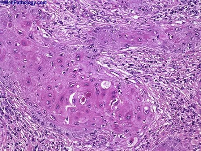
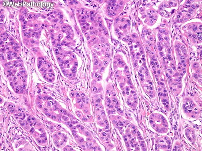
This histopathology image depicts a cutaneous squamous cell carcinoma on routine hematoxylin and eosin stained section. The biopsy shows nests of malignant squamous cells with keratinization and abundant keratin pearls, indicating squamous differentiation. The tumor exhibits moderate differentiation, evidenced by intercellular bridges, eosinophilic cytoplasm, nuclear pleomorphism, hyperchromasia, and mitotic activity. Invasion into the surrounding dermal stroma is evident, with desmoplastic-type fibrous response in some areas. The epidermis overlying the lesion shows dysplastic changes, and keratin pearl formation within tumor nests is a hallmark of keratinizing SCC. The architecture includes irregular cords and nests of polygonal cells, occasional central keratinization, and keratinized pearls of varying sizes. Clinically, these features suggest a malignant cutaneous neoplasm with potential for local invasion; staging would consider depth of invasion and perineural or lymphovascular involvement. This image is relevant for diagnostic pathology, histology education, and tumor biology research, illustrating characteristic squamous differentiation, keratin production, and invasion patterns. Differential considerations include well-differentiated keratinizing SCC versus moderately-to-poorly differentiated forms, verrucous carcinoma, and basal cell carcinoma with squamous features. The visual cues—keratin pearls, keratinization, and cohesive epidermal-derived tumor islands—assist in confirming diagnosis and guiding surgical management. Correlation with clinical data will inform prognosis and adjuvant therapy decisions and multidisciplinary care planning.
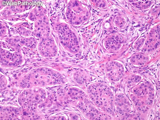
Imaging modality: light microscopy of an H&E–stained histology section from a recurrent submandibular gland mass. Primary subject: high‑grade mucoepidermoid carcinoma with scant cyst formation. Precise anatomical location: submandibular region (salivary gland), head and neck. Visual features: predominantly solid sheets and clusters of epidermoid (squamoid) and intermediate cells displaying cytologic atypia, marked nuclear pleomorphism, prominent nucleoli, and brisk mitotic activity. Keratinization is limited; extensive keratin pearls are not present; occasional small keratin pearls or individual cell keratinization may be seen. Mucicarmine-positive mucous cells are scattered throughout the tumor, indicating mixed differentiation with mucous-secreting components. The stroma is variable with embedded tumor nests and occasional duct-like structures; no prominent cystic spaces are described. Imaging perspective: micrographic cross-sections at high magnification highlight cellular morphology and intercellular bridges. Diagnostic interpretation: high-grade MEC with aggressive features, potential to mimic moderately differentiated squamous cell carcinoma but mucous cell component and mucicarmine positivity support MEC; absence of large keratin pearls argues against classic keratinizing SCC. Clinical relevance: biopsy morphology is essential for grading, prognosis, and therapeutic planning, as high‑grade MEC carries higher risk of recurrence and regional/metastatic spread; findings justify surgical management with consideration of adjuvant therapy and close follow-up.
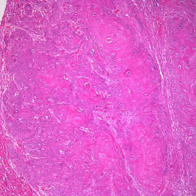
Imaging modality and technique: Not applicable in this field; however for description, histology uses light microscopy of hematoxylin and eosin stained vulvar biopsy tissue. Observed pattern: invasive keratinizing squamous cell carcinoma arising in vulvar epidermis with invasion into the dermis. Precise anatomy: vulvar external genitalia; epithelium of the labia majora/minora and adjacent hymenal region may be involved; depth of invasion is a key prognostic factor. Visual features: tumor nests and cords of malignant squamous cells with marked cellular pleomorphism, hyperchromatic nuclei, increased mitotic figures, and intercellular bridges. Keratinization with formation of keratin pearls is present; desmoplastic stromal reaction and chronic inflammatory infiltrate at invasion front. The lesion may display well to moderately differentiated features, with conspicuous keratinization characteristic of the keratinizing subtype. HPV-independent pathogenesis is typical for this vulvar carcinoma variant; the alternative HPV-associated basaloid/warty subtype occurs in younger women. Diagnostic significance: histology confirms diagnosis of vulvar squamous cell carcinoma and determines subtype, which has implications for HPV status, prognosis, and treatment planning (wide local excision, sentinel lymph node biopsy, radiotherapy). Potential clinical utility: aids differential diagnosis in elderly patients with vulvar lesions, informs prognosis, guides surgical planning, and supports educational or research use in gynecologic oncology.
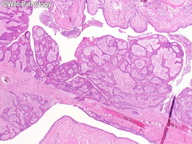
Modalities and techniques: Light microscopy of paraffin-embedded lung tissue stained with Hematoxylin and Eosin (H&E). The specimen comprises bronchial mucosa overlaid by invasive squamous cell carcinoma, arising centrally from a main or lobar bronchus with a conspicuous endobronchial polypoid component. Histologic features include nests and radiating cords of malignant squamous cells with intracellular bridges, variable keratinization, and occasional keratin pearls, infiltrating the bronchial wall and adjacent parenchyma. The tumor produces luminal obstruction, which commonly leads to postobstructive atelectasis, obstructive bronchopneumonia, and bronchiectasis downstream. The architecture shows a well to moderately differentiated squamous phenotype, with intercellular cohesion, desmosomal attachments, and brisk mitotic activity in solid and papillary regions. Lumen may be partially occluded by polypoid mass projecting into the bronchial lumen. Immunohistochemical stains optional to confirm squamous differentiation (p40, p63 positive; TTF-1 negative). Pathological significance: central endobronchial squamous cell carcinoma is associated with smoking history and presents with cough, hemoptysis, and recurrent infections; differential includes other central lung neoplasms and inflammatory polyps. Clinical correlation: radiology may show central mass with airway collapse; management includes endobronchial debulking and surgical resection with adjuvant therapy. This image is diagnostic for central airway obstruction by squamous histology and serves as valuable educational material for pathology, oncology, pulmonology, radiology, and thoracic surgery teams.
Histopathology image of a recurrent high-grade mucoepidermoid carcinoma arising in the submandibular gland. Specimen: salivary gland tumor, surgical resection. Modality: light microscopy; staining with Hematoxylin and Eosin (H&E), supplemented by mucicarmine highlighting mucous cells. The tumor shows solid architecture with sheets and clusters of epidermoid (squamoid) and intermediate cells, cytologic atypia, prominent nucleoli, and brisk mitotic activity. There is little to no cyst formation or conspicuous keratinization; keratin pearls are sparse or absent; intercellular bridges may be evident in epidermoid components. Scattered mucous-producing cells are mucicarmine-positive, supporting a diagnosis of mucoepidermoid carcinoma rather than pure squamous cell carcinoma. The lesion is high-grade by criteria: solid growth pattern, marked pleomorphism, high mitotic rate, and infiltrative borders within the submandibular region. Perineural or vascular invasion is not clearly demonstrated in this image. Differential considerations include moderately differentiated squamous cell carcinoma and adenosquamous carcinoma; however, mucous cells and mucin positivity favor MEC with salivary origin. Clinically relevant implications include aggressive behavior, potential for recurrence, and need for complete surgical excision with clear margins; adjuvant radiotherapy may be indicated. This image supports diagnostic histology, grading, and educational staging discussions for head and neck salivary gland neoplasms. Correlation with imaging helps assess regional spread and nodal involvement for staging.
sinonasal squamous cell carcinoma nasal cavity tumor
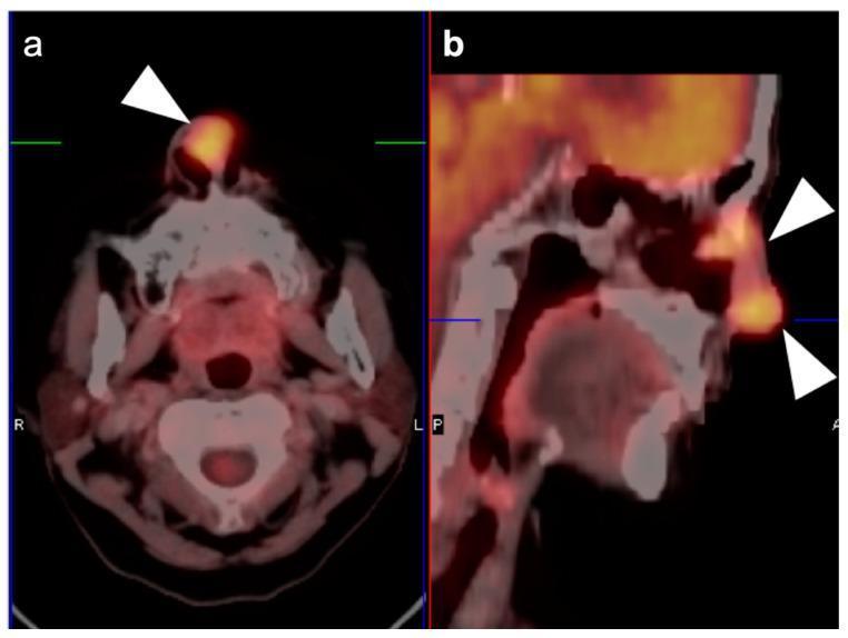
This diagnostic image displays FDG PET-CT scans of the head and neck, focusing on a sinonasal squamous cell carcinoma. Image (a) is an axial view showing a focal, intensely hypermetabolic tumor located at the anteroinferior nasal septum, indicated by a white arrowhead. The metabolic activity appears as a dense orange-to-yellow signal, signifying high glucose metabolism typical of malignancy. Image (b) is a sagittal reconstruction demonstrating the longitudinal spread of the tumor. The hypermetabolic activity extends throughout the entire nasal cavity, as highlighted by two white arrowheads, involving the nasal vestibule and spreading posteriorly along the nasal passages. This imaging modality combines the anatomical detail of computerized tomography (CT) with the functional metabolic data of positron emission tomography (PET), facilitating accurate staging and localization of sinonasal malignancies. The scans are critical for evaluating the extent of tumor invasion into adjacent sinonasal structures and planning surgical or radiation treatment.
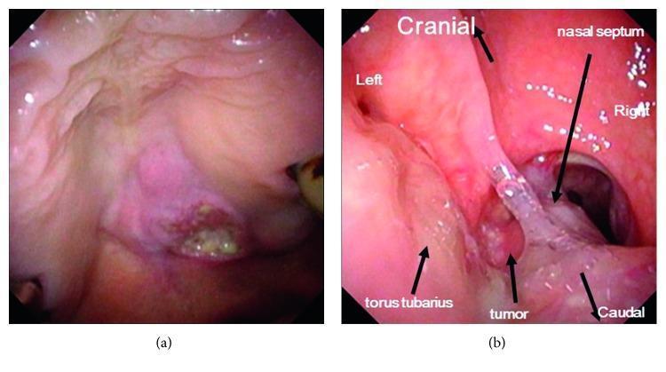
Clinical endoscopic views of a palatal squamous cell carcinoma. Image (a) shows an oral view of the tumor on the palate, presenting as a pinkish-red, irregular mass with an uneven texture and a central ulcerated area containing yellowish exudate. Image (b) provides a posterior nasal aperture view obtained via retrograde fiberscopy. This view demonstrates the tumor's superior extension into the left posterior nasal meatus with direct invasion and distortion of the nasal septum. Key anatomical landmarks are labeled, including the cranial-caudal orientation and the right torus tubarius, which appears unaffected. The tumor in the nasal cavity has a glistening, translucent, and irregular surface. This visual documentation illustrates a Stage T4b tumor, highlighting the clinical significance of assessing both oral and nasal involvement in advanced oropharyngeal and sinonasal malignancies.
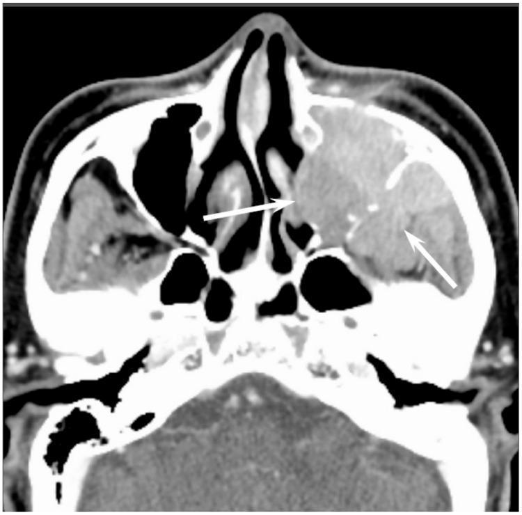
This contrast-enhanced axial CT scan of the midface demonstrates a large, homogeneously enhanced soft-tissue mass originating within the left maxillary sinus, consistent with diffuse large B-cell lymphoma (DLBCL). The lesion fills the entire sinus cavity and exhibits an infiltrative growth pattern. Notably, the remaining sinus walls are visible as thin, linear bony structures submerged within the tumor mass (indicated by white arrows), a characteristic finding of permeative-type tumor invasion often seen in sinonasal non-Hodgkin’s lymphoma. Unlike squamous cell carcinoma, which typically presents with aggressive, destructive bony changes and intratumoral necrosis, this mass shows uniform density and relative preservation of the skeletal framework. The contralateral right maxillary sinus and nasal cavity appear unremarkable. This diagnostic image is used to educate medical professionals on distinguishing sinonasal malignancies based on enhancement patterns and the nature of cortical bone involvement.
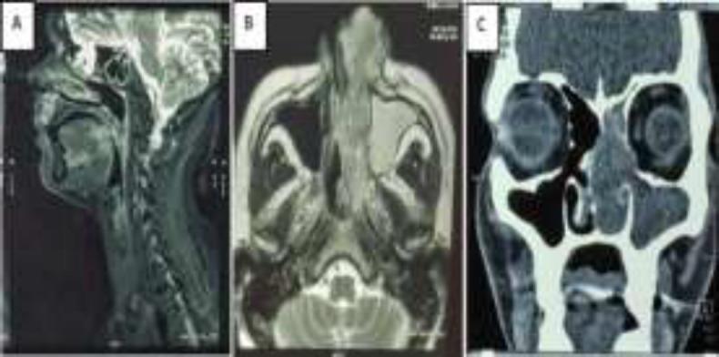
This composite of diagnostic images (MRI and CECT) illustrates a large sinonasal mass with aggressive features. Panel A (sagittal MRI) shows an enhancing lesion involving the base of the tongue. Panel B (axial MRI) reveals an irregular, heterogeneously lobulated soft tissue mass in the left nasal cavity. The mass extends into the deeper fascial planes at the medial orbital canthus and posteriorly into the left nasopharynx, accompanied by complete mucosal thickening of the left maxillary sinus. Panel C (coronal CECT) demonstrates the mass filling the left nasal cavity, causing significant mass effect evidenced by the rightward bowing of the nasal septum and widening of the left osteomeatal complex. Notable bony destruction includes erosion of the left lamina papyracea. These findings are clinically representative of a malignant process such as embryonal rhabdomyosarcoma or squamous cell carcinoma. The images are essential for understanding sinonasal anatomy, tumor staging, and the impact of large lesions on surrounding paranasal and orbital structures.
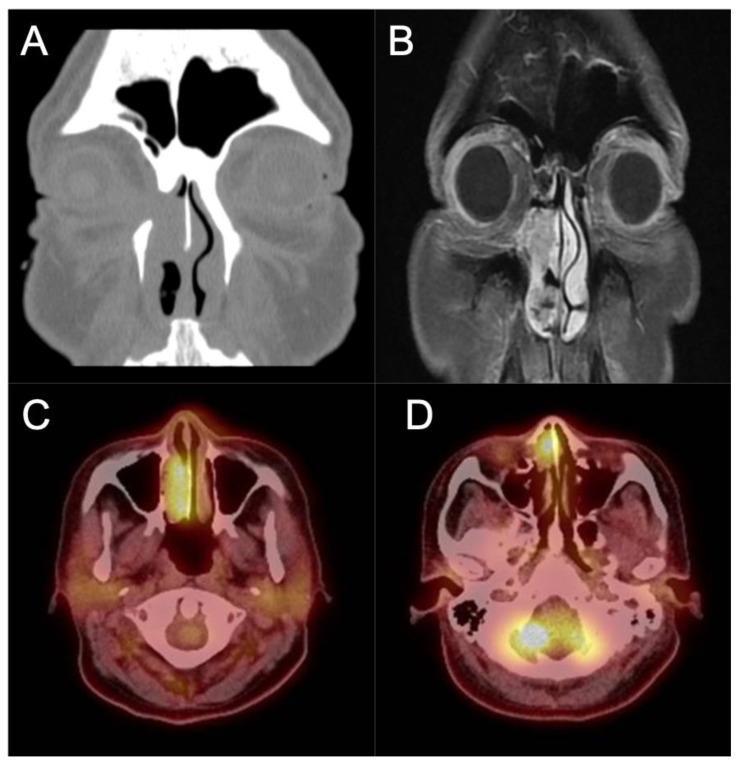
Multi-modal diagnostic imaging of inverted papilloma (IP) with malignant transformation into squamous cell carcinoma (SCC). (A) Coronal non-contrast CT scan shows a large, soft-tissue density mass occupying the right nasal cavity and ethmoid sinus. Visible bony dehiscence of the medial orbital wall (lamina papyracea) indicates aggressive local erosion. (B) Coronal T1-weighted contrast-enhanced MRI provides superior soft-tissue resolution, clearly delineating the tumor's invasion into the medial orbit and periorbita. (C, D) Axial PET/CT fusion images reveal intense FDG avidity (metabolic activity) localized to the right nasal mass, facilitating the differentiation between viable tumor and inspissated secretions. These images collectively illustrate the radiographic features of locally advanced sinonasal malignancy, emphasizing the importance of correlating CT (for bone assessment), MRI (for orbital/dural invasion), and PET/CT (for metabolic staging) in surgical planning and management of secondary malignant degeneration in chronic sinonasal disease.
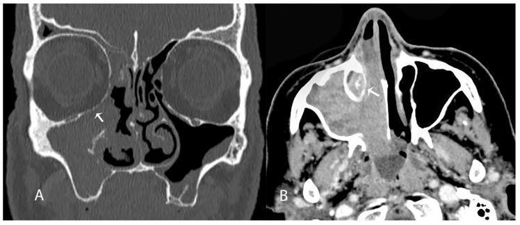
This diagnostic image displays two computed tomography (CT) scans of the paranasal sinuses demonstrating a carcinoma of the maxillary sinus and lateral nasal cavity wall. Panel A is a coronal (frontal) plane CT in a bone window, showing a soft tissue mass occupying the right maxillary sinus and nasal cavity. An arrow highlights focal bone erosion of the inferior orbital wall (orbital floor), indicating tumor extension beyond the sinus boundaries. Panel B is an axial plane CT showing the same soft tissue mass invading the right nasolacrimal duct region, marked by an arrow. The normally air-filled or clear anatomy is replaced by an infiltrative density, suggesting aggressive local progression typical of squamous cell carcinoma or similar sinonasal malignancies. These findings are clinically significant for surgical planning, such as medial maxillectomy, and for assessing the risk of postoperative complications like nasolacrimal duct stenosis.
"sinonasal squamous cell carcinoma"
mkdir -p /tmp/workspace/cpc-nasal-scc/assets /tmp/workspace/cpc-nasal-scc/output && ls -la /tmp/workspace/
cd /tmp/workspace/cpc-nasal-scc && python /tmp/skills/shared/scripts/fetch_images.py \ "https://cdn.orris.care/cdss_images/Pathology_1760055804037_9d6aada6-0dc4-409c-a972-34a7e682aaa7.jpg" \ "https://cdn.orris.care/cdss_images/pmc_clinical_VQA_18e31f29e5ff5a07635a59638f7a6206ab5999030497e15aab9626d125f96c9a.jpg" \ "https://cdn.orris.care/cdss_images/pmc_clinical_VQA_17f41fc0d7e2bd0dad3513e98738a5335db5b2240855177a7167c368bd8c01a5.jpg" \ "https://cdn.orris.care/cdss_images/pmc_clinical_VQA_2b4084779c31d7f83fb8db331ce64b8779d459c0f876070e99f3ad5200630e71.jpg" \ > assets/images.json python3 -c " import json data = json.load(open('assets/images.json')) for d in data: print(d['url'], 'OK' if d.get('base64') else d.get('error')) "
~/cpc-nasal-scc/build_ppt.py
#!/usr/bin/env python3
"""Build CPC presentation: Moderately Differentiated SCC of Nasal Cavity"""
import json, base64
from io import BytesIO
from pptx import Presentation
from pptx.util import Inches, Pt, Emu
from pptx.dml.color import RGBColor
from pptx.enum.text import PP_ALIGN, MSO_ANCHOR
from pptx.enum.shapes import MSO_SHAPE
from pptx.oxml.ns import qn

# ---------- palette ----------
NAVY = RGBColor(0x1B, 0x2A, 0x4A)
TEAL = RGBColor(0x0E, 0x6E, 0x6E)
LIGHT = RGBColor(0xF4, 0xF7, 0xF8)
WHITE = RGBColor(0xFF, 0xFF, 0xFF)
DARK = RGBColor(0x22, 0x22, 0x22)
ACCENT = RGBColor(0xC0, 0x39, 0x2B)
GREY = RGBColor(0x60, 0x60, 0x60)

images = json.load(open("assets/images.json"))
IMG = {d["url"]: d["base64"] for d in images if d.get("base64")}

def img_stream(url):
    b64 = IMG[url]
    raw = base64.b64decode(b64.split(",", 1)[1])
    return BytesIO(raw)

KERATIN_IMG = "https://cdn.orris.care/cdss_images/Pathology_1760055804037_9d6aada6-0dc4-409c-a972-34a7e682aaa7.jpg"
PET_IMG = "https://cdn.orris.care/cdss_images/pmc_clinical_VQA_18e31f29e5ff5a07635a59638f7a6206ab5999030497e15aab9626d125f96c9a.jpg"
IP_MALIG_IMG = "https://cdn.orris.care/cdss_images/pmc_clinical_VQA_17f41fc0d7e2bd0dad3513e98738a5335db5b2240855177a7167c368bd8c01a5.jpg"
CT_BONE_IMG = "https://cdn.orris.care/cdss_images/pmc_clinical_VQA_2b4084779c31d7f83fb8db331ce64b8779d459c0f876070e99f3ad5200630e71.jpg"

prs = Presentation()
prs.slide_width = Inches(13.333)
prs.slide_height = Inches(7.5)
BLANK = prs.slide_layouts[6]
SW, SH = prs.slide_width, prs.slide_height

def add_slide():
    return prs.slides.add_slide(BLANK)

def bg(slide, color=WHITE):
    slide.background.fill.solid()
    slide.background.fill.fore_color.rgb = color

def rect(slide, x, y, w, h, color, line=False):
    sh = slide.shapes.add_shape(MSO_SHAPE.RECTANGLE, x, y, w, h)
    sh.fill.solid()
    sh.fill.fore_color.rgb = color
    if line:
        sh.line.color.rgb = color
    else:
        sh.line.fill.background()
    sh.shadow.inherit = False
    return sh

def textbox(slide, x, y, w, h, text, size=18, color=DARK, bold=False, align=PP_ALIGN.LEFT,
            font="Calibri", anchor=None, line_spacing=1.0, italic=False):
    tb = slide.shapes.add_textbox(x, y, w, h)
    tf = tb.text_frame
    tf.word_wrap = True
    if anchor:
        tf.vertical_anchor = anchor
    lines = text.split("\n")
    for i, line in enumerate(lines):
        p = tf.paragraphs[0] if i == 0 else tf.add_paragraph()
        p.text = line
        p.alignment = align
        p.line_spacing = line_spacing
        for r in p.runs:
            r.font.size = Pt(size)
            r.font.color.rgb = color
            r.font.bold = bold
            r.font.name = font
            r.font.italic = italic
    return tb

def bullets(slide, x, y, w, h, items, size=15, color=DARK, bold_first=False, font="Calibri",
            space_after=8, line_spacing=1.05, bullet_char="\u2022 "):
    tb = slide.shapes.add_textbox(x, y, w, h)
    tf = tb.text_frame
    tf.word_wrap = True
    for i, item in enumerate(items):
        if isinstance(item, tuple):
            text, lvl = item
        else:
            text, lvl = item, 0
        p = tf.paragraphs[0] if i == 0 else tf.add_paragraph()
        prefix = ("    " * lvl) + (bullet_char if lvl == 0 else "- ")
        p.text = prefix + text
        p.space_after = Pt(space_after)
        p.line_spacing = line_spacing
        for r in p.runs:
            r.font.size = Pt(size - (2 if lvl else 0))
            r.font.color.rgb = color
            r.font.name = font
    return tb

def header(slide, kicker, title, num, total=18):
    rect(slide, 0, 0, SW, Inches(1.15), NAVY)
    rect(slide, 0, Inches(1.15), SW, Pt(3), ACCENT)
    textbox(slide, Inches(0.5), Inches(0.12), Inches(8), Inches(0.35), kicker.upper(),
            size=12, color=RGBColor(0x9F, 0xC9, 0xC9), bold=True, font="Calibri")
    textbox(slide, Inches(0.5), Inches(0.42), Inches(11.5), Inches(0.65), title,
            size=26, color=WHITE, bold=True, font="Calibri")
    textbox(slide, Inches(12.5), Inches(0.12), Inches(0.6), Inches(0.35), f"{num:02d}",
            size=13, color=RGBColor(0x9F,0xC9,0xC9), bold=True, align=PP_ALIGN.RIGHT)
    return

def footer(slide, text="CPC \u2022 Moderately Differentiated SCC \u2013 Nasal Cavity \u2022 Pathology Resident Presentation"):
    textbox(slide, Inches(0.5), Inches(7.15), Inches(12.3), Inches(0.3), text,
            size=9, color=GREY, italic=True)

def pic_framed(slide, url, x, y, w, h, caption=None):
    pic = slide.shapes.add_picture(img_stream(url), x, y, height=h)
    # center horizontally within w if narrower
    if pic.width > w:
        pic.width = w
        # keep aspect via python-pptx add_picture w/h both would distort; recompute
    frame = slide.shapes.add_shape(MSO_SHAPE.RECTANGLE, pic.left-Pt(2), pic.top-Pt(2), pic.width+Pt(4), pic.height+Pt(4))
    frame.fill.background()
    frame.line.color.rgb = TEAL
    frame.line.width = Pt(1.5)
    frame.shadow.inherit = False
    if caption:
        textbox(slide, pic.left, pic.top+pic.height+Pt(4), pic.width, Inches(0.4), caption,
                size=10, color=GREY, italic=True, align=PP_ALIGN.CENTER)
    return pic

def table_slide_table(slide, x, y, w, h, headers_row, rows, col_widths=None, header_color=NAVY,
                       font_size=13, header_size=13):
    n_rows = len(rows) + 1
    n_cols = len(headers_row)
    gtable = slide.shapes.add_table(n_rows, n_cols, x, y, w, h).table
    if col_widths:
        for i, cw in enumerate(col_widths):
            gtable.columns[i].width = cw
    for c, htext in enumerate(headers_row):
        cell = gtable.cell(0, c)
        cell.text = htext
        cell.fill.solid()
        cell.fill.fore_color.rgb = header_color
        for p in cell.text_frame.paragraphs:
            p.alignment = PP_ALIGN.CENTER
            for r in p.runs:
                r.font.bold = True
                r.font.size = Pt(header_size)
                r.font.color.rgb = WHITE
    for ri, row in enumerate(rows, start=1):
        for ci, val in enumerate(row):
            cell = gtable.cell(ri, ci)
            cell.text = str(val)
            cell.fill.solid()
            cell.fill.fore_color.rgb = LIGHT if ri % 2 == 0 else WHITE
            for p in cell.text_frame.paragraphs:
                for r in p.runs:
                    r.font.size = Pt(font_size)
                    r.font.color.rgb = DARK
    return gtable

TOTAL = 18

# ===================== SLIDE 1: TITLE =====================
s = add_slide(); bg(s, NAVY)
rect(s, 0, Inches(5.6), SW, Inches(1.9), TEAL)
rect(s, 0, Inches(5.55), SW, Pt(3), ACCENT)
textbox(s, Inches(0.9), Inches(1.6), Inches(11.5), Inches(0.5), "CLINICOPATHOLOGICAL CORRELATION (CPC)",
        size=20, color=RGBColor(0x9F,0xC9,0xC9), bold=True, align=PP_ALIGN.LEFT)
textbox(s, Inches(0.9), Inches(2.15), Inches(11.5), Inches(2.0),
        "Moderately Differentiated\nSquamous Cell Carcinoma\nof the Nasal Cavity",
        size=40, color=WHITE, bold=True, align=PP_ALIGN.LEFT, line_spacing=1.05)
textbox(s, Inches(0.9), Inches(5.8), Inches(11), Inches(0.5), "Department of Pathology",
        size=18, color=WHITE, bold=True)
textbox(s, Inches(0.9), Inches(6.25), Inches(11), Inches(0.9),
        "Presented by: Pathology Resident\nClinicoradiologic-Pathologic Case Discussion",
        size=14, color=RGBColor(0xE8,0xF3,0xF3), line_spacing=1.2)

# ===================== SLIDE 2: NOTE + OBJECTIVES =====================
s = add_slide(); bg(s, WHITE)
header(s, "Preliminary Note", "Case Source & Learning Objectives", 2, TOTAL)
rect(s, Inches(0.5), Inches(1.5), Inches(12.3), Inches(1.35), RGBColor(0xFD,0xF0,0xE8))
textbox(s, Inches(0.75), Inches(1.62), Inches(11.8), Inches(1.1),
        "Note: The submitted histopathology report (HP No. 4093-2026) documents a biopsy of the TONGUE "
        "showing Grade 1 (well-differentiated) SCC. As requested, this CPC session is built as a teaching "
        "case on Moderately Differentiated (Grade 2) SCC of the NASAL CAVITY, using standard "
        "sinonasal SCC literature and grading criteria \u2013 the site/grade differ intentionally from the attached report.",
        size=13, color=ACCENT, italic=True, line_spacing=1.15)
bullets(s, Inches(0.6), Inches(3.1), Inches(11.8), Inches(3.6), [
    "Correlate clinical presentation and imaging of a sinonasal mass with histopathological diagnosis",
    "Review the epidemiology, risk factors, and anatomic peculiarities of nasal cavity SCC",
    "Understand histological grading criteria distinguishing well / moderately / poorly differentiated SCC",
    "Discuss immunohistochemistry and key histological mimics in the sinonasal tract",
    "Apply AJCC/UICC TNM staging relevant to nasal cavity and paranasal sinus tumours",
    "Integrate findings into a unified clinicopathological diagnosis and management plan",
], size=16, space_after=14)
footer(s)

# ===================== SLIDE 3: ILLUSTRATIVE CLINICAL HISTORY =====================
s = add_slide(); bg(s, WHITE)
header(s, "Case Vignette (Illustrative)", "Clinical History", 3, TOTAL)
left_w = Inches(6.0)
rect(s, Inches(0.5), Inches(1.5), left_w, Inches(5.3), LIGHT)
textbox(s, Inches(0.7), Inches(1.65), Inches(5.6), Inches(0.4), "Patient Profile", size=16, bold=True, color=NAVY)
bullets(s, Inches(0.7), Inches(2.1), Inches(5.6), Inches(1.6), [
    "58-year-old male, chronic smoker (30 pack-years)",
    "History of occupational wood-dust / nickel exposure",
    "Presented with 4-month history of unilateral nasal obstruction",
], size=14)
textbox(s, Inches(0.7), Inches(3.7), Inches(5.6), Inches(0.4), "Presenting Complaints", size=16, bold=True, color=NAVY)
bullets(s, Inches(0.7), Inches(4.15), Inches(5.6), Inches(2.5), [
    "Blood-stained nasal discharge (epistaxis)",
    "Foul-smelling discharge, facial pain/pressure over left maxilla",
    "Progressive facial swelling and diplopia (late feature - orbital extension)",
    "No significant cervical lymphadenopathy on presentation",
], size=14)
rect(s, Inches(6.7), Inches(1.5), Inches(6.1), Inches(5.3), NAVY)
textbox(s, Inches(6.9), Inches(1.65), Inches(5.7), Inches(0.4), "Examination / Endoscopy", size=16, bold=True, color=WHITE)
bullets(s, Inches(6.9), Inches(2.15), Inches(5.7), Inches(4.4), [
    "Nasal endoscopy: friable, irregular, hemorrhagic mass filling left nasal cavity",
    "Mass bleeds on touch; surface ulceration with necrotic debris",
    "Septal deviation/bowing away from mass; possible extension into ethmoid/maxillary sinus",
    "Facial asymmetry if orbital or cheek involvement",
    "ENT differential at this stage: SCC, inverted papilloma (with malignant transformation), esthesioneuroblastoma, sinonasal lymphoma, adenoid cystic carcinoma",
], size=14.5, color=WHITE, space_after=12)
footer(s)

# ===================== SLIDE 4: EPIDEMIOLOGY & RISK FACTORS =====================
s = add_slide(); bg(s, WHITE)
header(s, "Background", "Epidemiology & Risk Factors", 4, TOTAL)
bullets(s, Inches(0.6), Inches(1.6), Inches(6.1), Inches(5.2), [
    "Sinonasal malignancies are rare - <1% of all cancers, ~3% of head & neck cancers",
    "SCC is the most common histology of the nasal cavity and paranasal sinuses (~50-80%)",
    "Peak incidence: 6th-7th decade; male predominance (~2:1)",
    "Maxillary sinus > nasal cavity > ethmoid sinus in frequency",
], size=15.5, space_after=14)
textbox(s, Inches(6.9), Inches(1.6), Inches(5.9), Inches(0.4), "Key Risk Factors", size=17, bold=True, color=TEAL)
bullets(s, Inches(6.9), Inches(2.1), Inches(5.9), Inches(4.6), [
    "Tobacco smoking and alcohol (synergistic effect, as in other head & neck SCC)",
    "Occupational exposure: wood dust, nickel, chromium, leather/textile dust",
    "Chronic sinonasal inflammation",
    "Human Papillomavirus (HPV) - implicated in a subset (non-keratinizing variant)",
    "Malignant transformation of inverted (Schneiderian) papilloma (~5-15% of cases)",
    "Prior radiation exposure to the region",
], size=15.5, color=DARK, space_after=12)
footer(s)

# ===================== SLIDE 5: IMAGING =====================
s = add_slide(); bg(s, WHITE)
header(s, "Investigations", "Radiological Correlation", 5, TOTAL)
textbox(s, Inches(0.6), Inches(1.35), Inches(11.8), Inches(0.4),
        "CT delineates bone erosion; MRI defines soft-tissue/orbital-dural extent; FDG-PET aids staging & recurrence surveillance",
        size=14, italic=True, color=GREY)
pic_framed(s, CT_BONE_IMG, Inches(0.55), Inches(1.9), Inches(4.0), Inches(3.0),
           "CT (bone window): soft-tissue mass in maxillary sinus/nasal cavity with orbital floor erosion")
pic_framed(s, PET_IMG, Inches(4.75), Inches(1.9), Inches(4.0), Inches(3.0),
           "FDG PET-CT: hypermetabolic tumour along nasal septum extending through nasal cavity")
pic_framed(s, IP_MALIG_IMG, Inches(8.95), Inches(1.9), Inches(4.0), Inches(3.0),
           "CT/MRI/PET: locally advanced sinonasal SCC with medial orbital wall erosion & periorbital invasion")
bullets(s, Inches(0.6), Inches(5.35), Inches(11.9), Inches(1.6), [
    "Findings driving suspicion of malignancy over benign polyp/papilloma: unilateral bone destruction, heterogeneous enhancement, orbital/skull-base invasion, FDG avidity",
    "Imaging guides biopsy site selection and pre-treatment staging (T-stage largely determined radiologically)",
], size=13.5, space_after=8)
footer(s)

# ===================== SLIDE 6: GROSS / BIOPSY =====================
s = add_slide(); bg(s, WHITE)
header(s, "Specimen", "Gross Examination & Biopsy", 6, TOTAL)
bullets(s, Inches(0.6), Inches(1.6), Inches(11.9), Inches(2.0), [
    "Nature of surgery: Endoscopic biopsy of nasal cavity mass / FFPE tissue block for histopathology",
    "Macroscopic findings (typical): multiple grey-white to grey-brown, firm-to-friable tissue fragments, "
    "ranging 0.2-1.5 cm, occasionally admixed with blood clot and necrotic debris",
    "Slide key: entire submitted tissue processed and embedded (as per standard small-biopsy protocol)",
], size=16, space_after=16)
rect(s, Inches(0.6), Inches(3.9), Inches(11.9), Inches(2.7), LIGHT)
textbox(s, Inches(0.85), Inches(4.05), Inches(11.4), Inches(0.4), "Clinical Diagnosis on Request Form", size=16, bold=True, color=NAVY)
textbox(s, Inches(0.85), Inches(4.55), Inches(11.4), Inches(1.8),
        "\"Fungating ulceroproliferative mass, left nasal cavity, under evaluation - r/o malignancy\"\n\n"
        "Grossing done by: Resident on duty     |     Reporting done by: Consultant Pathologist",
        size=15, color=DARK, line_spacing=1.3)
footer(s)

# ===================== SLIDE 7: MICROSCOPY - ARCHITECTURE =====================
s = add_slide(); bg(s, WHITE)
header(s, "Histopathology", "Microscopic Findings - Low Power", 7, TOTAL)
bullets(s, Inches(0.6), Inches(1.6), Inches(6.6), Inches(5.2), [
    "Sections show stratified squamous respiratory-type epithelium with an underlying infiltrating neoplasm",
    "Tumour arranged in irregular nests, cords, and islands infiltrating the submucosal stroma",
    "Nests show variable central keratinization; some show comedo-type necrosis",
    "Desmoplastic stromal reaction at the invasive front with chronic inflammatory infiltrate",
    "Areas of surface dysplasia / carcinoma in situ may be seen at the periphery",
    "Angiolymphatic and perineural spaces should be assessed for tumour emboli/invasion",
], size=15.5, space_after=13)
pic_framed(s, KERATIN_IMG, Inches(7.5), Inches(1.7), Inches(5.2), Inches(4.6),
           "H&E: nests of malignant squamous cells with keratin pearl formation and stromal invasion (representative keratinizing SCC morphology)")
footer(s)

# ===================== SLIDE 8: MICROSCOPY - GRADE 2 FEATURES =====================
s = add_slide(); bg(s, WHITE)
header(s, "Histopathology", "High Power - Features of Moderate Differentiation (Grade 2)", 8, TOTAL)
bullets(s, Inches(0.6), Inches(1.55), Inches(11.9), Inches(3.0), [
    "Tumour cells: polygonal, moderate-to-large size, with increased nuclear:cytoplasmic ratio",
    "Nuclei: vesicular to hyperchromatic chromatin, pleomorphic, prominent nucleoli, easily identifiable mitoses (including occasional atypical mitoses)",
    "Cytoplasm: moderate eosinophilic (glassy) cytoplasm; intercellular bridges discernible but less conspicuous than in well-differentiated SCC",
    "Keratinization: present but partial/patchy - individual cell keratinization and occasional keratin pearls, less abundant than Grade 1",
    "Architecture: infiltrative nests and cords with more irregular borders and single-cell infiltration compared to well-differentiated tumours",
    "Necrosis and mitotic activity intermediate between well- and poorly-differentiated tumours",
], size=15, space_after=10)
rect(s, Inches(0.6), Inches(4.85), Inches(11.9), Inches(1.85), RGBColor(0xE9,0xF3,0xF3))
textbox(s, Inches(0.85), Inches(4.98), Inches(11.3), Inches(0.4), "Impression", size=16, bold=True, color=TEAL)
textbox(s, Inches(0.85), Inches(5.45), Inches(11.3), Inches(1.1),
        "\"Sections show an infiltrating squamous cell carcinoma with intermediate nuclear atypia and "
        "patchy keratinization - features compatible with Moderately Differentiated (Grade 2) Squamous Cell Carcinoma, "
        "arising from the nasal cavity mucosa.\"", size=14.5, italic=True, color=DARK, line_spacing=1.2)
footer(s)

# ===================== SLIDE 9: GRADING TABLE =====================
s = add_slide(); bg(s, WHITE)
header(s, "Histological Grading", "Grade 1 vs Grade 2 vs Grade 3 SCC", 9, TOTAL)
headers = ["Feature", "Grade 1\n(Well differentiated)", "Grade 2\n(Moderately differentiated)", "Grade 3\n(Poorly differentiated)"]
rows = [
    ["Keratinization", "Abundant, prominent keratin pearls", "Focal / patchy keratin pearls, individual cell keratinization", "Minimal to absent"],
    ["Cell resemblance", "Closely resembles normal squamous epithelium", "Recognizable squamous origin, intermediate atypia", "Poor resemblance, marked pleomorphism"],
    ["Nuclear features", "Mild atypia, low N:C ratio", "Moderate atypia, vesicular/hyperchromatic nuclei, prominent nucleoli", "Marked atypia, high N:C ratio, bizarre nuclei"],
    ["Mitoses", "Few, typical", "Moderate number, occasional atypical", "Numerous, frequently atypical"],
    ["Intercellular bridges", "Easily seen", "Discernible but less conspicuous", "Sparse / absent"],
    ["Necrosis", "Minimal", "Focal", "Extensive"],
]
table_slide_table(s, Inches(0.4), Inches(1.5), Inches(12.5), Inches(4.7), headers, rows,
                   col_widths=[Inches(2.3), Inches(3.4), Inches(3.6), Inches(3.2)], font_size=12.5, header_size=13)
textbox(s, Inches(0.4), Inches(6.35), Inches(12.5), Inches(0.6),
        "Note: Grading systems (Broders-derived) are subjective with inter-observer variability; remains an adjunctive, non-mandatory component of TNM reporting per AJCC/UICC.",
        size=11.5, italic=True, color=GREY)
footer(s)

# ===================== SLIDE 10: IHC =====================
s = add_slide(); bg(s, WHITE)
header(s, "Ancillary Studies", "Immunohistochemistry", 10, TOTAL)
headers2 = ["Marker", "Expected Result in SCC", "Utility"]
rows2 = [
    ["p40 / p63", "Diffusely positive (nuclear)", "Confirms squamous lineage; most specific/sensitive squamous markers"],
    ["CK5/6, CK AE1/AE3", "Positive", "Confirms epithelial/squamous differentiation"],
    ["p16 / HPV testing", "Variable (positive in HPV-related subset)", "Identifies HPV-associated non-keratinizing SCC subtype; prognostic relevance debated at this site"],
    ["S100 / SOX10", "Negative", "Excludes melanoma / olfactory neuroblastoma / schwannian mimics"],
    ["Synaptophysin, chromogranin, CD56", "Negative", "Excludes neuroendocrine carcinoma / esthesioneuroblastoma / SNUC"],
    ["CD45 (LCA), CD3, CD20", "Negative", "Excludes lymphoma (esp. NK/T-cell lymphoma, a key sinonasal mimic)"],
]
table_slide_table(s, Inches(0.4), Inches(1.5), Inches(12.5), Inches(5.0), headers2, rows2,
                   col_widths=[Inches(2.8), Inches(4.2), Inches(5.5)], font_size=13, header_size=14)
footer(s)

# ===================== SLIDE 11: HISTOLOGICAL DIFFERENTIAL =====================
s = add_slide(); bg(s, WHITE)
header(s, "Differential Diagnosis", "Histopathological Mimics in the Sinonasal Tract", 11, TOTAL)
bullets(s, Inches(0.6), Inches(1.55), Inches(6.0), Inches(5.3), [
    "Sinonasal undifferentiated carcinoma (SNUC) - lacks squamous differentiation, more aggressive, negative p40 in most",
    "Inverted (Schneiderian) papilloma with dysplasia/malignant transformation - endophytic ribbons of epithelium; look for invasive component",
    "Esthesioneuroblastoma (olfactory neuroblastoma) - lobules of small round cells, Homer-Wright rosettes, synaptophysin+",
    "NUT-midline carcinoma - undifferentiated cells, NUT immunopositive, aggressive",
    "Sinonasal (extranodal NK/T-cell) lymphoma - destructive necrotic lesion, CD3+/CD56+/EBV+",
    "Adenoid cystic / mucoepidermoid carcinoma - glandular/cribriform or mixed mucinous-squamoid pattern",
    "Mucosal malignant melanoma - S100/SOX10/HMB45 positive, may be amelanotic",
], size=14, space_after=10)
rect(s, Inches(6.9), Inches(1.55), Inches(5.85), Inches(5.3), NAVY)
textbox(s, Inches(7.1), Inches(1.7), Inches(5.5), Inches(0.4), "Why it matters", size=16, bold=True, color=WHITE)
bullets(s, Inches(7.1), Inches(2.2), Inches(5.5), Inches(4.5), [
    "Sinonasal mimics can closely resemble SCC on H&E, especially in crushed or poorly-fixed small biopsies",
    "Misdiagnosis changes treatment radically (e.g., lymphoma - chemo/RT vs SCC - surgery + RT)",
    "IHC panel (p40, synaptophysin, CD45, S100) is essential before finalizing diagnosis",
    "Adequate, well-oriented biopsy with correlation to endoscopic and imaging findings reduces error",
], size=14.5, color=WHITE, space_after=12)
footer(s)

# ===================== SLIDE 12: TNM STAGING =====================
s = add_slide(); bg(s, WHITE)
header(s, "Staging", "AJCC/UICC TNM - Nasal Cavity & Ethmoid Sinus", 12, TOTAL)
headers3 = ["Stage", "T", "N", "M"]
rows3 = [
    ["T1", "Tumour confined to one subsite, with or without bony invasion", "-", "-"],
    ["T2", "Tumour invading two subsites in a single region, or extending to involve an adjacent region within the nasoethmoidal complex", "-", "-"],
    ["T3", "Tumour extends to invade medial orbit, maxillary sinus, palate, or cribriform plate", "-", "-"],
    ["T4a", "Moderately advanced: orbital contents, skin, minimal brain/dura extension, pterygoid plates, sphenoid/frontal sinus", "-", "-"],
    ["T4b", "Very advanced: orbital apex, brain, middle cranial fossa, cranial nerves other than V2, nasopharynx, clivus", "-", "-"],
    ["N stage", "Same as other head & neck sites - based on size/number/laterality of nodes and ENE", "N0-N3", "-"],
]
table_slide_table(s, Inches(0.4), Inches(1.5), Inches(12.5), Inches(4.9), headers3, rows3,
                   col_widths=[Inches(1.4), Inches(9.5), Inches(0.8), Inches(0.8)], font_size=12, header_size=13)
textbox(s, Inches(0.4), Inches(6.5), Inches(12.5), Inches(0.5),
        "Occult nodal metastasis in sinonasal SCC is relatively low (~10-15%) compared to oral/oropharyngeal SCC, influencing decisions on elective neck treatment.",
        size=11.5, italic=True, color=GREY)
footer(s)

# ===================== SLIDE 13: CPC SYNTHESIS TABLE =====================
s = add_slide(); bg(s, WHITE)
header(s, "Synthesis", "Clinicopathological Correlation", 13, TOTAL)
headers4 = ["Clinical / Radiological Finding", "Pathological Correlate"]
rows4 = [
    ["Unilateral nasal obstruction, blood-stained discharge", "Bulky infiltrative tumour with surface ulceration and friable, hemorrhagic mucosa"],
    ["Foul-smelling discharge", "Tumour necrosis and secondary infection within necrotic tumour nests"],
    ["CT: bone erosion of orbital floor / medial wall", "Deeply infiltrative growth pattern with destructive (non-pushing) invasion of nests into stroma and bone"],
    ["FDG-PET hypermetabolism", "High proliferative/mitotic activity and moderate-to-high cellularity of tumour"],
    ["No palpable cervical lymphadenopathy at presentation", "Relatively low rate of occult nodal metastasis characteristic of sinonasal SCC (unlike oral cavity SCC)"],
    ["Facial swelling / diplopia (late)", "Direct extension through eroded lamina papyracea into orbit/periorbita"],
    ["Friable, bleeds on touch (endoscopy)", "Increased vascularity, thin/absent surface keratin layer, desmoplastic stroma"],
]
table_slide_table(s, Inches(0.4), Inches(1.5), Inches(12.5), Inches(5.2), headers4, rows4,
                   col_widths=[Inches(6.0), Inches(6.5)], font_size=13, header_size=14)
footer(s)

# ===================== SLIDE 14: MANAGEMENT =====================
s = add_slide(); bg(s, WHITE)
header(s, "Management", "Treatment Overview", 14, TOTAL)
bullets(s, Inches(0.6), Inches(1.6), Inches(6.0), Inches(5.2), [
    "Primary modality: Surgical resection (endoscopic or open craniofacial resection) with clear margins",
    "Adjuvant radiotherapy for T3/T4, positive margins, perineural/lymphovascular invasion, or nodal disease",
    "Concurrent chemoradiotherapy considered for unresectable or advanced disease",
    "Elective neck treatment individualized given generally lower occult nodal metastasis rate",
    "Multidisciplinary tumour board input: ENT-head & neck surgery, radiation oncology, medical oncology, pathology, radiology",
], size=15.5, space_after=14)
textbox(s, Inches(6.9), Inches(1.6), Inches(5.9), Inches(0.4), "Prognostic Factors", size=17, bold=True, color=TEAL)
bullets(s, Inches(6.9), Inches(2.1), Inches(5.9), Inches(4.6), [
    "T-stage and skull base / orbital / dural invasion",
    "Histological grade (poorly differentiated -> higher metastatic risk & worse survival)",
    "Margin status after resection",
    "Perineural invasion and lymphovascular invasion",
    "Nodal metastasis at presentation",
    "5-year overall survival for sinonasal SCC overall: approximately 40-60%, worse with advanced T-stage",
], size=15, space_after=12)
footer(s)

# ===================== SLIDE 15: KEY TAKE-HOME =====================
s = add_slide(); bg(s, NAVY)
textbox(s, Inches(0.6), Inches(0.5), Inches(11.5), Inches(0.7), "Key Take-Home Points", size=30, bold=True, color=WHITE)
rect(s, Inches(0.6), Inches(1.3), Inches(3.2), Pt(3), ACCENT)
bullets(s, Inches(0.7), Inches(1.7), Inches(11.9), Inches(5.4), [
    "Nasal cavity/paranasal sinus SCC is uncommon but clinically important; unilateral obstruction with epistaxis in an older patient warrants biopsy",
    "Moderately differentiated (Grade 2) SCC shows intermediate features between well and poorly differentiated tumours: partial keratinization, moderate nuclear atypia, discernible but reduced intercellular bridges",
    "p40/p63/CK5-6 positivity confirms squamous lineage; a broader IHC panel excludes aggressive sinonasal mimics (SNUC, lymphoma, esthesioneuroblastoma, melanoma)",
    "Imaging (bone erosion, orbital/skull-base extension) closely mirrors the deeply infiltrative histological growth pattern - the essence of clinicopathological correlation",
    "Grade, margin status, and local extent (T-stage) - not grade alone - drive prognosis and treatment planning",
], size=17, color=WHITE, space_after=16)

# ===================== SLIDE 16: REFERENCES =====================
s = add_slide(); bg(s, WHITE)
header(s, "References", "Sources Consulted", 16, TOTAL)
bullets(s, Inches(0.6), Inches(1.7), Inches(11.9), Inches(5.0), [
    "Scott-Brown's Otorhinolaryngology, Head & Neck Surgery - \"Squamous Malignant Tumours\" and \"Histopathological Grading\" chapters",
    "Cummings Otolaryngology Head and Neck Surgery - Sinonasal malignancy, pathology and imaging sections",
    "Textbook of Family Medicine 9e - Sinonasal Tumours",
    "Poutoglidis A, et al. Occult nodal metastasis in sinonasal SCC - a systematic review. Eur Arch Otorhinolaryngol. 2024 (PMID: 38367074)",
    "Vasudevan SS, et al. Induction chemotherapy in sinonasal SCC - systematic review & meta-analysis. Acta Otolaryngol. 2025 (PMID: 40611588)",
    "Waters C, et al. Radiomics to predict recurrence in sinonasal SCC - systematic review & meta-analysis. Clin Otolaryngol. 2026 (PMID: 40910476)",
], size=15, space_after=14)
rect(s, Inches(0.6), Inches(5.9), Inches(11.9), Inches(0.9), RGBColor(0xFD,0xF0,0xE8))
textbox(s, Inches(0.8), Inches(6.0), Inches(11.5), Inches(0.7),
        "Recent systematic reviews (2024-2026) did not contradict the grading/staging framework used above; they primarily add data on nodal risk, radiomic recurrence prediction, and induction chemotherapy outcomes.",
        size=12.5, italic=True, color=ACCENT)
footer(s)

# ===================== SLIDE 17: DISCUSSION / QUESTIONS =====================
s = add_slide(); bg(s, TEAL)
textbox(s, Inches(0.8), Inches(2.6), Inches(11.7), Inches(1.0), "Discussion & Questions", size=40, bold=True, color=WHITE, align=PP_ALIGN.CENTER)
textbox(s, Inches(0.8), Inches(3.8), Inches(11.7), Inches(1.2),
        "Clinicopathological Correlation Session\nModerately Differentiated Squamous Cell Carcinoma - Nasal Cavity",
        size=18, color=RGBColor(0xE8,0xF3,0xF3), align=PP_ALIGN.CENTER, line_spacing=1.3)

prs.save("output/CPC_Moderately_Differentiated_SCC_Nose.pptx")
print("Saved. Slides:", len(prs.slides.__iter__.__self__._sldIdLst))