
Causes of reactive lymphadenitis
Reading File
Reactive lymphadenitis is lymph node enlargement due to a benign immune response to local or systemic stimuli - infections (bacterial, viral, fungal, parasitic) are the most common cause, but autoimmune diseases, drug reactions, and vaccine responses can also trigger it.
Searching sources...
Finding Sources
Finding Sources
Reading File
Reading File
Reading File
Loading Image
Reading File
Here is a comprehensive summary of the causes of reactive lymphadenitis, drawn from authoritative pathology and clinical textbooks.
Causes of Reactive Lymphadenitis
Reactive lymphadenitis is lymph node enlargement triggered when immune cells within the node are activated by foreign antigens - either infectious or non-infectious. The causes are best understood by examining the three main histological reaction patterns they produce.
1. Acute Nonspecific Lymphadenitis
This occurs either locally (draining a regional infection) or systemically. The node becomes swollen, gray-red, and engorged, with large germinal centers and neutrophilic infiltrates. With pyogenic organisms, abscess formation may follow.
Common causes:
- Bacterial infections - streptococci and staphylococci are the most frequent culprits
- Tonsillitis, dental infections, and bacterial pharyngitis (common causes of acute cervical lymphadenitis)
- Any regional pyogenic infection draining into a node group
2. Chronic Nonspecific Lymphadenitis - Three Patterns
A. Follicular Hyperplasia (B-cell driven)
Activated B cells migrate into follicles, creating enlarged, reactive germinal centers with tingible body macrophages.
Causes:
- Rheumatoid arthritis (and other autoimmune diseases)
- Toxoplasmosis
- Early HIV infection
B. Paracortical Hyperplasia (T-cell driven)
Activated parafollicular T cells transform into large immunoblasts, which can efface B-cell follicles.
Causes:
- Viral infections, especially Epstein-Barr virus (EBV) / infectious mononucleosis
- Vaccinations (post-immunization lymphadenopathy)
- Drug reactions, especially phenytoin (and other anticonvulsants)
C. Sinus Histiocytosis
Distension of lymphatic sinusoids by hypertrophied endothelial cells and infiltrating macrophages.
Causes:
- Lymph nodes draining carcinomas (represents an anti-tumor immune response)
3. Other Named Causes (Specific / Granulomatous)
| Cause | Key Features |
|---|---|
| Cat-scratch disease (Bartonella henselae) | Granulomatous or suppurative cervical adenopathy; history of cat scratch |
| Tuberculosis | Granulomatous lymphadenitis with caseating necrosis |
| Atypical mycobacteria (M. avium) | Subacute; violaceous overlying skin; common in children |
| Toxoplasmosis | Follicular hyperplasia with epithelioid histiocyte clusters blurring germinal centers; monocytoid B cells |
| EBV (infectious mononucleosis) | Generalized lymphadenopathy, paracortical pattern |
| CMV (cytomegalovirus) | Rare; chronic lymphadenopathy |
| Syphilis | Generalized or regional reactive adenopathy |
| Rubella | Generalized lymphadenopathy |
| HIV | Early: follicular hyperplasia; late: lymph node depletion |
| Kikuchi histiocytic necrotizing lymphadenitis | Benign self-limited; necrotizing without neutrophils |
| Kawasaki disease | Febrile illness with cervical lymphadenitis in children |
| Rosai-Dorfman disease | Massive sinus histiocytosis |
| Castleman disease | Benign lymph node hyperplasia |
4. Non-infectious / Non-microbial Causes
- Autoimmune diseases - rheumatoid arthritis, SLE, and others
- Drug reactions - phenytoin is classic; also other anticonvulsants
- Post-vaccination response
- Reaction to draining malignancy (sinus histiocytosis pattern)
Histological Summary
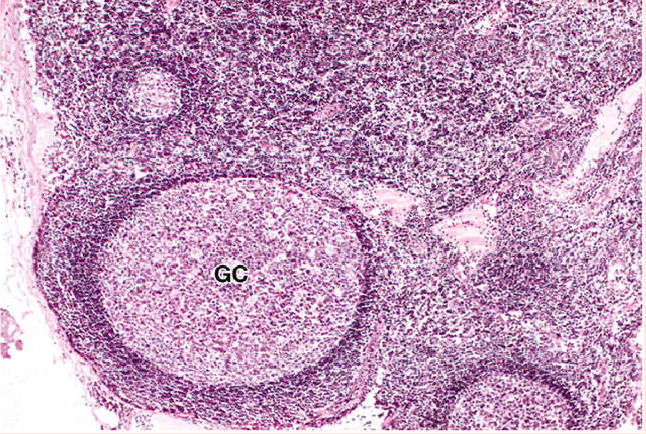
Reactive lymphadenitis: hyperplastic germinal center (GC) with B lymphocytes and macrophages inside; T lymphocyte mantle zone surrounding it. - Histology: A Text and Atlas (Pawlina)
Sources:
- Robbins & Kumar Basic Pathology (Robbins Pathology), pp. 2198-2230
- Histology: A Text and Atlas with Correlated Cell and Molecular Biology (Pawlina), pp. 1224-1225
- Henry's Clinical Diagnosis and Management by Laboratory Methods, p. 2776
- Cummings Otolaryngology Head and Neck Surgery, pp. 3889-3899
This is a shared conversation. Sign in to Orris to start your own chat.