
General examination for viva PICCLE
Reading File
General examination for viva (PICCLE) covers the systematic components assessed in a clinical exam. Let me pull the relevant textbook content right away.
Searching sources...
Finding Sources
Finding Sources
Reading File
Finding Sources
Searching the Web
PICCLE general examination viva pallor icterus cyanosis clubbing lymphadenopathy edema clinical
Finding Sources
Finding Sources
Finding Sources
Finding Sources
Reading File
Finding Sources
Finding Sources
Reading Web Page
https://www.studocu.com/in/document/rajiv-gandhi-university-…
I now have enough textbook + web search data to compile a thorough viva answer on PICCLE. Here it is:
General Examination for Viva - PICCLE
PICCLE is a systematic mnemonic for the general physical examination:
P - Pallor | I - Icterus | C - Cyanosis | C - Clubbing | L - Lymphadenopathy | E - Edema
P - Pallor
Definition: Paleness of skin and mucous membranes due to reduced hemoglobin, reduced blood flow, or vasoconstriction.
Sites to examine: Conjunctivae (lower palpebral), palmar creases, nail beds, tongue, buccal mucosa.
Best site: Lower palpebral conjunctiva - pallor here is most reliable.
Grading:
| Grade | Finding |
|---|---|
| + | Mild pallor - conjunctiva only |
| ++ | Moderate - conjunctiva + palmar creases |
| +++ | Severe - conjunctiva + palms + tongue |
Causes:
- Blood loss: GI bleed, trauma, menorrhagia
- Hemolysis: Malaria, sickle cell, autoimmune hemolytic anemia
- Decreased production: Iron deficiency, B12/folate deficiency, aplastic anemia, CKD (reduced erythropoietin), bone marrow infiltration (leukemia, myeloma)
- Hemodilution: Pregnancy, overhydration
Viva tip: Pallor ≠ anemia - pallor is a clinical sign; anemia is confirmed by Hb measurement. A person can look pale without anemia (shock, vasospasm) and can be anemic without visible pallor (dark skin).
I - Icterus (Jaundice)
Definition: Yellow discoloration of sclerae, skin, and mucous membranes due to elevated serum bilirubin (>2.5 mg/dL or >43 µmol/L).
Best site: Sclera under natural daylight. In dark-skinned individuals, examine the mucous membranes under the tongue.
Viva tip from Harrison's: Jaundice is rarely detectable if serum bilirubin is <2.5 mg/dL (43 µmol/L), but may remain detectable below this threshold during recovery due to tissue binding of conjugated bilirubin. - Harrison's Principles of Internal Medicine 22E
Classification:
| Type | Bilirubin | Urine color | Stool color | Causes |
|---|---|---|---|---|
| Pre-hepatic (hemolytic) | Unconjugated ↑ | Normal (pale/dark) | Normal/pale | Malaria, hemolysis, sickle cell |
| Hepatic (hepatocellular) | Both ↑ | Dark (conjugated in urine) | Pale | Hepatitis, cirrhosis, drugs |
| Post-hepatic (obstructive) | Conjugated ↑ | Dark (tea-colored) | Clay/pale | Gallstones, cholangiocarcinoma, pancreatic Ca, CBD stricture |
Signs accompanying jaundice:
- Pruritus (cholestasis - bile salts in skin)
- Pale stools + dark urine = obstructive
- Splenomegaly = hemolytic or hepatic
- Spider angiomata, palmar erythema = hepatocellular
C - Cyanosis
Definition: Bluish discoloration of skin/mucous membranes due to >5 g/dL of deoxyhemoglobin in capillary blood (not applicable in severe anemia - may not be detectable).
Types:
| Feature | Central Cyanosis | Peripheral Cyanosis |
|---|---|---|
| Site | Tongue, lips, buccal mucosa | Fingertips, toes, earlobes |
| Cause | Cardiorespiratory disease | Reduced blood flow/vasoconstriction |
| Tongue | Cyanosed | Normal |
| Oxygen test | Does NOT improve with O2 (if shunt) | Improves with O2 or warming |
| Common causes | CHD (R-to-L shunt), severe pneumonia, ARDS, pulmonary hypertension, high altitude | Heart failure, Raynaud's, shock, cold exposure |
- Barash Clinical Anesthesia: Peripheral cyanosis in fingers/toes should be distinguished from acrocyanosis; central cyanosis in buccal mucosa is usually secondary to arterial hypoxemia.
Special types:
- Differential cyanosis (cyanosis of feet but not hands): patent ductus arteriosus with pulmonary hypertension
- Reverse differential cyanosis (hands cyanosed, feet pink): transposition of great arteries with PDA
C - Clubbing
Definition: Bulbous enlargement of the terminal phalanges due to proliferation of connective tissue, associated with increased vascularity and edema.
Signs:
- Loss of nail fold angle (Lovibond's angle - normally <165°)
- Fluctuation of nail bed (spongy feel)
- Positive Schamroth's window test (diamond-shaped gap disappears when dorsal surfaces of two thumbs are apposed)
- Drumstick appearance of fingers
Grades (Schamroth / Clubbing Grading):
| Grade | Feature |
|---|---|
| 1 | Softening/fluctuation of nail bed |
| 2 | Obliteration of Lovibond's angle (hyponychial angle >180°) |
| 3 | Beaking/parrot-beak appearance, increased AP diameter of fingertip |
| 4 | Drumstick appearance |
| 5 | Periosteal new bone formation (hypertrophic pulmonary osteoarthropathy - HPOA) |
Causes - Mnemonic "ABCDE":
| System | Causes |
|---|---|
| Respiratory | Lung cancer (most common cause of unilateral), bronchiectasis, empyema, cystic fibrosis, pulmonary fibrosis, mesothelioma |
| Cardiac | Cyanotic congenital heart disease, infective endocarditis |
| GI/Hepatic | Cirrhosis, Crohn's disease, ulcerative colitis, celiac disease |
| Endocrine | Thyroid acropachy (thyrotoxicosis) |
| Congenital | Hereditary/familial (autosomal dominant, benign) |
| Unilateral | Arteriovenous fistula, subclavian artery aneurysm, Pancoast tumor (ipsilateral) |
| Pseudo-clubbing | Pachydermoperiostosis, acromegaly |
NOT a cause of clubbing: TB (uncomplicated), asthma, COPD, simple pneumonia.
L - Lymphadenopathy
Definition: Enlargement of lymph nodes (>1 cm in most regions; >1.5 cm in inguinal).
Examination: Palpate all nodal groups - cervical (anterior + posterior), submandibular, submental, supraclavicular, axillary, epitrochlear, inguinal, popliteal.
Key clinical sign: Virchow's node = left supraclavicular lymphadenopathy - classically suggests intra-abdominal malignancy (Troisier's sign). - Bailey & Love's Surgery, 28e
Character of nodes:
| Feature | Suggests |
|---|---|
| Soft, tender, fluctuant | Acute infection/abscess |
| Firm, rubbery, non-tender | Lymphoma |
| Hard, fixed, matted | Metastatic carcinoma |
| Matted, "bag of worms" | TB lymphadenitis (can suppurate/collar-stud abscess) |
Distribution:
- Localized: Look for a local cause in the drainage area
- Generalized: Lymphoma, leukemia, HIV, infectious mononucleosis, SLE, sarcoidosis, drugs (phenytoin)
Harrison's: Lymph nodes should be evaluated in multiple regions: popliteal, inguinal, epitrochlear, axillary, multiple cervical regions, with notation of size, consistency, mobility, and tenderness. - Harrison's 22E
E - Edema
Definition: Abnormal accumulation of interstitial fluid.
Types:
| Type | Feature | Causes |
|---|---|---|
| Pitting edema | Finger pressure leaves a pit | Cardiac failure, hypoalbuminemia (nephrotic, cirrhosis, malnutrition), CKD, drugs (CCBs) |
| Non-pitting edema | No pitting | Lymphedema, myxedema (hypothyroidism) |
Grading of pitting edema:
| Grade | Pit depth | Recovery time |
|---|---|---|
| 1+ | 2 mm | Rapid (<5 sec) |
| 2+ | 4 mm | <15 sec |
| 3+ | 6 mm | 15-30 sec |
| 4+ | 8 mm | >30 sec |
Examination sequence: Start at ankles/dorsum of foot. If edema present, go up to determine upper limit (tibial, pretibial, up to knee, up to thigh, sacral in bedridden, ascites = anasarca).
Causes by mechanism:
| Mechanism | Cause |
|---|---|
| ↑ Hydrostatic pressure | Right heart failure, venous obstruction, portal hypertension |
| ↓ Oncotic pressure | Nephrotic syndrome, cirrhosis, malnutrition (kwashiorkor), protein-losing enteropathy |
| ↑ Capillary permeability | Sepsis, burns, anaphylaxis, ARDS |
| Lymphatic obstruction | Malignancy, filariasis, post-surgery/radiation |
| Na+/water retention | CKD, Cushing's, drugs (NSAIDs, CCBs, corticosteroids) |
Anasarca: Generalized edema involving subcutaneous tissue throughout the body (face, extremities, trunk). Suggests severe hypoalbuminemia or severe right heart failure.
Quick Viva Summary Table
| Sign | Key Site | Threshold | Best Single Cause to Remember |
|---|---|---|---|
| Pallor | Lower palpebral conjunctiva | Hb <10 g/dL (visible) | Iron deficiency anemia |
| Icterus | Sclera (natural light) | Bilirubin >2.5 mg/dL | Hepatitis / CBD stone |
| Cyanosis | Tongue (central) | >5 g/dL deoxy-Hb | Cyanotic CHD / ARDS |
| Clubbing | Nail bed, Schamroth test | Grade 1 = nail bed spongy | Lung Ca / Bronchiectasis |
| Lymphadenopathy | All nodal groups | >1 cm | Lymphoma / metastasis |
| Edema | Ankles, sacrum | Pitting = fluid | Right heart failure / Nephrotic |
Sources: Harrison's Principles of Internal Medicine 22E; Bailey & Love's Short Practice of Surgery 28e; Barash Clinical Anesthesia 9e; Goldman-Cecil Medicine; Rosen's Emergency Medicine.
Insulin dose and type basal bolus
Reading File
Searching the medical library and current guidelines simultaneously.
Searching sources...
Finding Sources
Finding Sources
Searching the Web
insulin types basal bolus regimen dose calculation ADA 2025 guidelines
Finding Sources
Finding Sources
Reading File
Reading File
Reading File
Reading File
Reading File
Loading Image
Excellent - I have all the key textbook data and the insulin regimen diagram. Here is the complete viva answer:
Insulin - Types, Doses & Basal-Bolus Regimen
1. Classification of Insulin by Duration of Action
| Class | Insulin | Onset | Peak | Duration | Use |
|---|---|---|---|---|---|
| Ultra-rapid acting | Faster aspart, URLi | <15 min | 0.5–1 h | 3–4 h | Prandial (mealtime) |
| Rapid-acting | Aspart, Lispro, Glulisine | <15 min | 0.5–1.5 h | 3–5 h | Prandial (15 min before meal) |
| Short-acting | Regular (Soluble) | 30–60 min | 2–3 h | 4–8 h | Prandial (30 min before meal), IV infusions, DKA |
| Intermediate-acting | NPH (Isophane) | 2–4 h | 4–10 h | 10–16 h | Basal (twice daily) |
| Long-acting | Glargine (U-100/U-300) | 2–4 h | Peakless | 20–24 h | Basal (once daily) |
| Ultra-long acting | Degludec | 1–9 h | Peakless | ~42 h | Basal (once daily, flexible timing) |
| Inhaled | Technosphere insulin | <15 min | 1–2 h | ~3 h | Prandial (inhaled) |
| Premixed | 70/30 NPH:Regular; 75/25 protamine-lispro:lispro | <15 min–1 h | Dual | 10–18 h | Twice-daily regimens |
From Harrison's 22E (Table 416-5) and Goodman & Gilman's Pharmacology
Key pharmacology notes:
- Glargine differs from human insulin: Glycine replaces asparagine at A21, and two arginines are added to the B chain C-terminus → microprecipitates at physiological pH in subcutaneous tissue → slow, peakless release.
- Degludec forms multihexamers and binds albumin in subcutaneous tissue → ~42 h duration. Fewer nocturnal hypoglycemic events than glargine.
- Lispro has reversed B28/B29 amino acids (lysine↔proline) → less self-aggregation → faster absorption.
- Regular insulin is the only insulin that can be given IV (used in DKA, hyperkalemia, intraoperative management).
2. The Basal-Bolus Concept
The basal-bolus regimen physiologically mimics normal pancreatic insulin secretion:
| Component | Mimics | Insulin Used | Covers |
|---|---|---|---|
| Basal insulin | Background/fasting secretion | Glargine / Degludec / NPH | Hepatic glucose output overnight and between meals |
| Bolus (prandial) insulin | Mealtime surge | Rapid-acting (aspart/lispro/glulisine) | Postprandial glucose spikes |
| Correction dose | Rescue dose | Same rapid-acting insulin | Pre-meal hyperglycemia above target |
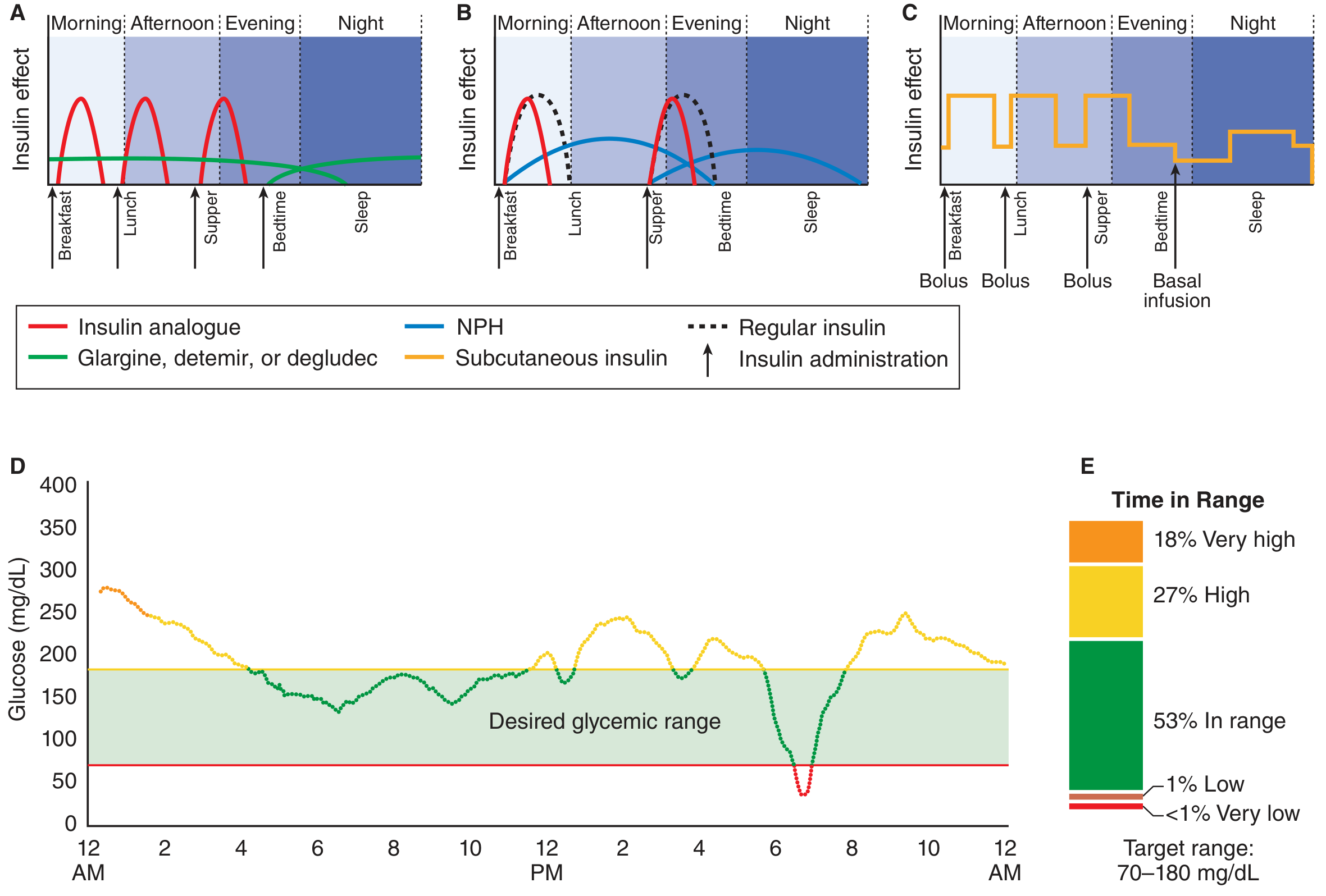
Figure: Panel A = ideal basal-bolus with glargine + rapid-acting analogue. Panel B = NPH-based regimen. Panel C = insulin pump. Panel D = glucose time-in-range profile. (Goodman & Gilman's Pharmacology, 9e)
3. Dose Calculation - Step by Step
Step 1: Total Daily Dose (TDD)
| Patient type | Formula |
|---|---|
| Type 1 DM (lean) | 0.4–0.7 units/kg/day |
| Type 2 DM, obese, pubertal adolescent | 0.7–1.0 units/kg/day (up to 1–2 units/kg due to insulin resistance) |
| Conservative initiation (ADA 2025) | 10 units/day OR 0.1–0.2 units/kg/day (basal only, to start) |
| Textbook of Family Medicine formula | TDD = weight (kg) × 0.7 |
Example: 70 kg patient → TDD = 70 × 0.7 = 49 units/day
Step 2: Split TDD into Basal and Bolus
| Component | Proportion of TDD | Example (TDD = 50 units) |
|---|---|---|
| Basal insulin | 50% of TDD (range 40–50%) | 25 units glargine once daily |
| Total bolus insulin | 50% of TDD (range 50–60%) | 25 units divided across meals |
"The basal dose is usually 40–50% of the total daily dose, with the remainder as prandial or premeal insulin." - Goodman & Gilman's Pharmacology
Step 3: Divide Bolus Across Meals
Option A - Equal split (3 meals):
- Bolus dose per meal = Total bolus ÷ 3
- Example: 25 units ÷ 3 = ~8 units per meal
Option B - Weight-based per meal:
- 0.1 units/kg/meal (e.g., 70 kg × 0.1 = 7 units per meal)
Option C - Carbohydrate counting (ICR - Insulin-to-Carb Ratio):
- Rule of 500 (for rapid-acting) or Rule of 450 (for regular insulin)
- ICR = 500 ÷ TDD
- Example: TDD = 50 → ICR = 500/50 = 10 → 1 unit covers 10 g of carbohydrate
Timing:
- Rapid-acting analogue: inject 15 minutes before a meal (unless pre-meal BG <80 mg/dL → inject at meal start)
- Regular insulin: inject 30 minutes before a meal
Step 4: Correction Dose (Insulin Sensitivity Factor)
Insulin Sensitivity Factor (ISF) = "Rule of 100" (or 1800 rule)
ISF = 1800 ÷ TDD (for rapid-acting insulin) ISF = 1500 ÷ TDD (for regular insulin)
| TDD | ISF (rapid-acting) | Meaning |
|---|---|---|
| 50 units/day | 1800 ÷ 50 = 36 mg/dL per unit | 1 unit lowers BG by ~36 mg/dL |
Correction dose formula:
Correction dose = (Current BG - Target BG) ÷ ISF Target BG for correction = typically 150 mg/dL (conservative, to avoid hypoglycemia) Use correction only if pre-meal BG >180 mg/dL
Step 5: Titration
Basal titration (ADA 2025):
- Increase by 2 units every 3 days until fasting plasma glucose (FPG) reaches target
- OR increase by 1–2 units/day or 10–15% twice weekly
- If hypoglycemia: reduce dose by 10–20%
Bolus titration:
- If 2-hour postprandial glucose is consistently >50 mg/dL above premeal: increase bolus by 1–2 units
- If negative delta (post-meal BG < pre-meal BG): reduce bolus by 1–2 units
4. Practical Regimen Examples
Type 1 DM (70 kg patient, TDD = 50 units)
- Basal: Glargine 25 units once at bedtime (or Degludec 25 units once daily)
- Bolus: Lispro/Aspart 8 units before each of 3 meals (24 units total)
- Correction: If pre-meal BG >180 mg/dL → add (BG - 150) ÷ 36 units
Type 2 DM - Starting Basal Only (ADA 2025)
- Start: 10 units/day of glargine at bedtime (or 0.1–0.2 units/kg/day)
- Titrate: +2 units every 3 days targeting FBG 80–130 mg/dL
- If A1C still above target after basal optimization → add prandial insulin
Type 2 DM - Intensifying to Full Basal-Bolus
- Add 1 dose of rapid-acting with the largest meal first
- Titrate that meal dose, then add second meal dose, then third (stepwise)
- ADA 2025: Preferred for non-critically ill hospitalized patients
5. Special Situations
| Situation | Adjustment |
|---|---|
| Fasting / NPO | Reduce bolus to zero; continue 50–80% of basal |
| Renal failure (CKD) | Reduce total dose (reduced insulin clearance → hypoglycemia risk) |
| Steroid-induced hyperglycemia | Predominantly postprandial → increase bolus insulin more than basal |
| Over-basalization | ADA 2025: Assess by HS-to-AM or pre-to-post prandial BG difference >50 mg/dL, hypoglycemia occurrence, and high glycemic variability (not just dose >0.5 U/kg) |
| Switching basal insulins | Usually 1:1 unit conversion; if high risk for hypoglycemia, reduce new dose by 10–20% |
6. Quick Summary Table for Viva
| Question | Answer |
|---|---|
| TDD formula | 0.5–0.7 U/kg/day (T1DM); 0.7–1 U/kg (T2DM); start 10 U/day |
| Basal:Bolus split | 50:50 (range 40:60 to 50:50) |
| Bolus per meal | TDD × 50% ÷ 3 meals OR 0.1 U/kg/meal |
| ISF | 1800 ÷ TDD |
| ICR | 500 ÷ TDD |
| Basal titration | +2 U every 3 days to FBG target |
| Best basal insulin | Glargine (once daily, peakless) or Degludec (once daily, ~42 h) |
| Best bolus insulin | Aspart / Lispro / Glulisine (rapid-acting, 15 min before meal) |
| Regular insulin advantage | Only insulin safe for IV use (DKA, hyperkalemia) |
| Correction target BG | 150 mg/dL (conservative) |
Sources: Harrison's Principles of Internal Medicine 22E; Goodman & Gilman's Pharmacological Basis of Therapeutics; Textbook of Family Medicine 9e; ADA Standards of Medical Care in Diabetes 2025
This is a shared conversation. Sign in to Orris to start your own chat.