
acute otitis media
acute otitis media tympanic membrane otoscopy
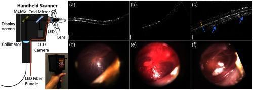
This composite educational graphic details a first-generation handheld optical coherence tomography (OCT) probe and its clinical application in otology. On the left, a schematic diagram labels the internal optics of the handheld scanner, including an LED fiber bundle, collimator, CCD camera, MEMS cold mirror, lens, and an integrated 5-inch display screen. Below the schematic is a photograph of the physical device being held. The right side presents comparative clinical data from pediatric subjects. Panels (a–c) show cross-sectional OCT images, while (d–f) provide corresponding digital otoscopy views. Column 1 (a, d) depicts a normal ear with a healthy tympanic membrane. Column 2 (b, e) illustrates acute otitis media, with otoscopy showing significant erythema and inflammation. Column 3 (c, f) demonstrates chronic otitis media, where the OCT image reveals a thickened tympanic membrane with complex multi-layered structures (indicated by arrows), and otoscopy shows visible white biofilms or middle ear effusion. This visual summarizes the integration of surface video and subsurface cross-sectional imaging for diagnosing middle ear pathologies in primary care settings.
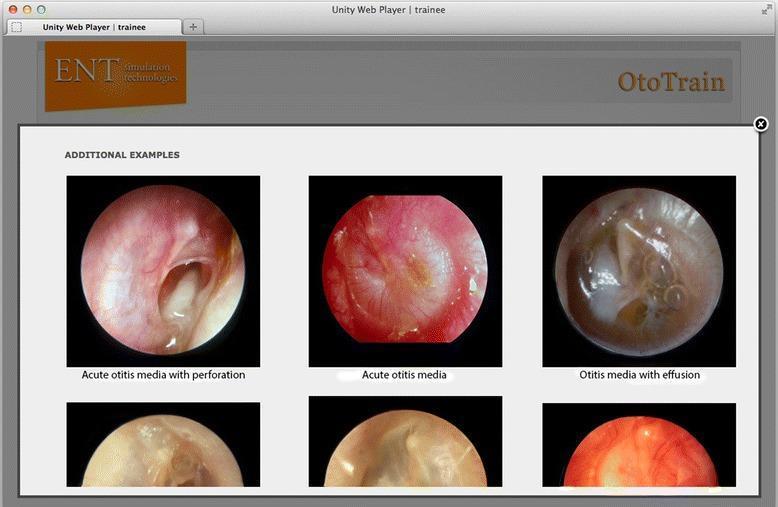
A comparison chart of otoscopic diagnostic images from a medical simulator, focusing on pathologies of the tympanic membrane (TM). The visual contains three labeled primary circular otoscopic views: 1) 'Acute otitis media with perforation,' showing an inflamed, erythematous TM with a large, irregular central perforation through which the middle ear space is visible; 2) 'Acute otitis media,' depicting an intensely hyperemic, opaque, and bulging TM with loss of normal landmarks and light reflex; and 3) 'Otitis media with effusion,' illustrating a translucent, amber-colored TM with visible air-fluid levels and bubbles in the middle ear space. These clinical photographs serve as educational examples for ENT trainees to differentiate between infectious stages (AOM) and non-purulent fluid accumulation (OME). The interface includes secondary supportive images below each category to demonstrate variability in presentation. This content is designed for medical students and residents to master diagnostic otoscopy and middle ear pathology recognition.
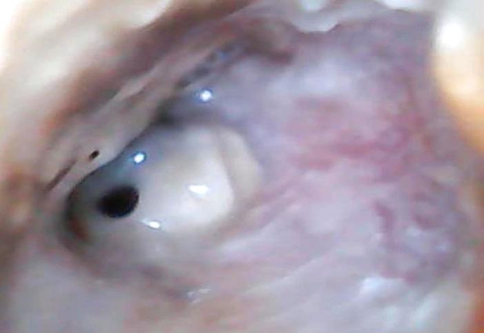
This clinical photograph displays an otoscopic view of the right ear, highlighting pathological findings of the external auditory canal (EAC) and tympanic membrane. The EAC shows significant inflammatory changes, characterized by diffuse erythema and edema of the canal walls. Visible purulent secretions are present within the lumen, suggesting an active infectious process such as chronic suppurative otitis media or acute exacerbation. The tympanic membrane is clearly visualized and exhibits a single, well-defined perforation located in the anterior-inferior quadrant. The perforation appears as a dark, circular opening amidst the remaining translucent, light-reflective portions of the membrane. Normal landmarks like the handle of the malleus are obscured by the surrounding inflammatory tissue and exudate. This image serves as a clinical example of suppurative middle ear disease with associated tympanic membrane rupture and secondary external canal inflammation, relevant for otolaryngology training in diagnostic otoscopy.
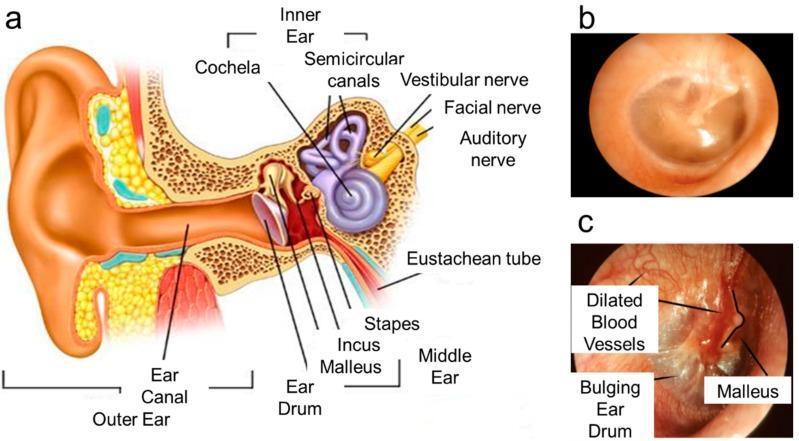
This medical educational graphic comprises an anatomical diagram and clinical otoscopic photographs illustrating the human ear in healthy and pathological states. Panel (a) is a cross-sectional anatomical diagram of the human ear, labeling key structures including the outer ear (pinna, ear canal), middle ear (eardrum/tympanic membrane, malleus, incus, stapes), and inner ear (cochlea, semicircular canals, vestibular and auditory nerves, Eustachian tube). Panel (b) shows a healthy tympanic membrane via white light otoscopy, characterized by a translucent, pinkish-gray appearance, a normal light reflex, and a concave shape. Panel (c) provides a comparative otoscopic view of acute otitis media. Pathological markers labeled include a bulging eardrum due to middle ear pressure, intense erythema (redness), and dilated blood vessels (vascular injection) across the membrane surface. The malleus remains visible but appears distorted by the inflammatory swelling. The image serves to teach the visual diagnostic criteria for middle ear infections compared to normal anatomy.
"acute otitis media"[MeSH Terms] AND management
Acute Otitis Media (AOM)
Definition
Epidemiology
- By 6 months of age, ~20% of children have had two or more AOM episodes
- By age 3 years, 50% have had three or more episodes; by age 6, ~60% have had at least one episode
- The global incidence is estimated at 10.8 new episodes per 100 people per year, ranging from 3.6 in central Europe to 43.4 in sub-Saharan West/central Africa
- Incidence peaks at 6-24 months of age
- Early onset of first AOM (before 12 months) - strongest predictor of recurrence
- Male sex, genetic predisposition (heritability 40-70%)
- Craniofacial abnormalities (e.g., cleft palate), immunodeficiency, adenoidal hypertrophy
- Day care attendance, older siblings, tobacco smoke exposure, pacifier use
- Low socioeconomic status, fall/winter season, recurrent URTIs
- Breastfeeding is protective
Pathophysiology
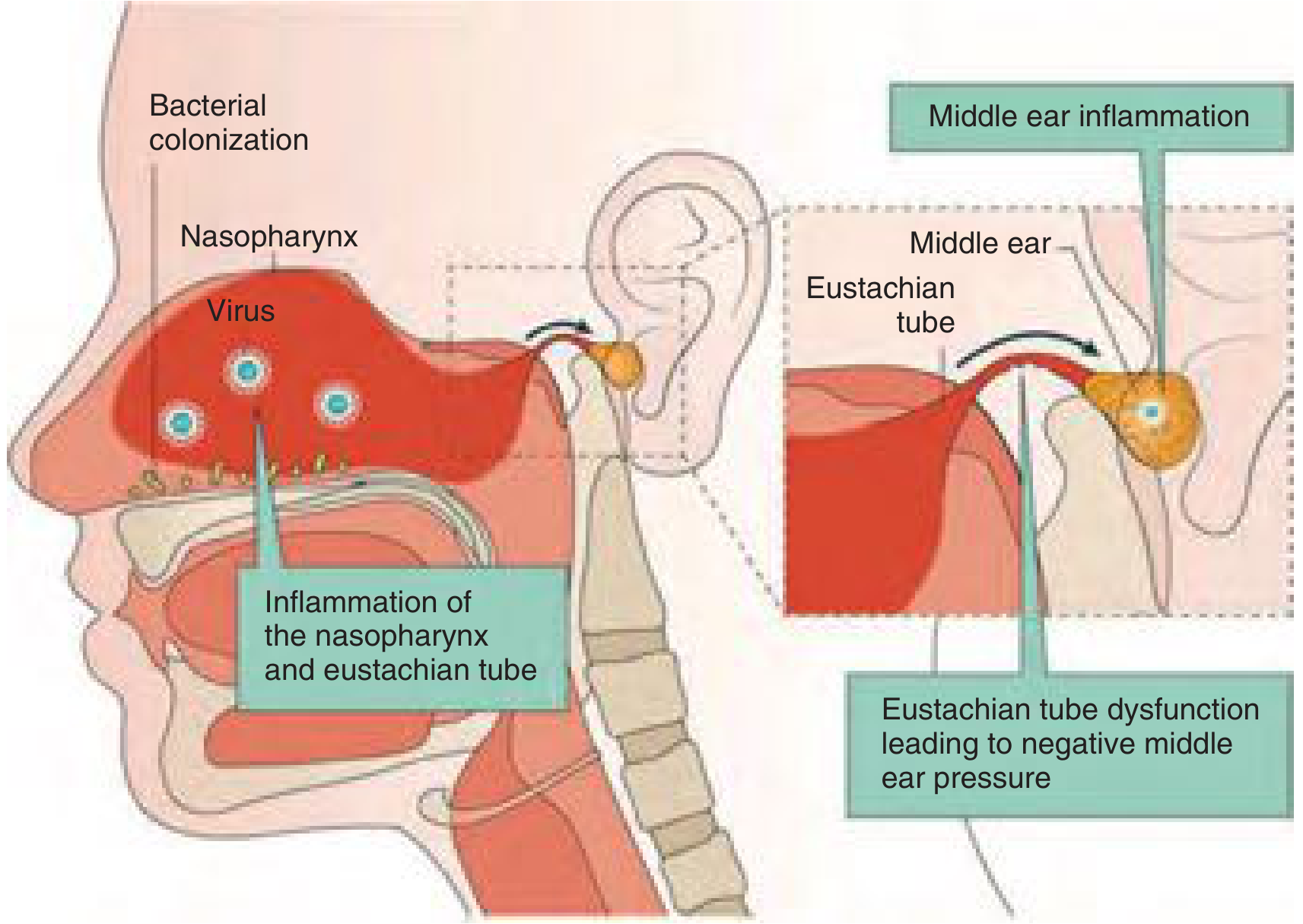
Eustachian Tube Anatomy in Children
- Protection - barrier against pathogens from nasopharynx
- Drainage - clears secretions via mucociliary flow
- Pressure equalization - maintains middle ear aeration
Pathogenic Sequence
- A viral URTI causes nasopharyngeal and ET edema/inflammation
- ET dysfunction leads to fluid accumulation in the middle ear
- Bacterial otopathogens colonizing the nasopharynx ascend and infect the stagnant fluid
- This triggers acute middle ear inflammation
Causative Organisms
| Organism | Notes |
|---|---|
| Streptococcus pneumoniae | Historically #1; pneumococcal vaccines (PCV7, PCV13) have significantly reduced its prevalence |
| Nontypeable Haemophilus influenzae | Now predominant in vaccinated populations |
| Moraxella catarrhalis | Common, often self-limiting |
Immunology
- Innate immunity: Mucociliary flow, lysozymes, defensins, complement, Toll-like receptor (TLR) activation
- Adaptive immunity: Middle ear secretory IgA and IgG; regulatory T-cells modulate inflammation
- Low IgA, IgG2 and mannose-binding lectin levels are found in chronic recurrent OM
- Cytokine polymorphisms (IL-6, IL-10, TNF) predict OM risk with RSV/rhinovirus co-infection
Clinical Features
Symptoms
- Otalgia (ear pain) - the cardinal symptom; in infants may manifest as irritability, tugging at the ear
- Fever
- Otorrhea (if tympanic membrane perforated)
- Hearing loss (conductive)
- Anorexia
Otoscopic Signs

- Bulging tympanic membrane - the most diagnostic sign (moderate-to-severe bulging = diagnostic)
- Erythema/hyperemia of the tympanic membrane
- Cloudiness/opacity of the membrane
- Obscured landmarks (handle of malleus, light reflex)
- Immobility on pneumatic otoscopy (also present in perforation, OME, blocked tube)
- New-onset otorrhea (if perforation)
- Moderate to severe bulging of TM, OR
- New-onset otorrhea not due to otitis externa, OR
- Mild bulging + recent onset (<48 h) of symptoms OR intense TM erythema
Management
Pain Control (all patients)
- NSAIDs (ibuprofen) or acetaminophen - effective for mild-to-moderate pain
- Topical agents (benzocaine, lidocaine) - brief additional benefit
Antibiotic Therapy
| Age | Indication |
|---|---|
| <6 months | Antibiotic treatment reasonable for all |
| 6 months - 2 years | Bilateral ear findings |
| ≥6 months | Otorrhea |
| >2 years | Symptoms worsening or not improving within 48-72 h |
| All ages | Severe otalgia, otalgia ≥2 days, or fever >102.2°F |
- Penicillin allergy: azithromycin or clarithromycin
- No improvement after 48-72 h → switch to amoxicillin-clavulanate, 2nd/3rd-gen oral cephalosporin, or IM ceftriaxone ×3 days
- Still failing → refer to specialist
Note: The number needed to treat (NNT) to prevent ONE case of mastoiditis is ~5000. The number needed to harm (NNH) for vomiting/diarrhea/rash is 14. Antibiotics reduce pain by day 2-3 with NNT of 20. (Harrison's 22E, p. 298)
Recurrent AOM
- Defined as ≥3 episodes in 6 months or ≥4 episodes in 1 year
- Consider tympanostomy tube insertion
- Middle ear effusions persist in 60-70% of AOM cases; should resolve within 3 months
Complications
Local Complications
- Tympanic membrane perforation with otorrhea
- Mastoiditis - rare; suggested by post-auricular tenderness, swelling, or ear protrusion. Requires CT scan and possibly cortical mastoidectomy
- Otitis media with effusion (OME) / "glue ear" - each AOM episode increases odds of OME by 12-fold
Facial Nerve Palsy
Intracranial Complications (rare)
- Meningitis
- Brain abscess
- Lateral sinus thrombophlebitis
- Labyrinthitis
Prevention
- Pneumococcal conjugate vaccines (PCV7, PCV13) - significantly reduce AOM due to vaccine-serotype pneumococcus
- Breastfeeding
- Avoiding tobacco smoke exposure
- Limiting pacifier use
- Reducing daycare group sizes
Recent Evidence
- A 2025 systematic review (PMID: 40717508) mapped the evidence base for AOM clinical practice guidelines, highlighting variability across international guidelines.
- A 2025 Cochrane review (PMID: 41307300) found no significant benefit of decongestants or antihistamines for AOM in children.
- A 2024 network meta-analysis of 89 RCTs (PMID: 37016201) confirmed amoxicillin/amoxicillin-clavulanate as optimal first-line antibiotics for AOM in children and adolescents; shorter courses (5 days) were comparably effective in children >2 years with non-severe disease.