
Peptic ulcer disease couse and treatment least guide line
"peptic ulcer disease"[MeSH Terms] AND "Helicobacter pylori"[MeSH Terms] AND treatment
peptic ulcer disease gastric duodenal ulcer endoscopy pathology
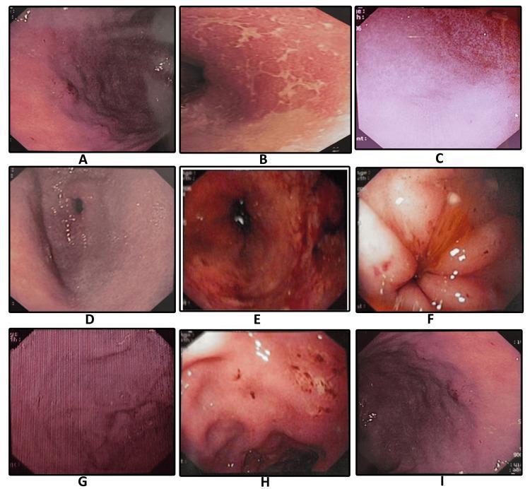
A composite of nine endoscopic images (labeled A-I) illustrating the diverse clinical manifestations of Helicobacter pylori-associated gastritis and peptic ulcer disease within the gastric and duodenal mucosa. The images provide a comparative overview of mucosal pathology: (A) demonstrates mucosal erosion with punctate dark lesions; (B) shows adherent sticky mucus appearing as whitish-yellow patches; (C) illustrates diffuse mucosal erythema; (D) depicts antral gastritis with generalized hyperemia; (E) highlights spotty redness across the mucosal surface; (F) reveals a duodenal ulcer with surrounding inflammatory changes; (G) displays classic nodular gastritis characterized by a uniform, 'cobblestone' or granular mucosal texture; (H) shows a gastric ulcer with evidence of active erosion; and (I) exhibits hypertrophic gastropathy with significantly enlarged rugal folds. This educational compilation is essential for gastroenterology training to differentiate specific morphological patterns such as nodularity, erosions, and fold thickening during diagnostic upper gastrointestinal endoscopy.
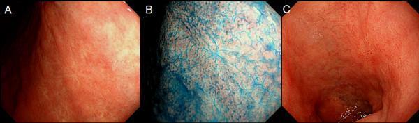
This set of three clinical endoscopic photographs illustrates the appearance of the upper gastrointestinal mucosa in the stomach and duodenal cap. Panel A shows the stomach lining under white light endoscopy, characterized by a smooth, reddish-pink mucosal surface with visible fine submucosal vascular patterns. Panel B demonstrates the same gastric area using chromoendoscopy with indigo-carmine dye. The blue contrast agent accumulates in the mucosal pits and crevices, highlighting the surface micro-architecture, mucosal folds, and subtle irregularities that may indicate previous pathology like healed peptic ulcer scars. Panel C provides a white light view of the duodenal cap, showing a similar pinkish hue but with more pronounced anatomical folds compared to the gastric body. The image set is designed for gastroenterology education to demonstrate the utility of chromoendoscopy in enhancing mucosal detail and identifying chronic changes or scars from previous peptic ulcer disease (PUD) that might be subtle under standard white light imaging.
PMID: 39626064
Peptic Ulcer Disease (PUD) - Causes and Treatment (Latest Guidelines 2024)
Definition
Epidemiology
- Lifetime prevalence: 8-14% of the general population
- Both incidence and prevalence have declined 31% worldwide from 1990-2019, largely due to better understanding and treatment of H. pylori and NSAID risks
- Peak incidence of complicated disease (hospitalization): ages 65-74
- PUD is the most common cause of upper GI bleeding
- Annual cost in the US: ~$6 billion USD - Yamada's Gastroenterology 7e; Sabiston Surgery, 21e
Pathophysiology: The Balance of Protective vs. Damaging Factors
| Protective Factors | Damaging Factors |
|---|---|
| Mucous layer (gel barrier) | Hydrochloric acid (HCl) + pepsin |
| Bicarbonate secretion (trapped in mucus) | H. pylori infection |
| Mucosal blood flow | NSAIDs / aspirin |
| Prostaglandin E2 (stimulates HCO3-, maintains barrier) | Alcohol |
| Growth factors and cell renewal | Smoking |
| Endogenous prostaglandins | Bile reflux, ischemia, stress |
Causes / Etiology
1. Helicobacter pylori (Most Common)
- Global prevalence of H. pylori ~43%
- Associated with 90% of duodenal ulcers and 70-90% of gastric ulcers
- H. pylori is a spiral-shaped, flagellate, microaerophilic gram-negative bacterium
- Mechanism: produces urease (splits urea → ammonia + bicarbonate), creating an alkaline microenvironment for survival in acidic gastric milieu
- Releases cytotoxins (e.g., CagA toxin) that destroy the mucosal barrier and underlying cells
- Its flagella allow movement through the mucus layer; mucolytic enzymes facilitate penetration
- When eradicated, ulcer recurrence becomes extremely rare - Sabiston Surgery 21e
2. NSAIDs / Aspirin (Second Most Common)
- NSAIDs inhibit cyclooxygenase (COX), reducing prostaglandin synthesis
- Prostaglandins are key mucosal protectants (stimulate mucus, bicarbonate, mucosal blood flow)
- Risk is dose-dependent and increased with advanced age, corticosteroid co-use, anticoagulants, prior PUD history
3. Zollinger-Ellison Syndrome (ZES)
- Gastrin-secreting tumor (gastrinoma), causing massive acid hypersecretion
- Think ZES when ulcers are multiple, refractory, or in unusual locations (distal duodenum/jejunum)
4. Other/Less Common Causes
- Systemic mastocytosis (histamine-driven)
- Crohn's disease
- Bisphosphonates (especially alendronate), SSRIs, potassium chloride, clopidogrel, mycophenolate
- Infections: HSV, CMV, EBV, tuberculosis, syphilis
- Ischemia, radiation, hypoxia, gastric bypass surgery
- Stress ulcers (ICU patients, burns, head trauma)
- Idiopathic ("non-H. pylori, non-NSAID ulcers" - increasing in developed countries)
Gastric vs. Duodenal Ulcers - Key Differences
| Feature | Gastric Ulcer | Duodenal Ulcer |
|---|---|---|
| Acid secretion | Normal or low (mucosal barrier defect) | Increased (excess acid production) |
| Gastrin | Elevated (H+ feedback reduced) | May be elevated due to G-cell hyperplasia |
| Pain timing | Shortly after eating (food stimulates acid) | 2-3 hours after eating; improves with food/antacids |
| H. pylori association | 70-90% | ~90% |
| Malignancy risk | Must biopsy to exclude cancer | Rarely malignant |
Clinical Presentation
- Most common: epigastric pain/burning ("dyspepsia"), often periodic
- Duodenal ulcer pain: often relieved by food, antacids; wakes patient at night
- Gastric ulcer pain: worsened by eating
- Nausea, early satiety, bloating
- Many patients are asymptomatic until a complication develops
Complications (the "4 Ps")
| Complication | Notes |
|---|---|
| Bleeding | Up to 15% of PUD patients; mortality up to 10% (esp. elderly) |
| Perforation | ~7% of PUD; free air on CXR; surgical emergency |
| Penetration | Into pancreas (pancreatitis-like pain radiating to back) |
| Pyloric obstruction | Gastric outlet obstruction; vomiting, weight loss |
Diagnosis
- Endoscopy (EGD) - gold standard; allows direct visualization, biopsy (mandatory for gastric ulcers to rule out malignancy), and treatment of bleeding
- 4-quadrant biopsy of gastric ulcers: sensitivity rises from 70% (1 biopsy) to 95% (4 biopsies) to 98% (7 biopsies)
- H. pylori testing (all PUD patients must be tested):
- Non-invasive: urea breath test (gold standard for diagnosis and test-of-cure), stool antigen test
- Invasive (via endoscopy): rapid urease test, histology, culture
- Plain abdominal X-ray: free air under diaphragm if perforation (upper GI contrast/barium is CONTRAINDICATED with perforation)
Treatment (2024 ACG & Current Guidelines)
Step 1: General Measures
- Stop NSAIDs if possible (or switch to COX-2 selective inhibitor + PPI)
- Smoking cessation (impairs ulcer healing)
- Avoid alcohol
- Avoid triggers
Step 2: Acid Suppression (Core of Therapy)
Proton Pump Inhibitors (PPIs) - First-Line
- Mechanism: irreversibly bind the H+/K+-ATPase (proton pump) at the parietal cell; require acid activation in secretory canaliculus
- Healing rate: 85% at 4 weeks, 96% at 8 weeks (superior to H2RAs)
- Examples: omeprazole, esomeprazole, pantoprazole, lansoprazole, rabeprazole
- Duration: 4-8 weeks for duodenal ulcers; 8-12 weeks for gastric ulcers
- Maintenance PPI: indicated for large ulcers (>2 cm), failed H. pylori eradication, or required ongoing NSAID/aspirin use
H2 Receptor Antagonists (H2RAs) - Second-Line
- Competitive inhibition of histamine H2 receptors on parietal cells
- Famotidine (most potent); cimetidine (weakest)
- Inferior to PPIs for ulcer healing; PPIs are preferred
- Note: Do NOT combine antacids or H2RAs with PPIs - the alkaline environment inhibits PPI activation
Antacids / Sucralfate
- Mucosal protectants; adjunctive role only
Step 3: H. pylori Eradication (Most Important for Long-Term Cure)
2024 ACG Clinical Guideline (Chey WD et al., Am J Gastroenterol 2024, PMID 39626064):
First-Line Regimens (Treatment-Naive, Unknown Susceptibility)
| Regimen | Drugs | Duration | Notes |
|---|---|---|---|
| Bismuth Quadruple Therapy (BQT) (PREFERRED) | PPI + bismuth subsalicylate + metronidazole + tetracycline | 14 days | Recommended as preferred first-line by 2024 ACG; avoids clarithromycin resistance |
| Rifabutin Triple Therapy | PPI + amoxicillin + rifabutin | 14 days | Suitable empiric alternative if no penicillin allergy |
| Potassium-Competitive Acid Blocker (P-CAB) Dual Therapy | Vonoprazan + amoxicillin | 14 days | Alternative; vonoprazan provides more potent acid suppression |
| Classic Clarithromycin Triple Therapy | PPI + clarithromycin + amoxicillin (or metronidazole) | 14 days | No longer first-line due to rising clarithromycin resistance (>15% in many regions) |
| Concomitant Quadruple | PPI + clarithromycin + amoxicillin + metronidazole | 14 days | May be used where clarithromycin resistance <15% |
Key 2024 update: Bismuth quadruple therapy for 14 days (not 10) is the preferred regimen when antibiotic susceptibility is unknown. Clarithromycin-containing regimens should only be used if susceptibility is confirmed. Salvage regimens with clarithromycin or levofloxacin require confirmed susceptibility.
Second-Line / Salvage (Treatment-Experienced, Persistent Infection)
| Scenario | Preferred Regimen |
|---|---|
| Not previously treated with optimized BQT | "Optimized" BQT for 14 days |
| Previously received BQT | Rifabutin triple therapy for 14 days |
| Susceptibility known | Tailored therapy based on culture/PCR |
Test-of-Cure
- All patients should have post-treatment confirmation of eradication
- Preferred: urea breath test or stool antigen (at least 4 weeks after completing therapy, with PPI held 2 weeks prior)
Step 4: NSAID-Related Ulcer Management
| Clinical Situation | Strategy |
|---|---|
| NSAID must be discontinued | PPI for ulcer healing (standard duration) |
| NSAID must be continued (e.g., cardiovascular) | Add PPI (full dose) and switch to COX-2 inhibitor if possible |
| High-risk patients on NSAIDs (age >65, prior PUD, corticosteroids, anticoagulants) | Prophylactic PPI (H2RA as alternative) |
Step 5: Surgical Treatment (Reserved for Complications)
- Perforation - exploratory laparoscopy/laparotomy; omental patch (Graham patch) for closure
- Uncontrolled hemorrhage - after failed endoscopic hemostasis
- Gastric outlet obstruction - refractory to medical management
- Malignancy - partial/total gastrectomy
- Highly selective vagotomy (parietal cell vagotomy): divides only acid-producing vagal branches; ulcer recurrence 10-15%; lowest dumping/diarrhea rates
- Truncal vagotomy + pyloroplasty: drainage required; higher dumping rate
- Truncal vagotomy + antrectomy: lowest recurrence (0-2%) but highest morbidity (20% postgastrectomy syndromes); reconstruction via Billroth I (gastroduodenostomy) or Billroth II (gastrojejunostomy)
Endoscopic Images of H. pylori-Associated Gastritis and PUD
Summary Treatment Algorithm
PUD confirmed (by endoscopy or clinical grounds)
        │
        ├── Test for H. pylori (ALL patients)
        │         │
        │         ├── POSITIVE → H. pylori eradication regimen (14 days)
        │         │              + PPI for 4-8 weeks (DU) or 8-12 weeks (GU)
        │         │              → Test-of-cure (UBT or stool antigen) 4+ weeks post-Rx
        │         │
        │         └── NEGATIVE → PPI therapy alone
        │                        Check for NSAID use, ZES, idiopathic
        │
        ├── NSAID-induced → Stop NSAID if possible + PPI
        │                   If NSAID needed → COX-2 inhibitor + PPI
        │
        ├── ZES → High-dose PPI + surgical/medical management of gastrinoma
        │
        └── Complications → Endoscopic hemostasis for bleeding
                             Surgery for perforation, obstruction, uncontrolled bleed
Key Sources
- Sabiston Textbook of Surgery, 21e - Chapters on Peptic Ulcer Disease and Gastric Surgery
- Yamada's Textbook of Gastroenterology, 7e - Chapter 49: Peptic Ulcer Disease
- Textbook of Family Medicine, 9e - Chapter 38
- Costanzo Physiology, 7e - Chapter 8 (Gastrointestinal Physiology)
- 2024 ACG Clinical Guideline: Chey WD et al., "ACG Clinical Guideline: Treatment of Helicobacter pylori Infection," Am J Gastroenterol 2024 (PMID 39626064) - Most current guideline available