
Femoral nerve.
Reading File
The femoral nerve is the largest branch of the lumbar plexus (L2-L4), supplying the anterior thigh muscles and skin. Let me pull detailed textbook content on this.
Searching sources...
Finding Sources
Finding Sources
Reading File
Reading File
Reading File
Reading File
Loading Image
Loading Image
Here is a complete overview of the femoral nerve drawn from Gray's Anatomy for Students, Bradley and Daroff's Neurology, Adams and Victor's Neurology, and Campbell's Operative Orthopaedics.
Femoral Nerve
Origin and Root Value
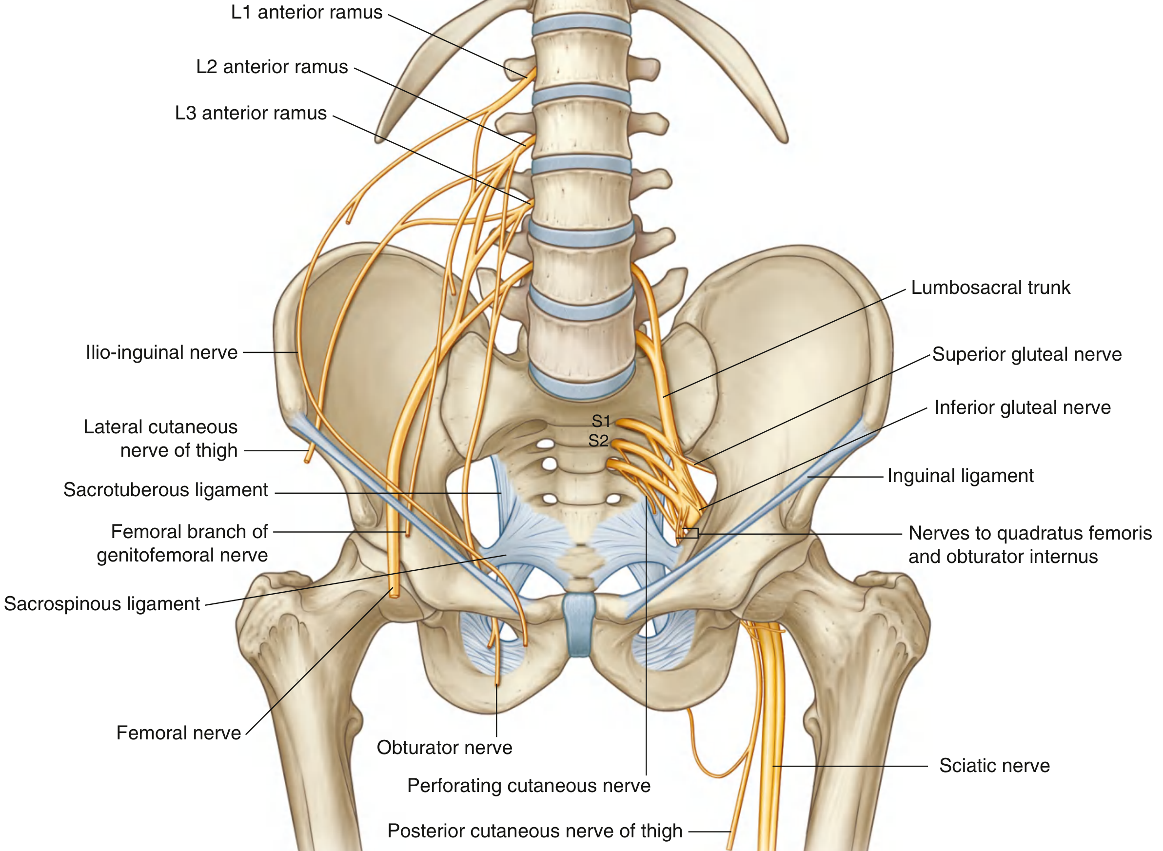
The femoral nerve is the largest branch of the lumbar plexus, formed from the posterior divisions of the ventral rami of L2, L3, and L4. It forms within the substance of the psoas major muscle on the posterior abdominal wall.
Course
- In the abdomen/pelvis: Descends within the iliacus compartment (between psoas major and iliacus), supplying both muscles as it travels downward. A motor branch to iliacus arises 4-5 cm before the inguinal ligament.
- Inguinal ligament: Passes under the inguinal ligament (not through the femoral canal) to enter the femoral triangle.
- Femoral triangle: Lies lateral to the femoral artery and outside the femoral sheath (which encloses only the artery and vein).
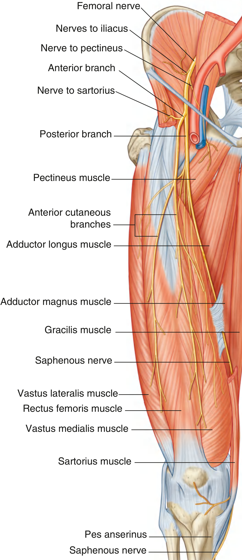
- Immediately below the inguinal ligament: Divides into anterior and posterior branches.
Branches and Distribution
Motor Branches
| Branch | Muscles Supplied |
|---|---|
| Branches in abdomen/pelvis | Psoas major, Iliacus |
| Nerve to pectineus | Pectineus |
| Anterior branch - motor | Sartorius |
| Posterior branch - motor | Rectus femoris, Vastus lateralis, Vastus intermedius, Vastus medialis (all 4 quadriceps) |
Cutaneous/Sensory Branches
| Branch | Area Supplied |
|---|---|
| Anterior cutaneous branches | Anterior thigh and anterior knee |
| Intermediate cutaneous nerve of thigh | Anterior and lateral thigh |
| Medial cutaneous nerve of thigh | Anteromedial thigh |
| Saphenous nerve (longest branch, from posterior division) | Anteromedial knee, medial leg, medial side of foot down to medial malleolus |
The saphenous nerve travels with the femoral artery through the adductor (subsartorial) canal but does NOT pass through the adductor hiatus. It instead pierces through the connective tissue near the end of the canal, emerging between sartorius and gracilis on the medial side of the knee. It is the only branch of the femoral nerve to reach below the knee.
Key Relations
- Lateral to femoral artery in the femoral triangle
- Outside the femoral sheath (important distinction - the sheath surrounds only the artery and vein)
- Medial to iliacus/iliopsoas proximally
The mnemonic for femoral triangle contents from lateral to medial: NAVY - Nerve, Artery, Vein, Y-fronts (lymphatics).
Femoral Nerve Injury - Causes
Pelvic / Proximal Lesions
- Self-retaining retractors compressing the nerve against the pelvic wall (during hysterectomy, prostatectomy, aortic repair, renal transplant)
- Retroperitoneal hematoma - iatrogenic (anticoagulants, femoral catheterization), hemophilia, ruptured AAA
- Pelvic tumors (lymphadenopathy, colorectal carcinoma), neurofibromas/schwannomas
- Lithotomy positioning with extreme hip flexion + external rotation (kinking at inguinal ligament)
Distal / Inguinal Lesions
- Penetrating wounds to the lower abdomen
- Total hip replacement (especially revisions)
- Pelvic fractures (stretching of branches)
- Hip hyperextension (dancers, yoga practitioners)
Clinical Features of Femoral Nerve Palsy
| Feature | Details |
|---|---|
| Weakness | Knee extension (quadriceps); hip flexion if lesion is in the pelvis |
| Reflex | Absent or depressed knee jerk (patellar reflex, L3/L4) |
| Sensory loss | Anterior thigh, anteromedial knee, medial leg (saphenous distribution) |
| Gait | Falls on stairs and slopes; can walk on flat surfaces using hip extensors and tensor fasciae latae to stabilize the knee |
| Autonomous zone | Small area just superior and medial to the patella |
| Pain | Usually absent - except in retroperitoneal hematoma cases |
Importantly, thigh adduction is preserved (obturator nerve, L2-L4 via a different pathway), which helps distinguish femoral neuropathy from upper lumbar radiculopathy.
Distinguishing Femoral Neuropathy from Lumbar Radiculopathy
| Feature | Femoral Neuropathy | L3/L4 Radiculopathy |
|---|---|---|
| Adductor weakness | Absent | Present |
| Knee jerk | Absent | Absent |
| EMG - iliacus | Normal (inguinal), abnormal (pelvic) | Abnormal |
| EMG - adductors | Normal | Abnormal |
| Sensory | Saphenous + anterior thigh | May follow dermatomal pattern |
Needle EMG of the quadriceps (denervated in femoral neuropathy) and thigh adductors (innervated by obturator nerve, L2-L4) is the key to distinguishing these entities. Femoral CMAP amplitude correlates with motor axonal loss and has prognostic value.
Investigation
- CT/MRI pelvis - urgently if retroperitoneal hematoma or pelvic mass is suspected
- Nerve conduction studies - femoral CMAP amplitude quantifies motor axonal loss
- Needle EMG - quadriceps (denervation), iliacus (pelvic vs. inguinal localization), adductors (exclude radiculopathy/plexopathy)
Management
- Retroperitoneal hematoma - may require emergent surgical drainage
- Conservative for most other causes - spontaneous recovery expected
- Knee orthosis (KO or KAFO) - for severe quadriceps weakness to prevent falls and aid walking
- Prevention: Avoid extreme hip flexion/external rotation in lithotomy; avoid self-retaining retractors in pelvic surgery; proper padding during prone positioning
Sources: Gray's Anatomy for Students, p. 666-667; Bradley and Daroff's Neurology in Clinical Practice, Ch. 27; Campbell's Operative Orthopaedics 15th Ed, p. 3877
This is a shared conversation. Sign in to Orris to start your own chat.