
Treatment of acute pneumonia
"community-acquired pneumonia" AND treatment
pneumonia chest X-ray consolidation lobar
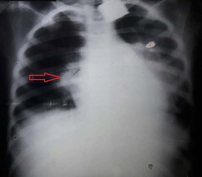
This diagnostic image is an anteroposterior (AP) chest X-ray of a pediatric patient demonstrating classic findings of lobar pneumonia. A prominent, dense, and homogenous opacification is localized to the right upper lobe, consistent with pulmonary consolidation. The opacification has a well-defined inferior border along the horizontal fissure, indicated by a red arrow, which is a hallmark of lobar involvement. Normal lung markings in this region are obscured, while the left lung field appears relatively clear and well-aerated. The cardiac silhouette and mediastinal structures appear within normal limits for this projection, with no significant shift or widening. The diaphragm and costophrenic angles are visible, though the right side is partially obscured by the adjacent consolidation. This image serves as an educational example of bacterial pneumonia presentation in a clinical setting, highlighting the radiographic appearance of alveolar space filling.

Multi-modal diagnostic imaging series illustrating community-acquired lobar pneumonia in a 30-year-old patient. (a) Posterior-anterior chest X-ray shows a clear area of consolidation in the left upper lobe. (b) B-mode lung ultrasound (LUS) reveals a complex, hypoechoic consolidation containing numerous hyperechoic punctate and linear structures, characteristic of a marked air bronchogram. (c-e) Contrast-enhanced ultrasound (CEUS) time-series demonstrates perfusion dynamics: (c) at 13 seconds, there is homogeneous pulmonary arterial enhancement; (d) by 1 minute, significant early washout occurs; (e) by 2 minutes, the washout is more pronounced. This specific CEUS pattern of rapid washout is indicative of pulmonary arterial vasoconstriction, often associated with the Euler–Liljestrand mechanism in response to local hypoxia in consolidated lung tissue. The series serves as an educational example of how multi-phase CEUS can supplement B-mode ultrasound and radiography in characterizing the hemodynamic response of pulmonary parenchyma during inflammatory processes.
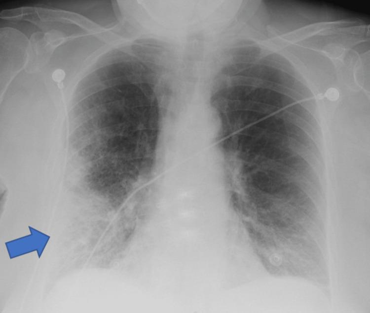
A frontal (anteroposterior) chest X-ray demonstrating clinical signs of pneumonia. A prominent blue arrow indicates a large area of increased opacity in the right lower lung field, suggestive of lobar consolidation or a dense pulmonary infiltrate. The opacity is heterogeneous with ill-defined borders, blending into the surrounding parenchyma. Beyond the focal area, there are diffuse bilateral reticulonodular interstitial markings, more pronounced in the mid and lower lung zones. The right costophrenic angle is relatively sharp, while the left shows mild blunting. Several medical devices are present: multiple radiopaque ECG leads/electrodes are positioned on the upper chest and shoulders, and a radiopaque enteric tube (nasogastric or feeding tube) is visible traversing the mediastinum toward the stomach. The image illustrates typical radiological findings of an infectious process in an acute clinical setting, highlighting focal consolidation against a background of generalized pulmonary congestion or inflammation.
PMID: 40679934
Treatment of Acute Pneumonia
Overview
Step 1: Severity Assessment and Site-of-Care Decision
CURB-65 Score (Harrison's, 22nd ed.)
- C - Confusion (new onset)
- U - Urea >7 mmol/L (BUN >20 mg/dL)
- R - Respiratory rate ≥30/min
- B - Blood pressure: systolic ≤90 or diastolic ≤60 mmHg
- 65 - Age ≥65 years
| Score | 30-day Mortality | Recommendation |
|---|---|---|
| 0 | 1.5% | Outpatient |
| 1-2 | ~9% | Hospitalize (or observation unit) |
| ≥3 | 22%+ | Hospitalize; consider ICU |
Pneumonia Severity Index (PSI)
Mandatory ICU admission regardless of score:
- Septic shock requiring vasopressors
- Acute respiratory failure requiring mechanical ventilation
- Three or more minor ATS/IDSA criteria (RR ≥30, PaO₂/FiO₂ ≤250, multilobar infiltrates, confusion, BUN ≥20, leukopenia, thrombocytopenia, hypothermia, hypotension requiring fluids)
Step 2: Empiric Antibiotic Regimens (Community-Acquired Pneumonia)
Outpatient (No Comorbidities)
- Amoxicillin 1000 mg PO q8h for ≥5 days, OR
- Doxycycline 100 mg PO q12h for ≥5 days, OR
- Azithromycin 500 mg on day 1, then 250 mg qday days 2-5 (use only where local macrolide resistance <25%)
Outpatient (With Comorbidities: DM, heart/lung/liver/renal disease, malignancy, asplenia, alcoholism)
- Beta-lactam (amoxicillin-clavulanate 875/125 mg q12h, cefpodoxime 200 mg q12h, or cefuroxime 500 mg q12h) PLUS doxycycline or azithromycin, OR
- Respiratory fluoroquinolone monotherapy: levofloxacin 750 mg PO qday or moxifloxacin 400 mg PO qday
Inpatient (Non-Severe)
- Beta-lactam (ampicillin-sulbactam 1.5-3 g IV q6h, ceftriaxone 1-2 g IV qday, cefotaxime 1-2 g IV q8h, or ceftaroline 600 mg IV q12h) PLUS macrolide (e.g., azithromycin), OR
- Respiratory fluoroquinolone monotherapy (levofloxacin, moxifloxacin)
Inpatient (Severe CAP / ICU)
- Beta-lactam PLUS macrolide, OR
- Beta-lactam PLUS fluoroquinolone
- Do NOT use fluoroquinolone monotherapy for severe CAP
Step 3: Coverage for Resistant Organisms
MRSA Risk (prior respiratory isolation OR recent hospitalization + parenteral antibiotics in last 90 days)
- Non-severe disease: Obtain cultures and nasal PCR for MRSA; do not expand empirically unless confirmed
- Severe disease: Expand empirically with:
- Vancomycin 15 mg/kg IV q12h (adjust by levels/renal function), OR
- Linezolid 600 mg IV/PO q12h
Pseudomonas aeruginosa Risk (same risk factors as above)
- Expand empirically with an anti-pseudomonal agent:
- Piperacillin-tazobactam 4.5 g IV q6h
- Cefepime 2 g IV q8h, or ceftazidime 2 g IV q8h
- Meropenem 1 g IV q8h, or imipenem 500 mg IV q6h
- Aztreonam 2 g IV q8h (for penicillin allergy)
Step 4: Key Pharmacological Principles
Step 5: Supportive Care
| Intervention | Details |
|---|---|
| Oxygen | Target SpO₂ ≥94% (≥88-92% in COPD with CO₂ retention) |
| Fluids | IV fluids for dehydration and hemodynamic support |
| Antipyretics | Paracetamol/acetaminophen for fever and discomfort |
| Nutrition | Early enteral nutrition in ICU patients |
| DVT prophylaxis | LMWH for hospitalized patients |
| Pleural effusion | Thoracentesis if significant; drain empyemas promptly |
| Corticosteroids | 2026 ATS guideline addresses this - evidence supports use in severe CAP with refractory shock; routine use is not standard outside this context |
Step 6: Hospital-Acquired Pneumonia (HAP) and VAP
- Non-severe, low MDR risk: Piperacillin-tazobactam OR cefepime OR ceftriaxone
- Severe or high MDR risk: Broaden to cover P. aeruginosa (two antipseudomonal agents) and MRSA (vancomycin or linezolid)
- Gram stain/culture of lower respiratory secretions should guide de-escalation
- VAP: treat at least 7-8 days; shorter courses (7 days) are favored when clinical response is adequate
Imaging: Typical Chest X-Ray Findings
Key Points Summary
- Assess severity first (CURB-65 or PSI) - this determines where and how aggressively to treat
- Start antibiotics promptly - delays beyond 4 hours worsen outcomes
- CAP outpatient: amoxicillin or doxycycline monotherapy for healthy patients; beta-lactam + macrolide or respiratory FQ for those with comorbidities
- CAP inpatient non-severe: beta-lactam + macrolide or FQ monotherapy
- Severe CAP/ICU: always dual therapy (beta-lactam + macrolide or FQ); never FQ monotherapy
- Consider MRSA/Pseudomonas coverage if prior respiratory isolation or recent hospitalization with IV antibiotics
- Duration: 5 days for responding non-severe CAP; 7 days for severe or slow responders
- De-escalate once culture data available
Recent Evidence Note: The 2026 ATS Clinical Practice Guideline (PMID: 40679934) addresses four updated areas: lung ultrasound for diagnosis, whether to modify antibiotics when a respiratory virus test is positive, antibiotic duration, and corticosteroid use. It may refine some of the above thresholds - check the full guideline for the latest recommendations.
Odcm full form
ODCM full form
Related common dosing abbreviations:
| Abbreviation | Meaning |
|---|---|
| OD | Once Daily |
| ODCM | Once Daily in the Morning |
| ODHS | Once Daily at Hour of Sleep (bedtime) |
| BD / BID | Twice Daily |
| TDS / TID | Three Times Daily |
| QID | Four Times Daily |
| PRN | As needed |
| AC | Before meals |
| PC | After meals |
Note: ODCM is commonly used in South Asian medical practice (India, etc.) as a local prescription shorthand. It is not a universally standardized abbreviation in Western pharmacopoeias, so always interpret it in the clinical context in which it appears.
Antibiotic of choice for tonsillitis
"tonsillitis" AND "antibiotic" AND "streptococcal"
Antibiotic of Choice for Tonsillitis
Causative Organism
First-Line Antibiotic
Penicillin V OR Amoxicillin - 10 days
| Drug | Dose (Adult) | Dose (Child) | Duration |
|---|---|---|---|
| Penicillin V | 500 mg PO BID-TID | 250 mg PO BID-TID | 10 days |
| Amoxicillin | 500 mg PO TID or 875 mg BID | 50 mg/kg/day divided | 10 days |
"A 10-day course of penicillin or amoxicillin has traditionally been first-line treatment for acute GABHS infection in most cases." - Cummings Otolaryngology, Head and Neck Surgery
"First line: Penicillin OR Amoxicillin - 10 days" - Red Book 2021 (AAP), Treatment Table 4.12
Penicillin Allergy Alternatives
| Drug | Notes |
|---|---|
| Cephalexin (1st-gen cephalosporin) | Preferred if non-anaphylactic penicillin allergy |
| Clindamycin | Effective, also used for penicillin failures |
| Azithromycin | 5-day course; use only if local macrolide resistance is low |
| Clarithromycin | Alternative macrolide |
From Red Book 2021: "Allergy: Cephalexin OR Clindamycin OR Azithromycin"
Penicillin Failure (7-37% of cases)
- Amoxicillin-clavulanate (covers beta-lactamase-producing co-pathogens in the tonsil)
- Clindamycin
- Cephalosporins (cefuroxime, cephalexin)
- Beta-lactamase protection by co-colonizing bacteria
- Poor penicillin penetration into pharyngeal tissue
- Loss of normal flora that inhibits GABHS virulence
- Emerging macrolide resistance in GAS M-protein variants
Key Points
- Never use antibiotics for viral tonsillitis - most cases are viral; use Centor/McIsaac score or rapid antigen test to identify bacterial cases before prescribing
- Full 10-day course is essential to prevent rheumatic fever and glomerulonephritis (post-streptococcal sequelae)
- Do not use amoxicillin if EBV (mononucleosis) is suspected - it causes a widespread maculopapular rash in these patients
- Symptomatic relief: Paracetamol or NSAIDs; aspirin should be avoided (Reye syndrome risk in children)
- Steroids: Not routinely indicated; a single dose of dexamethasone may help symptom relief in severe cases per some guidelines, but is not standard practice
- Tonsillectomy is considered for truly recurrent strep tonsillitis: ≥7 episodes/year, ≥5/year for 2 years, or ≥3/year for 3 years - Cummings Otolaryngology, Table 201.4
Recent Evidence
What is used for soothing
What context are you asking about soothing for?
Soothing Measures for Sore Throat / Tonsillitis
Pharmacological (Systemic)
| Agent | Role | Notes |
|---|---|---|
| Paracetamol (Acetaminophen) | Analgesic + antipyretic | First choice; safe in all ages |
| Ibuprofen / NSAIDs | Anti-inflammatory + analgesic | Often more effective than paracetamol for throat pain; avoid on empty stomach |
| Dexamethasone (single oral dose) | Reduces inflammation, speeds pain relief | Used in severe tonsillitis; not routine |
| Aspirin | Analgesic | Avoid in children (Reye syndrome risk) |
Local / Topical Soothing
| Agent | Form | Action |
|---|---|---|
| Benzydamine (Difflam) | Throat spray / gargle | Local anti-inflammatory + mild anaesthetic |
| Lidocaine / Benzocaine | Lozenges, sprays | Topical anaesthetic; numbs throat temporarily |
| Cetylpyridinium / Hexylresorcinol | Lozenges | Mild antiseptic + soothing |
| Glycerin + honey lozenges | Lozenges | Demulcent - coats and soothes mucosa |
| Warm salt water gargle | Gargle (0.9% NaCl) | Reduces swelling, clears debris, soothing |
| Povidone-iodine gargle | Gargle | Antiseptic; reduces bacterial load |
Home Remedies (Evidence-Supported)
| Remedy | Effect |
|---|---|
| Warm salt water gargle | Reduces mucosal oedema, removes exudate |
| Honey (1-2 tsp, especially in warm water/lemon) | Demulcent, mild antibacterial; well-supported in children >1 year |
| Warm fluids (tea, broth, warm water) | Soothes mucosa, maintains hydration |
| Ice chips / cold drinks / ice cream | Cold provides local anaesthesia and reduces swelling |
| Steam inhalation | Humidifies airway, provides comfort |
| Rest and adequate hydration | Essential for recovery |
What to Avoid
- Aspirin in children - risk of Reye syndrome
- Alcohol-based gargles - can irritate mucosa
- Dry, rough foods - worsen pain
- Smoking / secondhand smoke - delays healing
Quick Summary
- Ibuprofen or paracetamol - most effective systemic relief
- Benzydamine spray - best local topical option
- Warm salt water gargle - simple, effective, safe
- Cold fluids or ice cream - surprisingly effective local anaalgesia, especially in children post-tonsillectomy
Is IFA tablet given after birth of the baby to the mother
IFA tablet iron folic acid given postpartum mother after delivery WHO guidelines
https://www.ncbi.nlm.nih.gov/books/NBK379979
India RMNCH IFA tablet postpartum lactating mother 180 tablets program
IFA (Iron and Folic Acid) Tablets After Delivery - For the Mother
Short Answer
India's National Program (Anaemia Mukt Bharat / NHM)
| Phase | Tablet | Dose | Duration |
|---|---|---|---|
| Pregnancy (from 2nd trimester onward) | Red IFA tablet (60 mg iron + 500 mcg folic acid) | 1 tablet daily | Minimum 180 days during pregnancy |
| Postpartum / Lactating (0-6 months after delivery) | Red IFA tablet (60 mg iron + 500 mcg folic acid) | 1 tablet daily | 180 days post-delivery |
Why IFA is Given Postpartum
- Blood loss during delivery - average blood loss is 300-500 mL in normal vaginal delivery, more in caesarean - depletes iron stores significantly
- Breastfeeding demands - lactation increases nutritional requirements
- Prevention of postpartum anaemia - common cause of maternal morbidity and fatigue
- Folic acid - supports tissue recovery and cellular regeneration after delivery
- WHO recommendation - if a mother could not complete 6 months of IFA during pregnancy, she should continue supplementation postpartum to complete the full course
WHO Guideline
"If 6 months' duration cannot be achieved in pregnancy, continue to supplement during the postpartum period for 6 months, or increase the dose to 120 mg iron in pregnancy."
Key Points
- Yes, IFA is given postpartum - it is not stopped at delivery
- In India, it is given for 180 days post-delivery to lactating mothers
- The red-colored IFA tablet is used for pregnant and lactating women (distinguished from blue tablets for adolescents and pink tablets for children)
- ASHA workers are responsible for providing and counselling mothers about postpartum IFA supplementation at the community level
- Compliance is important - the tablets should be taken after food to reduce nausea/GI side effects