
Perineal body
"perineal body"
perineal body anatomy attachments function authoritative
Perineal body
Location
- Female: between the posterior vaginal opening and the anus
- Male: between the bulb of the penis and the anus
- It lies just anterior to the anal canal at the junction of the urogenital and anal triangles.
Muscular attachments
- Bulbospongiosus
- Superficial transverse perineal muscles
- Deep transverse perineal muscles
- External anal sphincter
- Fibers of levator ani, especially puborectalis/pubococcygeus contributions
Functions
- Supports the pelvic floor by acting as a central muscular anchor.
- Helps maintain the position of the vagina, rectum, and other pelvic viscera.
- Contributes to urinary and fecal continence through its relationships with the pelvic floor and external anal sphincter.
- Resists widening of the vaginal outlet and supports the posterior vaginal wall.
Clinical importance
- Pelvic-organ prolapse
- Perineal descent
- Anal sphincter dysfunction or fecal incontinence, particularly if an obstetric anal sphincter injury is present
Explain the perineal body’s anatomy with a labeled diagram
labeled anatomy diagram perineal body female perineal muscles external anal sphincter bulbospongiosus transverse perineal
"perineal body"
perineal body anatomy muscles attachments female male NCBI
Educational labeled anatomical diagram of the female perineal body, viewed inferiorly. Show the vulva anteriorly and anus posteriorly, and place a clearly labeled central perineal body between the posterior vaginal opening and anus. Show and label the muscle fibers attaching to it: bulbospongiosus, superficial transverse perineal muscles, deep transverse perineal muscles, external anal sphincter, and levator ani/puborectalis. Include the perineal membrane anteriorly and rectovaginal septum superiorly. Add a small inset comparing male location between bulb of penis and anus. Ensure labels are anatomically precise and legible.
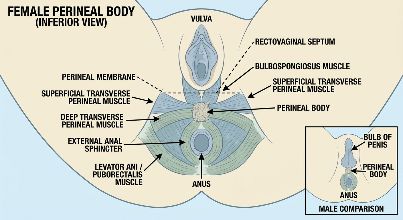
Perineal body: anatomical explanation
Position
- In females: between the posterior edge of the vaginal opening and the anus.
- In males: between the bulb of the penis and the anus.
- It is just posterior to the perineal membrane and connects to the posterior end of the urogenital hiatus of the levator ani.
Structures attaching to it
| Structure | Relationship to perineal body |
|---|---|
| Bulbospongiosus | Attaches anterolaterally. In females it helps narrow the vaginal opening; in males it assists urethral emptying and ejaculation. |
| Superficial transverse perineal muscles | Run from the ischial region medially to the body, stabilizing it. |
| Deep transverse perineal muscles | Meet/intersect at the perineal body. |
| External anal sphincter | Has anterior fibers that blend with the body, especially in females. |
| Levator ani, including puborectalis-related fibers | Connects with the body and contributes pelvic-floor support. |
| Perineal membrane | Attached anteriorly. |
| Rectovaginal septum in females | Joins superiorly. In males, the corresponding rectoprostatic septum attaches superiorly. |
| Sphincter urethrovaginalis | Also attaches in females. |
Functional significance
- Coordinates forces generated by the pelvic-floor and perineal muscles
- Supports the posterior vaginal wall in females
- Helps maintain the integrity of the pelvic floor
- Contributes indirectly to urinary and fecal continence through its association with the pelvic floor and external anal sphincter
Clinical relevance
Iron metabolism
iron metabolism hepcidin ferroportin
iron metabolism hepcidin ferroportin NIH fact sheet physiology
Iron metabolism
Overview
Dietary iron
     ↓
Duodenum absorbs iron
     ↓
Ferroportin exports iron into plasma
     ↓
Transferrin carries Fe3+ in blood
     ↓
Bone marrow uses iron for hemoglobin synthesis
     ↓
Red blood cells circulate for about 120 days
     ↓
Macrophages phagocytose old RBCs and recycle iron
     ↓
Ferroportin releases recycled iron back to transferrin

Liver stores excess iron as ferritin/hemosiderin
     ↑
Hepcidin controls ferroportin at intestine, macrophages, and liver
1. Absorption
Heme iron
- Found in meat, poultry, and fish.
- Absorbed as heme and subsequently broken down within enterocytes to release iron.
- Generally absorbed more efficiently than non-heme iron.
Non-heme iron
- Usually dietary ferric iron (Fe³⁺) from plant and fortified foods.
- It must be reduced to ferrous iron (Fe²⁺) at the intestinal brush border.
- Divalent metal transporter 1 (DMT1) transports Fe²⁺ into the enterocyte.
- Increases: vitamin C, gastric acid, iron deficiency, increased erythropoiesis, hypoxia.
- Decreases: phytates, polyphenols/tannins in tea or coffee, calcium in some settings, and inflammatory states.
- Stored as ferritin and lost when the enterocyte is shed.
- Exported into blood through ferroportin.
2. Transport in blood
- Each transferrin molecule can carry two Fe³⁺ ions.
- Transferrin delivers iron particularly to erythroid precursors in bone marrow via transferrin receptor 1.
- Cellular uptake occurs by receptor-mediated endocytosis.
3. Utilization
- Myoglobin in skeletal and cardiac muscle
- Cytochromes and other electron-transport enzymes
- Catalase, peroxidases, and other iron-containing enzymes
4. Recycling
- Senescent erythrocytes are phagocytosed by macrophages in the spleen, liver, and bone marrow.
- Hemoglobin is broken down.
- Heme is processed, releasing iron.
- Iron is either stored as ferritin or exported by ferroportin to bind plasma transferrin again.
5. Storage
- Hepatocytes
- Macrophages of liver, spleen, and bone marrow
- Skeletal muscle, to a lesser degree
Storage forms
- Ferritin: soluble, readily mobilized storage protein. Serum ferritin generally reflects iron stores but also rises as an acute-phase reactant.
- Hemosiderin: insoluble aggregated storage form, more prominent with iron excess.
6. Hepcidin: the master regulator
Mechanism
High hepcidin
     ↓
Less ferroportin on cell surface
     ↓
Less iron released from enterocytes, macrophages, and hepatocytes
     ↓
Low plasma iron availability
- Decreases intestinal iron absorption
- Traps recycled iron inside macrophages
- Reduces release of stored hepatic iron
- Lowers serum iron and transferrin saturation
Hepcidin increases with
- High body iron stores
- High transferrin saturation
- Inflammation, especially IL-6-mediated inflammation
- Infection
Hepcidin decreases with
- Iron deficiency
- Anemia
- Hypoxia
- Increased erythropoietic drive, partly through erythroferrone released from erythroblasts
7. Clinical correlations
| Condition | Hepcidin | Typical effect |
|---|---|---|
| Iron deficiency anemia | Low | Increased absorption and iron release |
| Anemia of chronic inflammation | High | Iron trapped in macrophages and reduced gut absorption |
| Hereditary hemochromatosis due to deficient hepcidin signaling | Inappropriately low | Excess absorption and tissue iron deposition |
| Ferroportin disease | Variable | Abnormal iron export and tissue distribution |
| Chronic kidney disease | Often high or inappropriately elevated | Functional iron restriction and impaired erythropoiesis |
Iron studies: common patterns
| Test | Iron deficiency anemia | Anemia of inflammation | Iron overload |
|---|---|---|---|
| Serum iron | Low | Low | High |
| Ferritin | Low | Normal or high | High |
| TIBC/transferrin | High | Low or normal | Low or normal |
| Transferrin saturation | Low | Low | High |
| Hepcidin | Low | High | Usually low in classic hemochromatosis |