
Fazekas scale
Fazekas scale white matter hyperintensities MRI grading
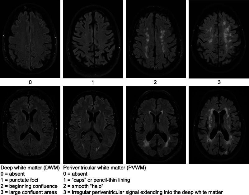
This diagnostic comparison chart illustrates the Fazekas scale for grading white matter hyperintensities (WMH) using axial FLAIR (Fluid-Attenuated Inversion Recovery) MRI sequences. The image is divided into two rows representing distinct anatomical regions: Deep White Matter (DWM) and Periventricular White Matter (PVWM). The DWM row (top) demonstrates progression from Grade 0 (absent) to Grade 1 (punctate foci), Grade 2 (beginning confluence), and Grade 3 (large confluent areas). The PVWM row (bottom) depicts clinical stages starting from Grade 0 (absent) to Grade 1 (periventricular 'caps' or pencil-thin lining), Grade 2 (smooth 'halo' around ventricles), and Grade 3 (irregular hyperintensities extending into the deep white matter). This radiological tool is essential in neurology and neuroradiology for quantifying chronic small vessel disease, vascular dementia, and age-related white matter changes. The visual highlights key morphological differences in signal intensity and distribution used to standardize clinical reporting and research outcomes.
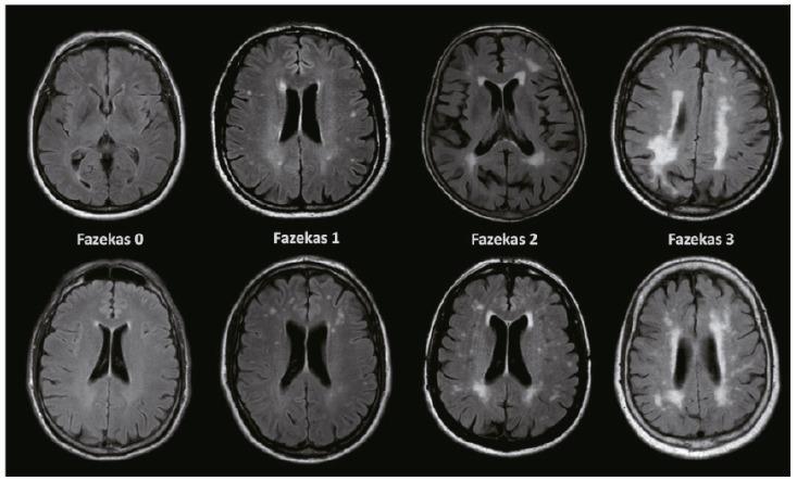
This diagnostic comparison chart features axial Fluid-Attenuated Inversion Recovery (FLAIR) MRI scans of the brain, illustrating the Fazekas scale for grading white matter hyperintensities (WMH). The image is organized into four columns representing Fazekas grades 0 through 3. Fazekas 0 (Normal) shows an absence of lesions. Fazekas 1 (Mild) displays isolated, punctate hyperintensities in the deep white matter or thin 'caps' at the ventricular horns. Fazekas 2 (Moderate) demonstrates larger, more numerous lesions beginning to coalesce, appearing as smooth 'halos' in periventricular regions. Fazekas 3 (Severe) reveals large, confluent hyperintensities that extensively involve both periventricular and deep white matter areas, creating irregular, irregular masses of high signal. These scans are critical for evaluating small vessel disease and its association with vascular cognitive impairment (VCI) and dementia. The educational focus is on identifying the morphology, distribution, and clinical progression of ischemic white matter changes.
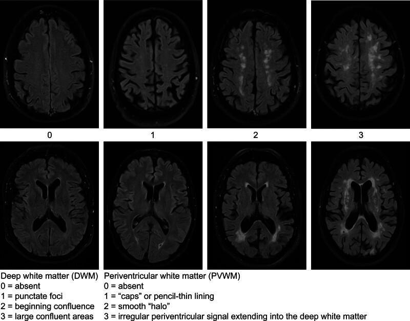
This diagnostic image provides a visual reference for the Fazekas scale, a radiological grading system used to quantify white matter hyperintensities (WMH) on brain MRI, typically seen in axial Fluid-Attenuated Inversion Recovery (FLAIR) sequences. The image is organized into two rows representing distinct anatomical regions of the white matter. The top row illustrates Deep White Matter (DWM) lesions progressing from Grade 0 (absent) to Grade 1 (punctate foci), Grade 2 (beginning confluence of lesions), and Grade 3 (large confluent areas). The bottom row illustrates Periventricular White Matter (PVWM) lesions, progressing from Grade 0 (absent) to Grade 1 (thin 'caps' or pencil-thin lining at the ventricular horns), Grade 2 (a smooth 'halo' surrounding the ventricles), and Grade 3 (irregular periventricular signals extending deep into the white matter). This comparison chart is a critical educational tool for neurology and radiology for assessing small vessel disease, leukoaraiosis, and cerebral vascular damage.
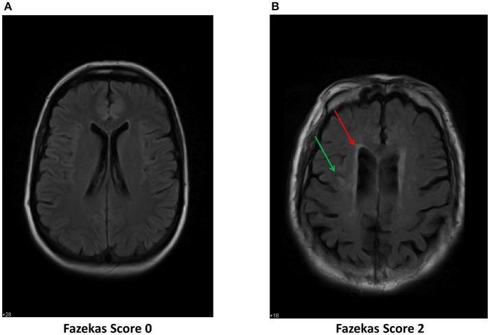
This diagnostic image consists of two side-by-side axial MRI scans of the brain using Fluid-Attenuated Inversion Recovery (FLAIR) sequences, illustrating the Fazekas scale for grading white matter hyperintensities (WMH). Figure A displays a normal brain scan categorized as Fazekas Grade 0, characterized by a homogenous appearance of the white matter with no visible lesions. Figure B displays clinical findings categorized as Fazekas Grade 2, demonstrating moderate leukoaraiosis. Specifically, red arrows highlight periventricular hyperintensities (caps or 'halos' surrounding the lateral ventricles), while green arrows point to punctate or beginning confluent hyperintensities within the deep white matter. These visual markers represent pathological tissue changes such as demyelination, gliosis, and axonal loss, often associated with small vessel disease, age-related cognitive decline, or risk factors for postoperative delirium (POD). The comparison serves as an educational tool for neuroradiological assessment of white matter disease progression and cerebral vascular health.
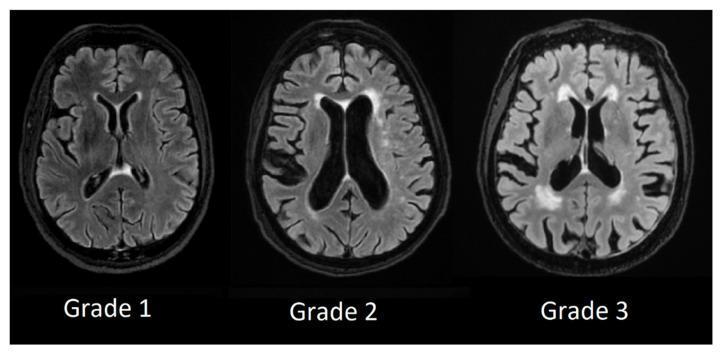
Comparison of axial MRI brain scans (T2-weighted FLAIR sequence) demonstrating the Fazekas scale for periventricular white matter hyperintensities (PVWMH). The image illustrates three levels of disease progression: Grade 1 displays mild, thin hyperintensities limited to the 'caps' of the anterior and posterior ventricular horns. Grade 2 shows a more pronounced 'smooth halo' of hyperintensity surrounding the lateral ventricles. Grade 3 depicts severe, irregular, and confluent hyperintensities extending significantly from the ventricles into the deep white matter. These visual findings are critical diagnostic markers for cerebral small vessel disease, often associated with vascular dementia and cognitive decline. The scans provide a clear visual reference for neuroradiological grading and the assessment of white matter integrity.
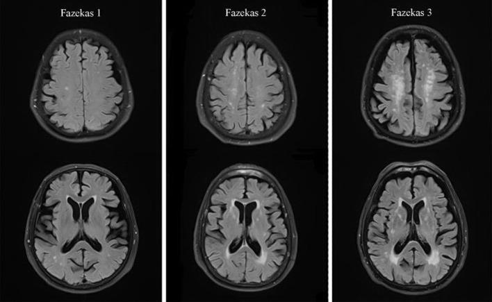
This diagnostic image consists of axial T2-weighted Fluid-Attenuated Inversion Recovery (FLAIR) MRI scans of the human brain, illustrating the Fazekas scale for grading white matter hyperintensities (WMH). The image is organized into three columns representing Fazekas grades 1, 2, and 3. Grade 1 (left) shows mild punctiform lesions in the deep subcortical white matter (DSWMH) and thin, cap-like or pencil-thin hyperintensities along the periventricular white matter (PVWMH). Grade 2 (middle) demonstrates moderate progression, characterized by the initial fusion of subcortical lesions and a smooth, halo-like thickening around the lateral ventricles. Grade 3 (right) represents severe disease, showing massive confluence of hyperintense lesions in the deep white matter and irregular periventricular signals that extend deeply into the brain parenchyma. This comparison chart is essential for clinical neurology and radiology to assess small vessel disease, leukoaraiosis, and age-related cognitive decline markers.
Fazekas scale white matter hyperintensities
Fazekas scale white matter hyperintensities grading clinical significance
Fazekas Scale
Grading System
Periventricular White Matter (PVWM)
| Grade | Description |
|---|---|
| 0 | Absent |
| 1 | "Caps" or pencil-thin lining at ventricular horns |
| 2 | Smooth "halo" surrounding the lateral ventricles |
| 3 | Irregular periventricular WMH extending into deep white matter |
Deep White Matter (DWM)
| Grade | Description |
|---|---|
| 0 | Absent |
| 1 | Punctate foci |
| 2 | Beginning confluence of foci |
| 3 | Large confluent (fused) areas |
Reference Images
Clinical Significance
- Stroke: Higher Fazekas scores predict future stroke risk and worse functional outcomes after stroke
- Cognitive decline and dementia: Strong correlation with vascular cognitive impairment (VCI); also associated with Alzheimer's disease (amyloid angiopathy)
- Depression and disability
- Increased mortality
Pathophysiology of WMH
- Chronic cerebral hypoperfusion / disrupted cerebrovascular autoregulation
- Blood-brain barrier breakdown
- Demyelination, gliosis, and axonal loss
- Inflammation
- Amyloid angiopathy
Practical Notes
- Assessed on axial T2-weighted FLAIR sequences
- In clinical reporting, the terms mild (grade 1), moderate (grade 2), and severe (grade 3) are generally preferred over numeric grades
- The scale is subjective and semi-quantitative; automated deep learning models are now being validated as alternatives (per Nature Scientific Reports, 2025)
- A 2026 systematic review (PMID: 41297452) further links higher WMH burden (higher Fazekas scores) with frailty in older adults