
lateral condyle consists of of elbow
Reading File
The lateral condyle of the humerus consists of the capitulum and the lateral part of the trochlea, along with the lateral epicondyle. Let me pull the exact details from the medical library.
Searching sources...
Finding Sources
Finding Sources
Finding Sources
Finding Sources
Finding Sources
Finding Sources
Reading File
Reading File
Loading Image
Finding Sources
Here is a complete breakdown of the lateral condyle of the humerus at the elbow:
Lateral Condyle of the Humerus - Composition
The lateral condyle is the lateral part of the distal humerus. It consists of the following components:
1. Capitulum (Capitellum)
- The rounded, hemispherical articular surface on the anterolateral aspect of the distal humerus
- Articulates with the head of the radius (radiohumeral joint)
- Faces anteriorly and inferiorly
- Separated from the trochlea by a shallow groove (capitulo-trochlear sulcus)
2. Lateral Part of the Trochlea (Lateral Trochlear Ridge)
- The trochlea extends medially, but its lateral ridge/wall is part of the lateral condyle
- This is clinically important: in Milch Type I lateral condyle fractures, the lateral trochlear ridge remains with the main humeral shaft; in Milch Type II, it goes with the fractured fragment
3. Lateral Epicondyle
- The non-articular bony prominence just proximal to the capitulum
- Site of attachment for the common extensor origin (extensor carpi radialis brevis, extensor digitorum, extensor digiti minimi, extensor carpi ulnaris)
- Also gives attachment to the lateral (radial) collateral ligament
4. Radial Fossa
- A small depression on the anterior surface of the lateral condyle just above the capitulum
- Accommodates the anterior rim of the radial head during full elbow flexion
Summary Table
| Component | Type | Articulates with |
|---|---|---|
| Capitulum | Articular | Radial head |
| Lateral trochlear ridge | Articular (partial) | Ulnar trochlear notch (lateral wall) |
| Lateral epicondyle | Non-articular | - (muscle/ligament attachment) |
| Radial fossa | Non-articular (depression) | Radial head rim (in flexion) |
Anatomy Image (3D CT reconstruction)
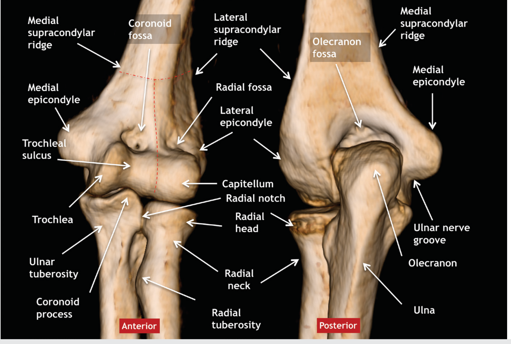
The red dashed lines in the image above show the approximate borders dividing the medial and lateral condyles. The capitellum and lateral epicondyle are clearly visible on the lateral side.
Clinical Notes
- Lateral condyle fracture is the 2nd most common pediatric elbow fracture (17%), after supracondylar fractures, most common between ages 4-6 years
- Fractures typically involve both the articular (capitulum + lateral trochlear ridge) and non-articular (epicondyle) portions
- Mechanism: fall on outstretched hand causing valgus stress, or avulsion
- Complication of missed/malunited lateral condyle fracture: cubitus valgus deformity, which can cause tardy ulnar nerve palsy (the nerve gets stretched around the medial elbow over time)
Sources: Imaging Anatomy: Bones, Joints, Vessels and Nerves, p. 497; Miller's Review of Orthopaedics 9th Ed., p. 903; Campbell's Operative Orthopaedics 15th Ed.
This is a shared conversation. Sign in to Orris to start your own chat.