Post traumatic brain injury. Riding a two-wheeler. Fell down on the left side of the face. Had a laceration on the temporal region. Uh, examination. Atrophy of the left eye. Not reacting. Uh, and, uh, I only literally left us download moment is there. And, uh, the facial, mild facial palsy on the left side present. But upward, uh, eye, upper word normal. Only deviation of angle of mouth and loss of nasolabial fold is present. Vastest sensations are normal. All other cranial nerves are normal. Where can be the most probable lesion present?
- Left eye: atrophy + non-reactive pupil (optic nerve/CN II damage)
- Left facial palsy: lower motor neuron pattern BUT with sparing of the forehead (upper face spared, only angle of mouth deviation + loss of nasolabial fold)
- Normal taste, normal other cranial nerves
- Mechanism: trauma to left temporal/facial region
post traumatic brain injury optic nerve palsy facial nerve UMN pattern temporal region lesion localization
It's not atrophy. Left titosis.
You will not meiosis. It's dilated. And the pupil is not seen, not EOM, right side restricted.
- Ptosis (left)
- Dilated pupil, non-reactive (left)
- EOM restricted (likely "down and out" position)
- Lower facial palsy (UMN pattern - forehead spared, angle of mouth deviated, nasolabial fold lost)
- Normal taste, normal other CNs
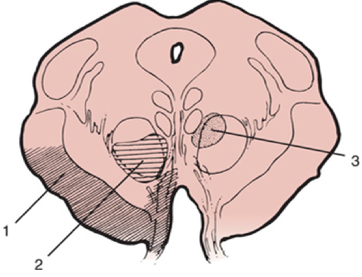
Lesion Localization: Left Ventromedial Midbrain (Weber Syndrome)
Reconstructed Clinical Signs
| Sign | Side | Structure Damaged |
|---|---|---|
| Ptosis | Left | CN III (levator palpebrae) |
| Dilated, non-reactive pupil | Left | CN III parasympathetic (Edinger-Westphal nucleus fascicle) |
| Restricted EOM (down and out) | Left | CN III motor fascicle |
| Lower facial palsy (forehead SPARED) | Left face / Right UMN | Corticobulbar tract crossing in cerebral peduncle |
Why This Is Weber Syndrome
-
The CN III fascicle exits the ventromedial midbrain (cerebral peduncle). A lesion here hits CN III before it exits the skull, causing ipsilateral (left) CN III palsy with:
- Ptosis (Müller + levator palpebrae denervation)
- Dilated, fixed pupil (parasympathetic Edinger-Westphal fibers run on the outside of CN III - maximally vulnerable to compression/trauma)
- "Down and out" eye (unopposed CN IV + CN VI)
-
The corticospinal and corticobulbar tracts run through the same cerebral peduncle and have not yet crossed. So a left peduncle lesion damages the left corticobulbar fibers heading to the right facial nucleus (lower face only, because upper face has bilateral cortical supply). This causes contralateral UMN lower facial palsy - which in this case is on the... left face.
Wait - the facial palsy is also on the LEFT. This means the corticobulbar involvement is right-sided, producing a left lower facial UMN palsy, meaning the lesion is in the right cerebral peduncle - OR -
Reconciling the laterality
- Ipsilateral CN III palsy (left) = lesion is on the left midbrain peduncle
- Contralateral UMN facial palsy = would normally be on the right...
Revised - Most Probable Single Lesion: Left Cerebral Peduncle / Posterior Limb of Internal Capsule
- Left CN III palsy (ptosis + dilated pupil + EOM restriction)
- Left UMN facial palsy (forehead spared - corticobulbar)
- Normal taste, normal sensation
- Direct damage to the left CN III fascicle as it exits - giving ipsilateral CN III palsy
- Damage to left corticobulbar fibers which control the contralateral (right) facial nucleus lower face - but here the left facial lower palsy may reflect right peduncle involvement OR this could represent an uncal herniation pattern where:
Most Likely Final Answer: Uncal Herniation / CN III Compression at the Tentorial Edge
Left Temporal Lobe Contusion with Uncal Herniation
- The expanding left temporal hematoma/contusion pushes the uncus (medial temporal lobe) over the tentorial notch
- This compresses the left CN III as it runs along the tentorial edge - causing ipsilateral ptosis + dilated fixed pupil + EOM restriction (the parasympathetic fibers on the outside of CN III are hit first)
- As herniation progresses, it compresses the left cerebral peduncle - disrupting the corticobulbar tract to the right facial nucleus lower face
- The left UMN lower facial palsy (forehead spared) is the corticobulbar sign from the contralateral peduncle compression
Summary Table
| Feature | Explanation |
|---|---|
| Left ptosis | Left CN III compression at tentorial notch |
| Left dilated, fixed pupil | Parasympathetic fibers of CN III (outermost, compressed first) |
| Left EOM restricted | CN III motor fascicle involvement |
| Left UMN lower facial palsy | Corticobulbar tract involvement (contralateral peduncle) |
| Forehead spared | UMN pattern - bilateral cortical supply to upper face |
| Normal taste, sensation | CN V, VII LMN, IX, X all intact |
| Mechanism | Left temporal blow → extradural/subdural hematoma → uncal herniation |