
Acute labyrinthitis clinical features, investigation and treatment protocol
Reading File
Now let me search the library and PubMed in parallel for comprehensive coverage.
Finding Sources
Finding Sources
Searching PubMed
acute labyrinthitis treatment vestibular neuritis
Good results. Now let me read the key textbook sections in parallel:
Reading File
Reading File
Reading File
Searching PubMed
vestibular neuritis labyrinthitis corticosteroids treatment
Good data. Let me now read the comprehensive Scott-Brown's LABYRINTHITIS section and the Shambaugh Surgery block, plus search for viral labyrinthitis specifically:
Reading File
Reading File
Finding Sources
Excellent. Now let me read the Cummings section on labyrinthitis infections and the Shambaugh acute section, plus the viral/treatment details from Scott-Brown's:
Reading File
Reading File
Reading File
Let me now read the Cummings Otolaryngology suppurative labyrinthitis block for management details, and also the Family Medicine / Rosen's Emergency block for clinical protocols:
Reading File
Reading File
I now have comprehensive data from multiple authoritative textbooks. Let me also fetch the MRI image that was referenced and get the treatment details from KJ Lee:
Reading File
Loading Image
I now have comprehensive information from multiple authoritative sources. Let me compile the full, detailed answer.
Acute Labyrinthitis - Clinical Features, Investigations, and Treatment Protocol
Definition and Classification
Labyrinthitis is inflammation of the membranous labyrinth (both cochlear and vestibular components of the inner ear). It is distinguished from vestibular neuritis by the presence of cochlear symptoms (hearing loss, tinnitus) in addition to vestibular dysfunction. Otolaryngologists generally recognize three main types (Schuknecht's classification):
- Serous labyrinthitis - inflammation without pus formation; bacterial toxins/mediators enter via the oval window, round window, or labyrinthine fistula without bacterial invasion of the labyrinth itself
- Suppurative (otogenic or meningitic) labyrinthitis - actual bacterial infection within the labyrinth
- Viral labyrinthitis - presumed viral etiology, no bacterial source identifiable
- Shambaugh Surgery of the Ear, pp. 3938-3941
- K.J. Lee's Essential Otolaryngology, pp. 867-927
- Scott-Brown's Otorhinolaryngology Vol. 2, pp. 3901-3925
Etiology
Bacterial Causes
- Serous labyrinthitis: most commonly complicates acute otitis media (AOM). Bacterial toxins (especially streptococcal) increase round window membrane permeability, allowing entry without direct bacterial invasion. Common pathogens: Streptococcus pneumoniae, H. influenzae, M. catarrhalis
- Suppurative (otogenic): bacteria enter via labyrinthine fistula (most commonly from cholesteatoma), surgical defect, or congenital anomaly. Most commonly associated with cholesteatoma in the modern era
- Suppurative (meningitic): bacteria enter via cochlear aqueduct or IAC from the subarachnoid space. Important route in children with cochlear implants and congenital inner ear malformations
Viral Causes
-
Presumed viral etiology when acute-onset vertigo + SNHL occur without identifiable otologic or meningitic cause
-
Implicated viruses: HSV type 1 (reactivation model), CMV, mumps, measles, rubella, varicella-zoster (Ramsay Hunt)
-
Definitive viral isolation from perilymph is rarely achievable; identification remains elusive in most cases
-
Cummings Otolaryngology, pp. 1836-1843
Clinical Features
Symptoms
| Feature | Serous Labyrinthitis | Suppurative Labyrinthitis | Viral Labyrinthitis |
|---|---|---|---|
| Vertigo | Mild to moderate | Severe, sudden onset | Acute, severe |
| Hearing loss | Mild-moderate SNHL (reversible) | Profound SNHL (permanent) | Moderate-severe SNHL |
| Tinnitus | Present | Present | Present |
| Nausea/Vomiting | Yes | Severe | Severe |
| Fever/Toxicity | Mild (in context of AOM) | May be present | Usually absent |
| Meningeal signs | Absent | May be present (meningitic type) | Absent |
| Recovery | Complete and rapid | Poor for hearing; vestibular compensates over weeks-months | Variable |
Key differentiating point from vestibular neuritis: labyrinthitis has associated cochlear symptoms (hearing loss, tinnitus, aural fullness, otalgia). Vestibular neuritis spares cochlear function. - Shambaugh Surgery, p. 3941
Nystagmus pattern:
- Direction-fixed, horizontal nystagmus beating away from the affected ear (inhibitory type)
- Nystagmus beating toward the affected ear is an ominous "irritative" sign indicating acute inner ear injury - Scott-Brown's Vol. 2, p. 3910-3911
Vestibular timeline:
- Acute phase: 48-72 hours of severe vertigo
- Subacute phase: dysequilibrium and unsteadiness for 4-6 weeks
- Chronic recovery/compensation: may persist several months
Investigations
Bedside Assessment
- Otoscopy - middle ear effusion, TM perforation, cholesteatoma (purulent AOM or COM with cholesteatoma suggests suppurative type)
- Head impulse test (HIT) - positive (catch-up saccade) in peripheral vestibulopathy; helps confirm peripheral origin
- HINTS exam (Head Impulse - Nystagmus - Test of Skew) - to rule out central cause (normal HIT + direction-changing nystagmus + skew deviation = central; always exclude posterior circulation stroke)
- Dix-Hallpike - to exclude BPPV
Audiological
- Pure tone audiogram - SNHL, often in high frequencies; may have additional conductive component if middle ear disease present
- Speech audiometry - assess word recognition score
- Tympanometry - assess middle ear status
- DPOAE/TEOAE - may show outer hair cell loss in basal turn
- Histopathology (if available): loss of outer hair cells primarily in basal turn, with significant incidence of endolymphatic hydrops - K.J. Lee, p. 908
Vestibular Testing (when necessary)
- Caloric testing / VNG/ENG - canal paresis on affected side
- VEMP (cervical/ocular) - saccular/utricular assessment
- Video head impulse test (vHIT) - quantifies VOR gain deficits per canal
Microbiology
- Culture of middle ear effusion when present (to guide antibiotic therapy)
- Blood cultures if febrile/systemically unwell
- CSF analysis if meningitis is suspected
Imaging
- MRI with gadolinium contrast (T1 post-contrast): prominent enhancement of the cochlea and vestibule - the gold standard for diagnosing acute labyrinthitis radiologically. Enhancement reflects blood-labyrinth barrier disruption due to capillary endothelial damage
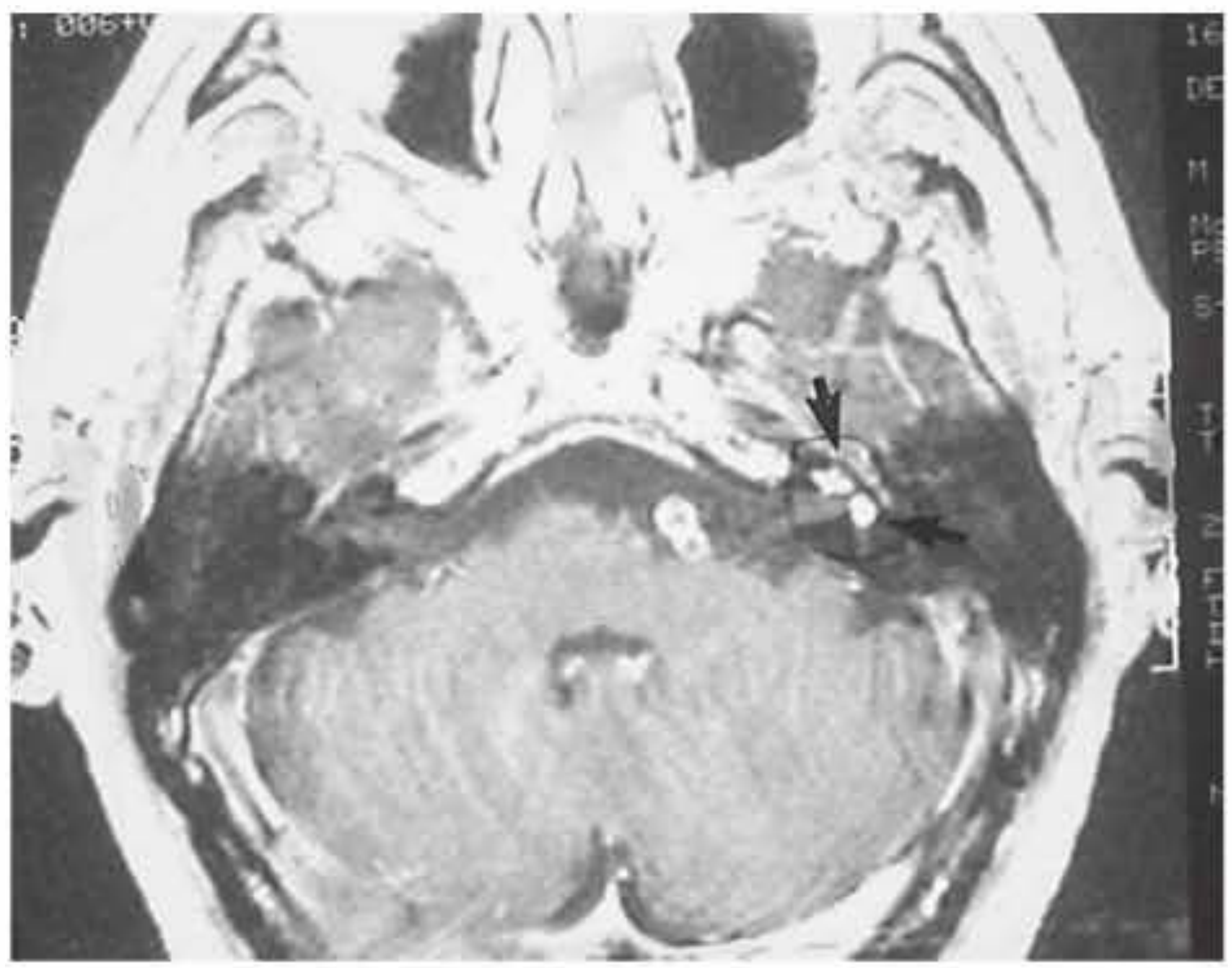
Acute labyrinthitis: axial MRI T1 post-gadolinium image demonstrating prominent enhancement of the left cochlea and vestibule (arrows). - Shambaugh Surgery of the Ear, p. 291
- CT temporal bones - not helpful in diagnosis of labyrinthitis itself, but delineates underlying anatomical abnormalities (cholesteatoma fistula, cochlear malformation, ossification in chronic cases)
- MRI/CT is not indicated in straightforward serous or viral labyrinthitis without other suspected complications - K.J. Lee, p. 906
- For labyrinthitis ossificans (late complication): serial CT and MRI to monitor for fibrous/bony obliteration of the labyrinth
Treatment Protocol
1. Viral / Idiopathic (Serous without identified bacterial source)
Vestibular suppressants (acute phase, max 3-5 days - prolonged use delays central compensation):
- Prochlorperazine 12.5 mg IM or 5-10 mg PO/PR
- Promethazine 25 mg IM/PO
- Dimenhydrinate 50 mg PO
- Betahistine (for longer-term labyrinthine compensation support)
Antiemetics:
- Ondansetron 4-8 mg IV/IM
- Metoclopramide 10 mg IV/IM
Corticosteroids:
- Prednisolone 1 mg/kg/day tapering over 7-14 days - evidence for benefit in vestibular neuritis/viral labyrinthitis is moderate but supported in many guidelines; improves hearing recovery outcomes particularly in cases with SNHL. Cochlear NF-κB suppression before/with treatment is the proposed mechanism - Scott-Brown's, p. 924-925
Antivirals:
- Not routinely recommended. Aciclovir or valaciclovir can be considered in Ramsay Hunt syndrome (VZV) or suspected HSV reactivation
Vestibular rehabilitation:
- Early mobilization and balance exercises after the acute phase (days 3-5 onward)
- Supervised vestibular rehabilitation therapy (VRT) to accelerate central compensation
2. Serous Labyrinthitis (complicating AOM)
- Oral antibiotics targeting AOM pathogens (S. pneumoniae, H. influenzae, M. catarrhalis): amoxicillin-clavulanate (first-line), or amoxicillin 80-90 mg/kg/day in children
- Adjuvant corticosteroids (oral ± intratympanic) - data are lacking but may improve hearing outcomes - K.J. Lee, p. 922
- Myringotomy if middle ear infection unresolved with antibiotics
- Vestibular suppressants / antiemetics as above
- Symptoms are typically reversible with appropriate antibiotic treatment
3. Suppurative (Otogenic / Meningitic) Labyrinthitis - URGENT
This is a medical and surgical emergency.
Antimicrobials:
- Parenteral antibiotics with CSF penetration - e.g., ceftriaxone 2g IV BD (covers S. pneumoniae, gram-negatives)
- Add metronidazole if cholesteatoma/anaerobic source suspected
- Duration: typically 4-6 weeks for severe otogenic infection
Steroids:
- Dexamethasone IV - shown to improve hearing outcomes in meningitic labyrinthitis due to H. influenzae and S. pneumoniae - K.J. Lee, p. 1004
- May prevent labyrinthitis ossificans if given in the initial phase - Scott-Brown's, p. 3923-3925
Surgical management:
| Source | Procedure |
|---|---|
| Non-cholesteatoma otogenic (AOM) | Myringotomy + ventilation tube |
| Cholesteatoma | Tympanomastoidectomy (urgent) |
| Cochleotomy | In select cases as per literature |
Vestibular suppressants (same as above)
Monitoring for complications:
- Meningitis - LP if suspected, cover with appropriate antibiotics
- Labyrinthitis ossificans - serial CT/MRI if SNHL present
- Cochlear implantation (CI): early intervention if bilateral SNHL with signs of ossification developing. Once ossification progresses, the scala tympani is obliterated and CI becomes technically very difficult - K.J. Lee, p. 1008
4. Specific Etiologies
| Etiology | Specific Treatment |
|---|---|
| Syphilitic labyrinthitis | Penicillin G IV; steroids for cochleovestibular symptoms |
| CMV labyrinthitis (pediatric) | Valganciclovir (for congenital CMV with SNHL within 1 month of birth) |
| Ramsay Hunt syndrome | Aciclovir/valaciclovir + prednisolone |
| Measles/mumps (preventable) | MMR vaccination (primary prevention) |
Complications
- Labyrinthitis ossificans - fibrous then bony obliteration of the membranous labyrinth; most common after suppurative labyrinthitis, especially meningitic. Cochlear T2 signal lost on MRI. May lead to profound bilateral deafness if cochlear implantation delayed
- Meningitis - bidirectional: labyrinthitis can cause or result from meningitis; risk elevated with cochlear implants, perilymph fistula, and congenital inner ear malformations
- Permanent profound SNHL - 2-20% (average ~10%) following bacterial labyrinthitis
- Persistent vestibulopathy - chronic dysequilibrium lasting months; requires VRT
- Brain abscess / intracranial extension - particularly from cholesteatoma-associated suppurative labyrinthitis
Key Diagnostic Distinctions
| Feature | Viral Labyrinthitis | Vestibular Neuritis | Ménière's Disease | BPPV |
|---|---|---|---|---|
| Hearing loss | Yes (SNHL) | No | Fluctuating low-frequency SNHL | No |
| Tinnitus | Yes | Rarely | Yes | No |
| Aural fullness | Yes | Rare | Yes (classic) | No |
| Duration | Days-weeks (single episode) | Days-weeks (single episode) | Minutes-hours (episodic) | Seconds (positional) |
| Nystagmus | Spontaneous, direction-fixed | Spontaneous, direction-fixed | Spontaneous during attack | Geotropic/apogeotropic, fatigable |
| Nausea/vomiting | Yes | Yes | Yes | Mild |
- Rosen's Emergency Medicine, p. 3628; Scott-Brown's Vol. 2, Table 62.4
Sources:
- Shambaugh Surgery of the Ear (6e), pp. 3938-3948
- Cummings Otolaryngology Head & Neck Surgery, pp. 1835-1843
- Scott-Brown's Otorhinolaryngology Head & Neck Surgery Vol. 2, pp. 3888-3925
- K.J. Lee's Essential Otolaryngology (12e), pp. 867-1008
- Rosen's Emergency Medicine, p. 3628
- Textbook of Family Medicine 9e, pp. 537-539
This is a shared conversation. Sign in to Orris to start your own chat.