
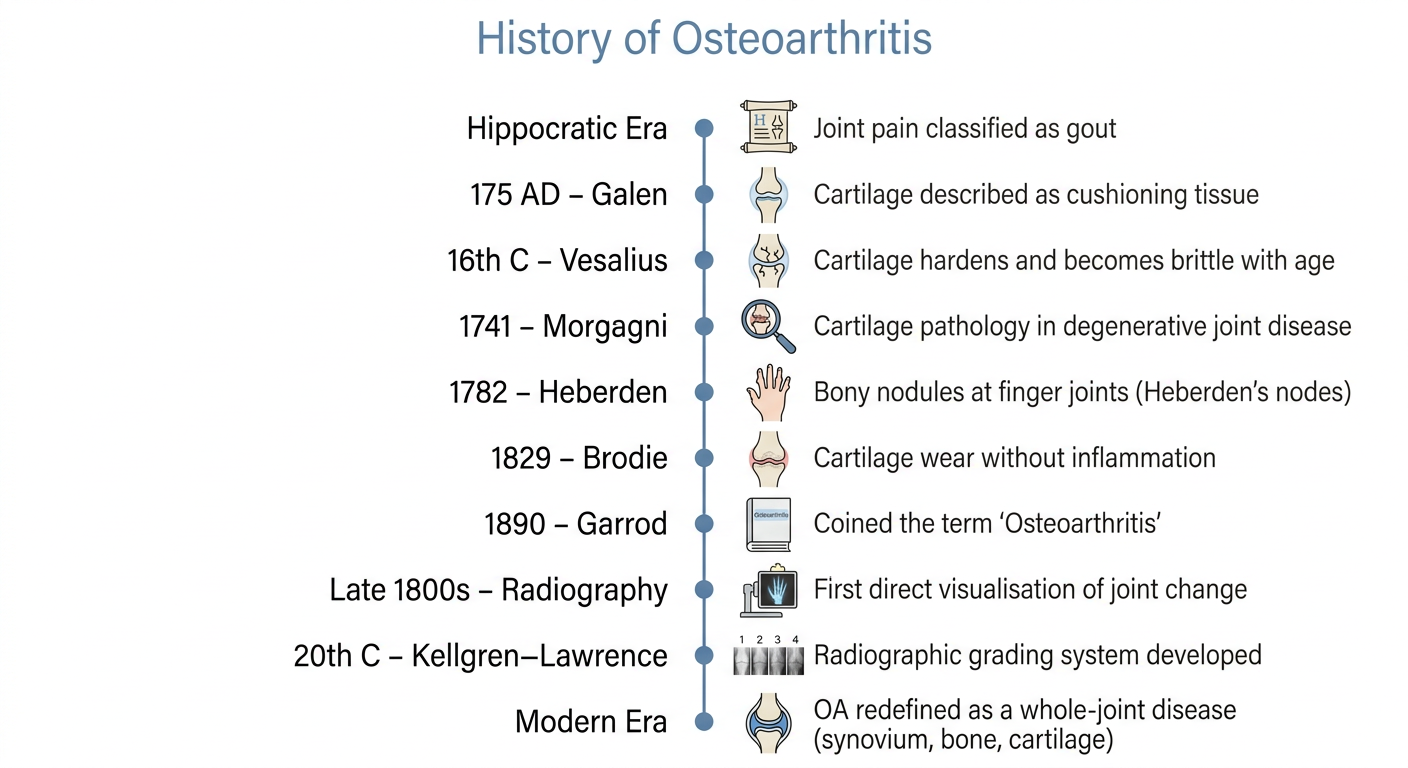
HISTORY Osteoarthritis (OA) as a separate disease is a relatively recent concept. Through much of recorded medical history, dating to Hippocratic medicine, physicians lacked a reliable basis for distinguishing among the causes of chronic joint pain and swelling, and such joint complaints were commonly classified as gout. [8]. William Heberden, in 1782, identified small, hard, bony nodules at the distal interphalangeal joints that were inconsistent with a gouty aetiology and proposed them as a separate condition; these nodules were subsequently named Heberden's nodes [9]. Benjamin Brodie, in 1829, observed that cartilage could undergo wear and tear in older individuals in the absence of inflammatory signs, initiating the conceptual separation of degenerative joint disease from inflammatory arthritis. Robert Smith, in 1835, provided one of the earliest detailed clinical descriptions of hip osteoarthritis [10]. The term "osteoarthritis" was coined by A.E. Garrod in 1890, although the underlying intra-articular pathology remained incompletely understood for some time thereafter [8]. The introduction of radiography in the late nineteenth century allowed direct visualisation of structural joint change for the first time, rather than inference from symptoms alone [8]. Kellgren and Moore subsequently demonstrated a genuine association between Heberden's nodes and osteoarthritis, a finding that contributed to the distinction between generalised OA and OA secondary to other joint pathology [11]. This radiographic approach was later formalised in the Kellgren–Lawrence grading system, which remains among the most widely applied tools for research and clinical staging of OA [12]. The conceptual history of articular cartilage existed before the disease concept of OA by nearly two millennia. Galen, circa 175 AD, described cartilage as a smooth cushioning tissue protecting joint surfaces [13]. Andreas Vesalius, in the sixteenth century, noted that cartilage undergoes progressive hardening and increased brittleness with age [13]. The eighteenth century contributed two further landmarks in close succession: Morgagni's 1741 account of cartilage pathology in degenerative joint disease, and William Hunter's investigations two years later, which remain foundational to the scientific study of articular cartilage [13]. Progress in this area subsequently stalled, largely because cartilage is avascular and aneural, properties that impeded study with the methods then available [13]. Advances in enzyme chemistry and biochemistry eventually permitted characterisation of cartilage synthesis and degradation at the molecular level; by the early twentieth century, this allowed better understanding of cartilage's layered structure and the identification of hyaluronic acid as central to joint lubrication [13]. The more substantial conceptual shift has occurred through subsequent advances in immunology, molecular biology, and collagen research [13], which have repositioned OA from a disease confined to cartilage toward a disease of the whole joint [14]. Current understanding implicates the synovium [15], ligaments [16], bone marrow [17], and subchondral bone, with damage distributed across these structures underlying the shock-absorption failure, pain, and functional loss characteristic of the disease [18]. ....GENERATE IMAGE WITH MINIMAL POINTS
A clean, minimal medical timeline infographic titled "History of Osteoarthritis" with the following key milestones listed vertically along a timeline line: • Hippocratic Era: Joint pain classified as gout • 175 AD – Galen: Cartilage described as cushioning tissue • 16th C – Vesalius: Cartilage hardens and becomes brittle with age • 1741 – Morgagni: Cartilage pathology in degenerative joint disease • 1782 – Heberden: Bony nodules at finger joints (Heberden's nodes) • 1829 – Brodie: Cartilage wear without inflammation • 1890 – Garrod: Coined the term "Osteoarthritis" • Late 1800s – Radiography: First direct visualisation of joint change • 20th C – Kellgren–Lawrence: Radiographic grading system developed • Modern Era: OA redefined as a whole-joint disease (synovium, bone, cartilage) Style: clean white background, simple vertical timeline with dots, minimal icons, medical/academic aesthetic, muted blue and grey color palette, sans-serif font
CAN U CHK THESIS?
What kind of review do you need?
Too much time taking
too much time consuming