
tachycardia
Reading File
"Tachycardia" is broad - let me clarify before diving deep.
Asking for Preferences
What aspect of tachycardia are you looking for?
Now let me search the medical library for a comprehensive overview.
Finding Sources
Searching the Web
tachycardia classification overview types 2024
Good - I have strong results. Let me now read the key sections from Braunwald's (gold standard cardiology text) and Rosen's Emergency Medicine for the classification overview, in parallel.
Reading File
Reading File
Finding Sources
There's a classification flowchart in Frameworks for Internal Medicine - let me grab that and also read more of the Rosen's content on the various tachycardia types.
Reading File
Reading File
Excellent content gathered. Let me also fetch the tachycardia classification flowchart image from Frameworks for Internal Medicine.
Loading Image
Reading File
I now have everything needed for a thorough overview. Let me compile the full response.
Tachycardia - Overview & Classification
Definition: Heart rate ≥ 100 beats/min. It can be a normal physiological response (sinus tachycardia) or reflect underlying pathology (arrhythmic tachycardia).
Classification Framework
The primary ECG-based framework divides all tachycardias by QRS width and rhythm regularity:
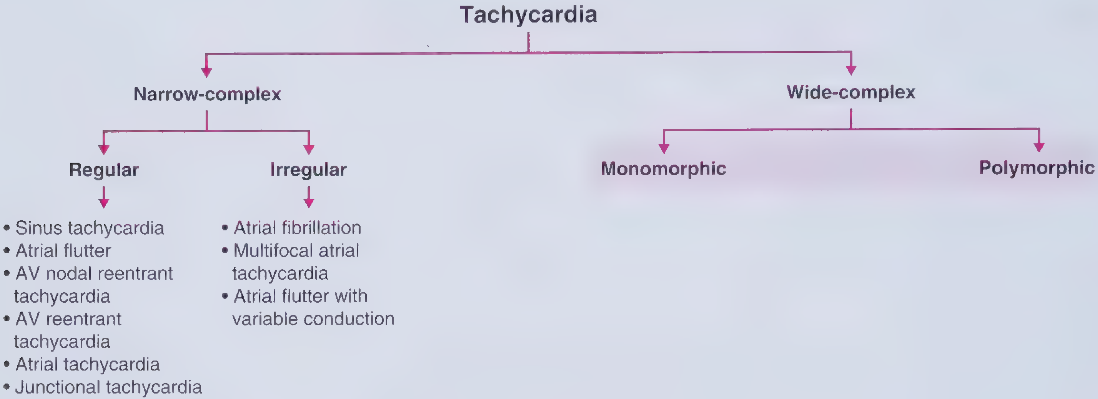
1. Narrow-Complex Tachycardia (QRS < 120 ms)
Reflects rapid activation via the normal His-Purkinje system, so the origin is at or above the His bundle (supraventricular).
A. Regular Narrow-Complex Tachycardias
| Type | Key Features |
|---|---|
| Sinus Tachycardia | Normal P waves before each QRS; rate rarely >170 bpm in adults; gradual onset/offset; usually a physiological response |
| Atrial Flutter | Sawtooth flutter waves ~300/min; 2:1, 3:1, or 4:1 AV block gives regular ventricular rate |
| AVNRT (AV nodal reentrant tachycardia) | Most common paroxysmal SVT; reentry circuit within AV node; P waves buried in or just after QRS |
| AVRT (AV reentrant tachycardia) | Requires accessory pathway (e.g., WPW); P wave after QRS in the ST segment |
| Atrial Tachycardia | Abnormal P wave morphology; focus outside sinus node; rate 100-240/min |
| Junctional Tachycardia | Origin in AV junction; inverted or absent P waves |
B. Irregular Narrow-Complex Tachycardias
| Type | Key Features |
|---|---|
| Atrial Fibrillation | Most common sustained arrhythmia; irregularly irregular rhythm; no distinct P waves; 300-600 atrial impulses/min; ventricular rate typically ≤150-170/min |
| Multifocal Atrial Tachycardia (MAT) | ≥3 distinct P-wave morphologies; classically seen with COPD |
| Atrial Flutter with variable conduction | Flutter waves with changing AV block ratio |
2. Wide-Complex Tachycardia (QRS ≥ 120 ms)
Indicates abnormally slow ventricular activation. Three major causes:
- Origin below the His bundle (ventricular tachycardia)
- SVT with pre-existing bundle branch block
- SVT with rate-dependent aberrant conduction
A. Monomorphic Wide-Complex (uniform QRS morphology)
Regular:
- Monomorphic VT - most often from myocardial scar (ischemic heart disease); H-V interval shorter than normal on EPS
- SVT with BBB - same SVT as above but QRS widens due to baseline or rate-related bundle branch block
- Pacemaker-mediated tachycardia
Irregular:
- AF with aberrancy (BBB or accessory pathway)
- AF with WPW - ventricular rate can exceed 200/min; AV nodal blockers are contraindicated
B. Polymorphic Wide-Complex (variable QRS morphology)
- Torsades de Pointes - associated with prolonged QT interval; twisting QRS axis around baseline
- Polymorphic VT - often in setting of acute ischemia or channelopathies (Brugada, catecholaminergic)
- Ventricular fibrillation - chaotic, no organized QRS; cardiac arrest
Mechanism Categories
| Mechanism | Examples |
|---|---|
| Reentry | AVNRT, AVRT, atrial flutter, most VT |
| Enhanced automaticity | Sinus tachycardia, atrial tachycardia, accelerated idioventricular rhythm |
| Triggered activity | Torsades de Pointes (EADs), digoxin toxicity (DADs) |
Physiological vs. Pathological Sinus Tachycardia
Sinus tachycardia is almost always a compensatory response - treating it without addressing the underlying cause is counterproductive. Common causes include:
- Sympathetic excess: pain, anxiety, fever, hyperthyroidism, drugs/stimulants
- Reduced perfusion: hypovolemia, anemia, hypoxemia, sepsis
- Cardiac: decompensated heart failure, PE, tamponade
Key Diagnostic Tool: QRS Width + Regularity on ECG
- Is QRS narrow or wide? → Narrows to SVT vs. VT
- Is the rhythm regular or irregular? → Identifies AF, flutter, MAT
- What do the P waves look like? → Determines atrial origin
- What is the P:QRS relationship? → Distinguishes AVNRT, AVRT, AT
- EPS (electrophysiology study): H-V interval distinguishes SVT (normal or prolonged H-V) from VT (short or absent H-V) when ECG is equivocal - per Braunwald's Heart Disease
Sources: Frameworks for Internal Medicine | ROSEN's Emergency Medicine 10e | Braunwald's Heart Disease
This is a shared conversation. Sign in to Orris to start your own chat.