Case 1: A 55-year-old male patient presents with an white patch on the left lateral border of his tongue since 4 months. Patient is chronic alcoholic and smoker. On examination, a well-demarcated 2 cm x 1.5 cm homogeneous white plaque with raised margin is present which cannot be peeled off. 1.What is the diagnosis and aetiological factor of the given condition? 2. Enumerate three common differential diagnosis for this condition. 3. What are the important risk factors for this condition ? 4. Define epithelial dysplasia and list four key microscopic features indicative of dysplastic changes in this epithelium. 5. What are management strategy for above given condition

Reading File
Finding Sources
Finding Sources
Searching PubMed

oral leukoplakia malignant transformation

Reading File
Reading File
Reading File
Reading File
Finding Sources
Finding Sources
Finding Sources
Reading File
Reading File
Finding Sources
Reading File
Searching Images

oral leukoplakia white patch lateral border of tongue
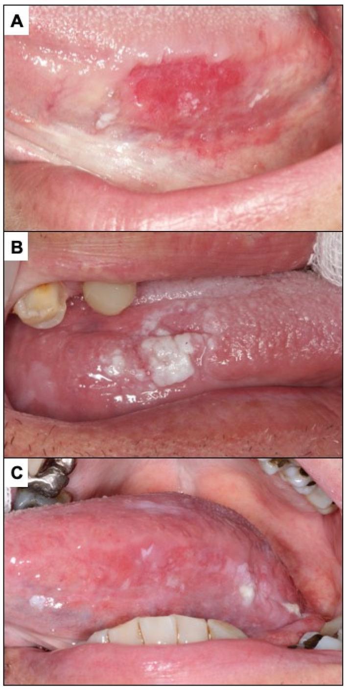
A series of three clinical photographs (A, B, and C) illustrating various clinical presentations of oral potentially malignant disorders and oral squamous cell carcinoma (OSCC) on the lateral border of the tongue.
A series of three clinical photographs (A, B, and C) illustrating various clinical presentations of oral potentially malignant disorders and oral squamous cell carcinoma (OSCC) on the lateral border of the tongue.

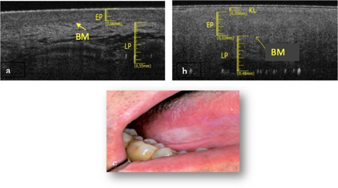
This composite image provides a diagnostic and clinical comparison of healthy tongue mucosa versus oral leukoplakia. Panels (a) and (b) present Optical Coherence Tomography (OCT) scans. Panel (a) shows healthy lateral tongue tissue with a clearly defined, continuous basement membrane (BM), an epithelium (EP) thickness of 0.16 mm, and a deep lamina propria (LP) extending to 0.55 mm. In contrast, panel (b) displays a leukoplakia lesion characterized by a thinner EP (0.05 mm), the presence of a superficial keratin layer (KL), and a disrupted, poorly defined basement membrane with a shallower LP depth of 0.32 mm. Panel (c) is a clinical photograph showing an irregular, opaque white patch on the lateral border of the tongue, consistent with the clinical appearance of leukoplakia. The image demonstrates the use of OCT as a non-invasive tool to visualize microscopic changes in tissue architecture, specifically epidermal thinning and basement membrane fragmentation, which are critical in assessing potentially malignant oral disorders.
This composite image provides a diagnostic and clinical comparison of healthy tongue mucosa versus oral leukoplakia. Panels (a) and (b) present Optical Coherence Tomography (OCT) scans. Panel (a) shows healthy lateral tongue tissue with a clearly defined, continuous basement membrane (BM), an epithelium (EP) thickness of 0.16 mm, and a deep lamina propria (LP) extending to 0.55 mm. In contrast, panel (b) displays a leukoplakia lesion characterized by a thinner EP (0.05 mm), the presence of a superficial keratin layer (KL), and a disrupted, poorly defined basement membrane with a shallower LP depth of 0.32 mm. Panel (c) is a clinical photograph showing an irregular, opaque white patch on the lateral border of the tongue, consistent with the clinical appearance of leukoplakia. The image demonstrates the use of OCT as a non-invasive tool to visualize microscopic changes in tissue architecture, specifically epidermal thinning and basement membrane fragmentation, which are critical in assessing potentially malignant oral disorders.

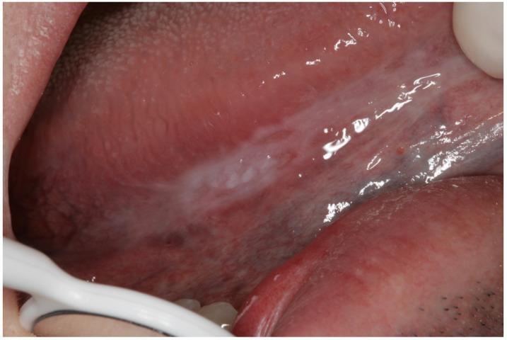
This clinical photograph shows a close-up intraoral view of the lateral border of the tongue. A distinct, predominantly white, plaque-like lesion (leukoplakia) is visible, characterized by an irregular shape and non-homogeneous texture. The lesion appears as a thickened, slightly raised white patch with varying degrees of opacity. The surrounding oral mucosa displays a typical pinkish-red hue with visible minor vascularity and small dark punctate spots nearby. Such lesions are classified as oral potentially malignant disorders (OPMDs) and require clinical evaluation and biopsy to rule out incipient oral squamous cell carcinoma. The image serves as an educational example of how early-stage oral cancer or high-risk dysplasia can manifest as a persistent white plaque on the tongue's lateral surface.
This clinical photograph shows a close-up intraoral view of the lateral border of the tongue. A distinct, predominantly white, plaque-like lesion (leukoplakia) is visible, characterized by an irregular shape and non-homogeneous texture. The lesion appears as a thickened, slightly raised white patch with varying degrees of opacity. The surrounding oral mucosa displays a typical pinkish-red hue with visible minor vascularity and small dark punctate spots nearby. Such lesions are classified as oral potentially malignant disorders (OPMDs) and require clinical evaluation and biopsy to rule out incipient oral squamous cell carcinoma. The image serves as an educational example of how early-stage oral cancer or high-risk dysplasia can manifest as a persistent white plaque on the tongue's lateral surface.

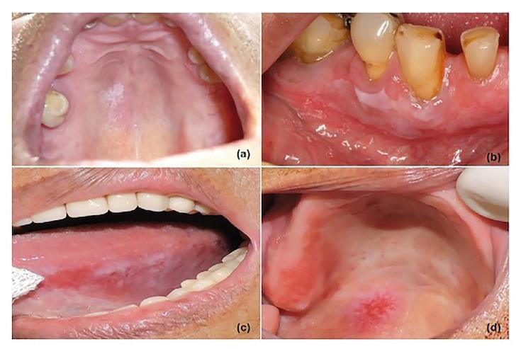
This composite of four clinical photographs illustrates various presentations of oral leukoplakia (OL) across different intraoral anatomical sites. Image (a) shows a single, homogeneous white patch on the hard palate. Image (b) demonstrates a flat, homogeneous white lesion involving the right inferior gingiva. Image (c) presents a nonhomogeneous leukoplakia on the left lateral border of the tongue, characterized by extensive white and reddish patches (erythroleukoplakia) with irregular surface texture, indicating a high risk of malignancy. Image (d) displays a mixed red and white lesion on the palate, featuring a central granular erythematous area surrounded by diffuse white leukoplakic changes. These images serve as educational tools for distinguishing between homogeneous and nonhomogeneous clinical subtypes of oral potentially malignant disorders (OPMDs). The collection highlights the importance of morphological assessment (color variation, texture, and border definition) and anatomical location in evaluating the risk of dysplasia and malignant transformation in oral lesions.

This composite of four clinical photographs illustrates various presentations of oral leukoplakia (OL) across different intraoral anatomical sites. Image (a) shows a single, homogeneous white patch on the hard palate. Image (b) demonstrates a flat, homogeneous white lesion involving the right inferior gingiva. Image (c) presents a nonhomogeneous leukoplakia on the left lateral border of the tongue, characterized by extensive white and reddish patches (erythroleukoplakia) with irregular surface texture, indicating a high risk of malignancy. Image (d) displays a mixed red and white lesion on the palate, featuring a central granular erythematous area surrounded by diffuse white leukoplakic changes. These images serve as educational tools for distinguishing between homogeneous and nonhomogeneous clinical subtypes of oral potentially malignant disorders (OPMDs). The collection highlights the importance of morphological assessment (color variation, texture, and border definition) and anatomical location in evaluating the risk of dysplasia and malignant transformation in oral lesions.

Searching Images

oral epithelial dysplasia histology microscopic
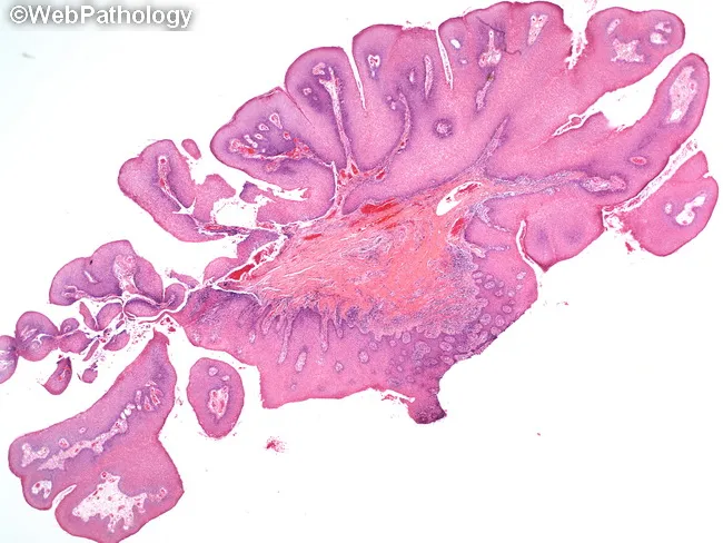
Low-magnification light microscopic image of an oral mucosal lesion with a pedunculated, papillomatous architecture. Finger‑like papillary fronds project from a surface epithelium, each projection containing a fibrovascular core. The lining is keratinized stratified squamous epithelium with a preserved maturation pattern to the surface. Hyperkeratosis may give the lesion a white clinical appearance. Koilocytes can be present in superficial layers, indicating human papillomavirus–associated change, while basilar hyperplasia and mild basilar mitotic activity may occur without true dysplasia. There is no invasion or marked architectural disarray. The subepithelial stroma is minimally inflamed, and the connective tissue darkly stained at higher power due to its fibrovascular composition. Overall, features are compatible with an oral squamous papilloma, a benign exophytic lesion. Differential considerations include verruca vulgaris and condyloma acuminatum, but the absence of significant koilocytic atypia or epithelial dysplasia supports a benign diagnosis. Clinically, such lesions are usually pedunculated, exophytic, and white when keratinized, often occurring in the oral cavity of children or young adults. Surgical excision is typically curative with low recurrence risk. This image emphasizes classic papillomatous growth, keratinized surface, and benign histology, useful for teaching oral pathology, differential diagnosis, and HPV-related mucosal lesions, including diagnostic pitfalls in distinguishing from papillary carcinomas.
Low-magnification light microscopic image of an oral mucosal lesion with a pedunculated, papillomatous architecture. Finger‑like papillary fronds project from a surface epithelium, each projection containing a fibrovascular core. The lining is keratinized stratified squamous epithelium with a preserved maturation pattern to the surface. Hyperkeratosis may give the lesion a white clinical appearance. Koilocytes can be present in superficial layers, indicating human papillomavirus–associated change, while basilar hyperplasia and mild basilar mitotic activity may occur without true dysplasia. There is no invasion or marked architectural disarray. The subepithelial stroma is minimally inflamed, and the connective tissue darkly stained at higher power due to its fibrovascular composition. Overall, features are compatible with an oral squamous papilloma, a benign exophytic lesion. Differential considerations include verruca vulgaris and condyloma acuminatum, but the absence of significant koilocytic atypia or epithelial dysplasia supports a benign diagnosis. Clinically, such lesions are usually pedunculated, exophytic, and white when keratinized, often occurring in the oral cavity of children or young adults. Surgical excision is typically curative with low recurrence risk. This image emphasizes classic papillomatous growth, keratinized surface, and benign histology, useful for teaching oral pathology, differential diagnosis, and HPV-related mucosal lesions, including diagnostic pitfalls in distinguishing from papillary carcinomas.

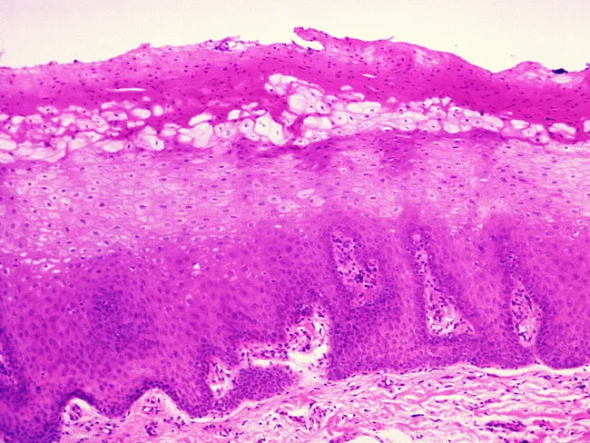
Hairy leukoplakia histology on oral mucosa. Imaging modality: light microscopy of formalin-fixed, paraffin-embedded biopsy stained with Hematoxylin and Eosin (H&E). The lesion arises in stratified squamous epithelium of the oral mucosa, typically the lateral tongue, presenting as a corrugated white plaque clinically. Microscopic features include epidermal acanthosis with marked parakeratosis and surface corrugation. In the upper spinous layer, there is a band of vacuolated, balloon-shaped keratinocytes with abundant pale cytoplasm. These cells display nuclear clearing (Cowdry type A inclusions) with peripheral condensation of chromatin along the nuclear membrane, creating a beaded rim. The lower epidermis shows intact basal layer without overt dysplasia. Candida hyphae are detected in approximately 80% of cases, often without acute inflammatory infiltrate. Importantly, there is no dysplasia or malignant transformation in typical hairy leukoplakia. Immunohistochemistry, polymerase chain reaction (PCR), and in situ hybridization can be used to confirm Epstein-Barr Virus (EBV) infection within keratinocyte nuclei if clinically indicated. The findings, in the proper clinical context (immunosuppression or HIV infection), support a benign EBV-driven epithelial lesion. This image is valuable for education and differential diagnosis with other white oral lesions such as candidiasis, leukoplakia, and lichen planus, and for teaching pathophysiology of EBV-related mucosal disease.
Hairy leukoplakia histology on oral mucosa. Imaging modality: light microscopy of formalin-fixed, paraffin-embedded biopsy stained with Hematoxylin and Eosin (H&E). The lesion arises in stratified squamous epithelium of the oral mucosa, typically the lateral tongue, presenting as a corrugated white plaque clinically. Microscopic features include epidermal acanthosis with marked parakeratosis and surface corrugation. In the upper spinous layer, there is a band of vacuolated, balloon-shaped keratinocytes with abundant pale cytoplasm. These cells display nuclear clearing (Cowdry type A inclusions) with peripheral condensation of chromatin along the nuclear membrane, creating a beaded rim. The lower epidermis shows intact basal layer without overt dysplasia. Candida hyphae are detected in approximately 80% of cases, often without acute inflammatory infiltrate. Importantly, there is no dysplasia or malignant transformation in typical hairy leukoplakia. Immunohistochemistry, polymerase chain reaction (PCR), and in situ hybridization can be used to confirm Epstein-Barr Virus (EBV) infection within keratinocyte nuclei if clinically indicated. The findings, in the proper clinical context (immunosuppression or HIV infection), support a benign EBV-driven epithelial lesion. This image is valuable for education and differential diagnosis with other white oral lesions such as candidiasis, leukoplakia, and lichen planus, and for teaching pathophysiology of EBV-related mucosal disease.

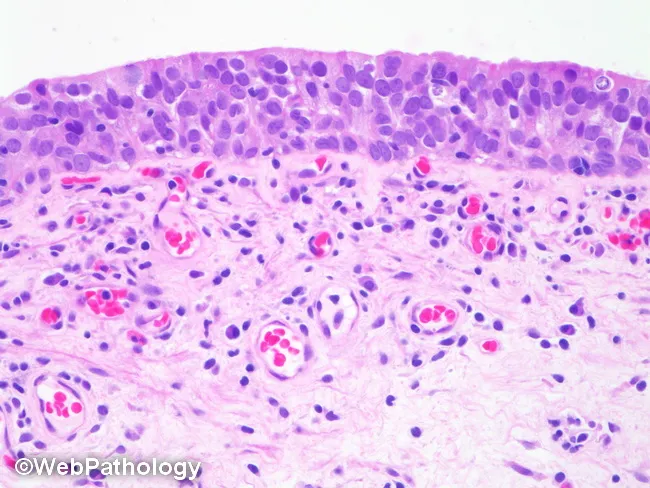
Image depicts light microscopic evaluation of oral mucosa with hematoxylin and eosin staining. Surface stratified squamous epithelium sits above a vascular lamina propria containing mixed inflammatory cells dominated by lymphocytes and plasma cells. The subepithelial tissue shows mild edema and capillary congestion without epithelial dysplasia, necrosis, or fungal granulomas. Overall pattern suggests nonspecific chronic mucosal inflammation or mild mucositis, potentially from irritation, infection, or autoimmune process. Clinically relevant for confirming inflammatory changes, guiding management of chronic mucositis, trauma related lesions, or infectious stomatitis, and differentiating from premalignant or malignant processes. Useful in educational, diagnostic, and research contexts to illustrate mucosal inflammatory histology and to support differential diagnosis in patients with oral mucosal lesions. Sampling technique, fixation, and stain quality influence interpretation and should be correlated with clinical findings and additional tests if indicated. Basement membrane zone appears intact with no subepithelial clefting or dyskeratosis evident at this magnification. This image serves as a reference for educational comparison to inflammatory conditions such as glossitis, stomatitis, lichen planus, candidiasis, or irritant contact lesions. In translational research, similar sections permit evaluation of infiltrate patterns, microvascular density, and matrix remodeling relevant to mucosal immunology and pathology.
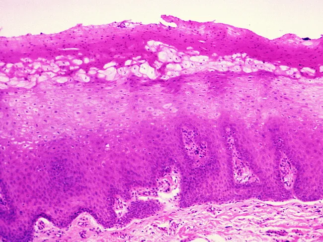
Image depicts light microscopic evaluation of oral mucosa with hematoxylin and eosin staining. Surface stratified squamous epithelium sits above a vascular lamina propria containing mixed inflammatory cells dominated by lymphocytes and plasma cells. The subepithelial tissue shows mild edema and capillary congestion without epithelial dysplasia, necrosis, or fungal granulomas. Overall pattern suggests nonspecific chronic mucosal inflammation or mild mucositis, potentially from irritation, infection, or autoimmune process. Clinically relevant for confirming inflammatory changes, guiding management of chronic mucositis, trauma related lesions, or infectious stomatitis, and differentiating from premalignant or malignant processes. Useful in educational, diagnostic, and research contexts to illustrate mucosal inflammatory histology and to support differential diagnosis in patients with oral mucosal lesions. Sampling technique, fixation, and stain quality influence interpretation and should be correlated with clinical findings and additional tests if indicated. Basement membrane zone appears intact with no subepithelial clefting or dyskeratosis evident at this magnification. This image serves as a reference for educational comparison to inflammatory conditions such as glossitis, stomatitis, lichen planus, candidiasis, or irritant contact lesions. In translational research, similar sections permit evaluation of infiltrate patterns, microvascular density, and matrix remodeling relevant to mucosal immunology and pathology.

Imaging modality: Light microscopy of an oral mucosa biopsy stained with Hematoxylin and Eosin. The specimen shows classic hairy leukoplakia histology. Microscopically, there is epithelial acanthosis and parakeratotic surface with pronounced corrugation. In the upper spinous layer, a band of vacuolated, ballooned cells with abundant pale cytoplasm is evident. These dyskeratotic-appearing keratinocytes display nuclear clearing with a characteristic Cowdry type A intranuclear inclusion pattern and peripheral chromatin that appears in a beaded rim against the nuclear membrane. The basement membrane and basal cell layer remain intact, with no cytologic dysplasia. Candida hyphae infiltrate the superficial stratum corneum in roughly 80% of cases and may occur without a conspicuous inflammatory response. The lesion's erythematous-keratotic surface on mucosa may be observed macroscopically as a white-to-gray white plaque with irregular surface. Although the microscopic features are highly suggestive, confirmation can be achieved with ancillary techniques, including EBV-encoded RNA in-situ hybridization, immunohistochemistry for EBV latent membrane protein, or PCR, to demonstrate EBV within keratinocyte nuclei. Clinically, hairy leukoplakia occurs in the setting of immunosuppression (notably HIV infection) and recognition is important to guide management. No dysplasia indicates benign-viral keratinocytic change rather than malignant transformation. This image underscores the classic viral cytopathic pattern for education.

Imaging modality: Light microscopy of an oral mucosa biopsy stained with Hematoxylin and Eosin. The specimen shows classic hairy leukoplakia histology. Microscopically, there is epithelial acanthosis and parakeratotic surface with pronounced corrugation. In the upper spinous layer, a band of vacuolated, ballooned cells with abundant pale cytoplasm is evident. These dyskeratotic-appearing keratinocytes display nuclear clearing with a characteristic Cowdry type A intranuclear inclusion pattern and peripheral chromatin that appears in a beaded rim against the nuclear membrane. The basement membrane and basal cell layer remain intact, with no cytologic dysplasia. Candida hyphae infiltrate the superficial stratum corneum in roughly 80% of cases and may occur without a conspicuous inflammatory response. The lesion's erythematous-keratotic surface on mucosa may be observed macroscopically as a white-to-gray white plaque with irregular surface. Although the microscopic features are highly suggestive, confirmation can be achieved with ancillary techniques, including EBV-encoded RNA in-situ hybridization, immunohistochemistry for EBV latent membrane protein, or PCR, to demonstrate EBV within keratinocyte nuclei. Clinically, hairy leukoplakia occurs in the setting of immunosuppression (notably HIV infection) and recognition is important to guide management. No dysplasia indicates benign-viral keratinocytic change rather than malignant transformation. This image underscores the classic viral cytopathic pattern for education.

I have enough to give a complete answer. This case describes classic homogeneous oral leukoplakia of the lateral tongue border.
Oral leukoplakia - white plaque on lateral border of tongue

1. Diagnosis and Aetiological Factor

Diagnosis: Oral Leukoplakia (homogeneous type) — a well-demarcated, non-scrapable white plaque of the lateral tongue border.
Leukoplakia is defined clinically as "a white patch or plaque that cannot be characterized as any other definable lesion" — it is a diagnosis of exclusion, made clinically, with the histologic range spanning hyperkeratosis/acanthosis to varying degrees of epithelial dysplasia, carcinoma in situ, and even invasive squamous cell carcinoma (Cummings Otolaryngology, p. 1579; K J Lee's Essential Otolaryngology, p. 677). It is the most common oral potentially malignant (premalignant) lesion, though most lesions remain clinically benign over time.
Aetiological factor: In this patient the key driver is tobacco use (smoking) combined with chronic alcohol use — the two most closely associated habits with leukoplakia, acting synergistically (multiplicatively rather than additively) to cause DNA damage via carcinogens (e.g., polynuclear aromatic hydrocarbons in tobacco) and via alcohol's role as a carcinogen solvent, nutritional deficiency induction, and acetaldehyde generation (Scott-Brown's Otorhinolaryngology, p. 477; Cummings, p. 1579).

2. Three Common Differential Diagnoses

  1. Oral lichen planus (reticular/plaque type) — presents as white striae/plaques, but classically bilateral with a lacy (Wickham's striae) pattern rather than a solitary unilateral plaque.
  2. Frictional/traumatic keratosis (linea alba or morsicatio) — a reactive hyperkeratosis from chronic cheek/tongue biting or ill-fitting dentition; regresses on removal of the traumatic stimulus, unlike true leukoplakia.
  3. Chronic hyperplastic (candidal) leukoplakia / oral candidiasis — a white plaque due to Candida superinfection that also cannot always be wiped off in the hyperplastic form; other candidal forms are removable.
(Other reasonable considerations include hairy leukoplakia (EBV-associated, seen in immunosuppression, and typically bilateral/corrugated) and white sponge nevus, which is a hereditary condition presenting since childhood.)

3. Risk Factors

  • Tobacco use in all forms — smoking, chewing tobacco, and smokeless tobacco (strongest association)
  • Chronic alcohol consumption — synergistic with tobacco, multiplying cancer risk
  • Areca/betel nut chewing (with or without tobacco) — especially relevant in South Asian populations
  • Chronic mechanical irritation/friction (though controversial for true transformation risk)
  • Actinic (UV) exposure — relevant for lip lesions (actinic cheilitis)
  • Age and sex — more common in middle-aged to older men
  • HPV infection (implicated in a subset of oral potentially malignant lesions)
  • Nutritional deficiencies (iron, vitamin A) and immunosuppression
  • Sanguinaria-containing oral care products (less common cause)

4. Epithelial Dysplasia — Definition and Key Microscopic Features

Definition: Epithelial dysplasia is the abnormal maturation and differentiation of squamous mucosal epithelium, characterized by an accumulation of architectural and cytological abnormalities that fall short of frank invasive carcinoma. It is graded as mild, moderate, or severe (or low-grade/high-grade), based on how much of the epithelial thickness is involved by atypia — the more severe the dysplasia, the greater the risk of malignant transformation (mean transformation rate around 12%, mean time to transformation ~4.3 years) (Scott-Brown's Otorhinolaryngology, p. 477; Cummings, p. 1656).
Four key microscopic (cytological/architectural) features of dysplasia:
  1. Increased nuclear-to-cytoplasmic ratio with nuclear pleomorphism and hyperchromasia
  2. Loss of normal cellular polarity / architectural disorganization (basal cell crowding, disordered maturation)
  3. Increased and abnormal mitotic activity (mitoses above the basal layer, atypical mitotic figures)
  4. Loss of intercellular adherence / cohesion, along with individual cell keratinization (dyskeratosis) and prominent nucleoli
Grading correlates with the proportion of epithelial thickness affected — mild dysplasia shows atypia confined to the lower third (basal/parabasal layers), moderate dysplasia extends into the middle third, and severe dysplasia shows full-thickness atypia without invasion through the basement membrane (Cummings, Fig. 87.7, p. 1581).

5. Management Strategy

Management is dictated by histopathology obtained on incisional biopsy — leukoplakia is a clinical term requiring microscopic confirmation before treatment planning. For extensive or heterogeneous lesions, multiple biopsies are taken, prioritizing the most suspicious areas (erythematous, granular, ulcerated, or indurated zones) to avoid sampling error.
Stepwise approach:
  1. Risk factor elimination — mandatory counseling and cessation of smoking and alcohol use (this alone can lead to regression in a substantial proportion of lesions).
  2. Incisional/punch biopsy to grade dysplasia.
  3. Based on histology:
    • No dysplasia or mild dysplasia: periodic clinical observation (every 3-6 months) with photographic documentation, OR elective conservative excision if the clinician/patient prefer definitive management.
    • Moderate to severe dysplasia or carcinoma in situ: complete surgical removal is required — options include scalpel excision, CO2 laser ablation, electrocautery, or cryoablation, with clear margins.
    • Invasive carcinoma on biopsy: managed per oral squamous cell carcinoma protocols (wide surgical excision +/- neck dissection +/- adjuvant radiotherapy).
  4. Long-term follow-up is essential regardless of grade, given the possibility of recurrence, multifocal/field cancerization effect, and the fact that dysplasia grading has inter-observer variability.
  5. Chemoprevention (retinoids, antioxidants, COX-2 inhibitors) has been studied but no agent has proven efficacy in preventing malignant transformation in randomized trials — so it is not currently standard of care.
Recent high-quality evidence supports this framework: a 2024 systematic review and meta-analysis (Pimenta-Barros et al., Oral Diseases, PMID: 39314164) confirms malignant transformation risk correlates with dysplasia grade and site (tongue/floor of mouth carry higher risk), and a 2023 meta-analysis (PMID: 37224426) found transformation rates of oral leukoplakia have remained relatively stable (~role of non-homogeneous morphology and dysplasia grade as the strongest predictors) over the past 20 years, reinforcing the need for biopsy-guided grading and close surveillance rather than a "watch only" approach, especially given this patient's tongue location (a higher-risk site) and combined smoking/alcohol exposure.
This is a shared conversation. Sign in to Orris to start your own chat.