
What is a dentigerous cyst
A dentigerous cyst (also called a follicular cyst) is the most common developmental odontogenic cyst. It forms around the crown of an unerupted tooth, attached at the cementoenamel junction, and expands by accumulating fluid between the reduced enamel epithelium and the tooth crown. It most commonly affects the mandibular third molars, followed by maxillary canines and mandibular premolars.
Searching sources...
Reading File
Finding Sources
Searching PubMed
"dentigerous cyst" diagnosis treatment
Reading File
Reading File
Finding Sources
Reading File
Now let me fetch the radiograph image for visual reference:
Loading Image
Here is a complete overview of the dentigerous cyst:
Dentigerous Cyst (Follicular Cyst)
Definition
A dentigerous cyst is the most common developmental odontogenic cyst and the most common jaw cyst in children. By definition, it must be associated with the crown of an unerupted tooth, a developing tooth, or an odontoma. The cyst wall attaches at the cementoenamel junction (CEJ), encasing the crown of the affected tooth.
- Cummings Otolaryngology Head and Neck Surgery
- Scott-Brown's Otorhinolaryngology Head & Neck Surgery
Pathogenesis
Dentigerous cysts form when fluid accumulates between the reduced enamel epithelium and the tooth crown. This fluid accumulation may be surrounded partially or largely by connective tissue as well as epithelium. The longer a tooth remains impacted, the greater the chance of developing a dentigerous cyst.
Epidemiology
- Most common in teenagers and young adults
- Most frequently associated with:
- Mandibular third molars (most common - impacted wisdom teeth)
- Maxillary canines (second most common impacted tooth)
- Mandibular premolars
- Any impacted tooth carries increased risk
Radiographic Features
The classic appearance is a well-demarcated, well-corticated unilocular radiolucency surrounding the crown of an unerupted tooth, with the radiolucency originating from the cementoenamel junction.
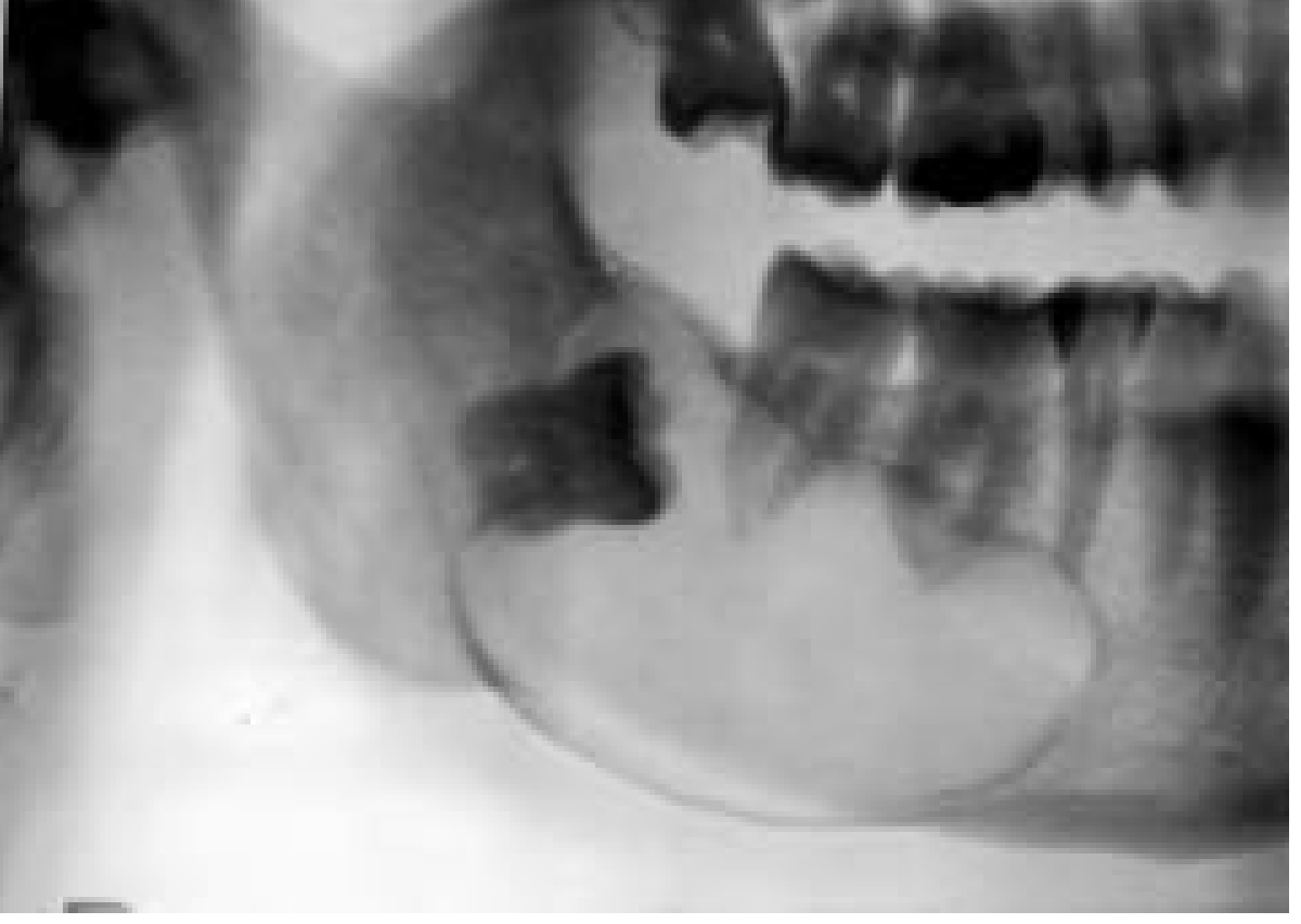
Key radiographic points:
- A dental follicle-related radiolucency around an unerupted tooth is considered normal if < 4 mm from the tooth surface to the outer edge of the follicle; anything larger raises suspicion for cystic change
- The border may show sclerosis or rarifying osteitis if secondary infection is present
- Large cysts will still show evidence of origin from the CEJ
- There is considerable overlap between small dentigerous cysts and hyperplastic follicles
Microscopic Features
Histologically, the specimen shows:
- Variably dense fibro-collagenous connective tissue (some areas loose and myxomatous)
- Odontogenic epithelial rests scattered in the connective tissue
- Luminal lining of nonkeratinized stratified squamous epithelium
- Mucous prosoplasia within the lumen (not uncommon)
- Cholesterol slits and multinucleated giant cells may be present in inflamed cysts
Neoplastic Potential
Dentigerous cysts retain the ability to transform into true neoplasms. One study reported that 17% of ameloblastomas were associated with an existing dentigerous cyst. Both squamous cell carcinoma and mucoepidermoid carcinoma have been reported in association with dentigerous cysts - this is a key reason why complete excision and histopathological examination are essential.
Important diagnostic caution: Odontogenic tumors such as ameloblastoma and odontogenic keratocyst (OKC) may also involve unerupted teeth and can present a diagnostic dilemma, as they can mimic a dentigerous cyst radiographically.
Treatment
| Cyst Size | Preferred Approach |
|---|---|
| Small/moderate | Enucleation at time of tooth extraction |
| Large | Decompression first, then subsequent enucleation |
- Marsupialization or enucleation are both accepted options
- Bone grafting (allogeneic or xenogeneic) may be used after removal; autogenous bone harvest should be reserved for large cysts where bone substitutes are unavailable
- Enucleation (rather than mere biopsy) is preferred because it allows the entire specimen to be examined histopathologically
Subtype: Eruption Cyst
The eruption cyst is a subtype of dentigerous cyst confined only by the overlying alveolar mucosa. It appears as a soft-tissue swelling over an erupting tooth, sometimes with a blue hue. It almost always resolves without intervention - a simple "deroofing" procedure suffices in cases where tooth eruption is delayed or impeded.
Recent Evidence
A 2025 systematic review (PMID 39406309) specifically examined conservative treatment of dentigerous cysts in pediatric patients, supporting the approach of marsupialization/decompression as a viable conservative option before definitive enucleation in children - important because aggressive early enucleation in pediatric patients can damage developing tooth buds.
This is a shared conversation. Sign in to Orris to start your own chat.