
Pharmacological actions of phynatoin
phenytoin mechanism of action sodium channel antiepileptic
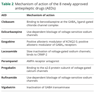
Table 2 Mechanism of action of the 8 newly approved antiepileptic drugs (AEDs) <table><thead><tr><th>AED</th><th>Mechanism of action</th></tr></thead><tbody><tr><td>Clobazam</td><td>Binding to benzodiazepine at the GABA<sub>A</sub> ligand-gated chloride channel complex</td></tr><tr><td>Eslicarbazepine</td><td>Use-dependent blockage of voltage-sensitive sodium channels</td></tr><tr><td>Ezogabine</td><td>Positive allosteric modulator of KCNQ2-5; positive allosteric modulator of GABA<sub>A</sub> receptors</td></tr><tr><td>Lacosamide</td><td>Slow inactivation of voltage-gated sodium channels; binds to CRMP-2</td></tr><tr><td>Perampanel</td><td>AMPA receptor antagonist</td></tr><tr><td>Pregabalin</td><td>Binding to the α2-δ protein subunit of voltage-gated calcium channels</td></tr><tr><td>Rufinamide</td><td>Use-dependent blockage of voltage-sensitive sodium channels</td></tr><tr><td>Vigabatrin</td><td>Inactivation of GABA transaminase</td></tr></tbody></table>
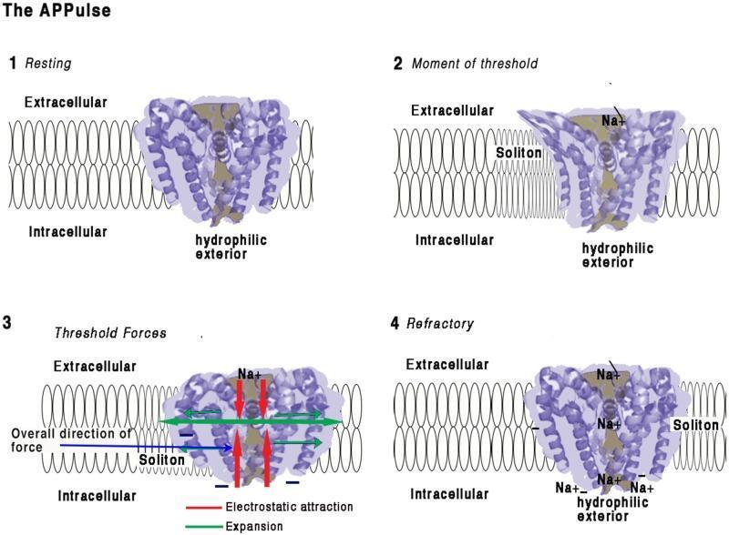
This pathophysiology diagram illustrates the four-stage mechanism of a voltage-gated sodium (Na+) ion channel embedded in a lipid bilayer, focusing on the mechanical and electrostatic changes during depolarization. (1) Resting: The channel's coiled helices are closed, maintaining an electrostatic insulating seal across the 10 nm membrane. (2) Moment of Threshold: An approaching membrane soliton (mechanical wave) exerts pressure, displacing helices and disrupting the electrical insulation. This allows Na+ ions to enter the pore and interact with hydrophilic, negatively charged interior surfaces. (3) Threshold Forces: Inward Na+ influx creates electrostatic attraction (red arrows) toward the intracellular space. Mechanical 'iris-like' opening and contraction occur, causing lateral membrane expansion (green arrows) and a net intracellular force vector (blue arrow). (4) Refractory: Positive charge equalization occurs on the intracellular side, leading to pore closure. The channel remains in a refractory state with localized Na+ accumulation until charges are cleared by diffusion, preventing immediate reactivation.
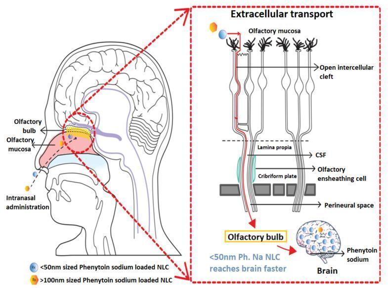
A medical schematic diagram illustrating the direct nose-to-brain delivery of phenytoin sodium loaded Nanostructured Lipid Carriers (NLCs) via the olfactory epithelial pathway. The illustration is divided into a sagittal anatomical view of the human head and a detailed inset of the cellular transport mechanism. The primary anatomical view shows the route of intranasal administration with NLCs depositing on the olfactory mucosa. The inset highlights the extracellular transport mechanism through the olfactory epithelium. It depicts NLCs of different sizes (<50nm and >100nm) moving through open intercellular clefts in the olfactory mucosa, traversing the lamina propria, and crossing the cribriform plate. The diagram shows the particles being transported through the perineural space and along olfactory ensheathing cells to reach the olfactory bulb and eventually the brain. Annotations specify that the smaller <50nm phenytoin sodium NLCs reach the brain faster than larger particles. This educational visual explains the pathophysiology and pharmacology of bypass-blood-brain-barrier drug delivery systems for acute seizure management.
Pharmacological Actions of Phenytoin
1. Central Nervous System (CNS) Actions
- At therapeutic concentrations: Selectively inhibits voltage-activated Na+ channels with no changes to spontaneous activity or responses to GABA/glutamate.
- At toxic concentrations (5-10x therapeutic): Produces excitatory signs, CNS depression, reduces spontaneous activity, and enhances GABA responses.
- At lethal doses: Can produce a type of decerebrate rigidity.
2. Primary Mechanism of Action - Sodium Channel Blockade
| Feature | Detail |
|---|---|
| Target | Voltage-activated Na+ channels |
| Effect | Slows the rate of recovery from inactivation |
| Dependency | Voltage-dependent - greater effect when membrane is depolarized |
| Dependency | Use-dependent - greater effect at higher firing frequencies |
| Net result | Limits sustained repetitive firing of action potentials |
3. Effects on Peripheral Neurons & Cardiac Tissue
- Antiarrhythmic effect: Stabilizes cardiac cell membranes via the same Na+ channel blockade - historically used for digitalis-induced arrhythmias.
- Rapid IV administration risks: Cardiac arrhythmias, hypotension, and AV block (especially in elderly or those with cardiac disease). IV rate must not exceed 50 mg/min (fosphenytoin: <150 mg phenytoin equivalents/min).
- Bradycardia and AV conduction depression can occur.
4. Seizure Specificity
| Seizure Type | Effect |
|---|---|
| Focal onset seizures | Effective |
| Generalized tonic-clonic (primary or secondary) | Effective |
| Status epilepticus (IV/fosphenytoin) | Effective |
| Absence seizures | Ineffective |
| Juvenile myoclonic epilepsy | May worsen |
| Dravet syndrome | May worsen |
5. Other Pharmacological Effects
6. Pharmacokinetic Features Influencing Actions
- Protein binding: ~90% bound to serum albumin; only free drug is pharmacologically active. Displacement by other drugs (e.g., valproate) can abruptly increase free phenytoin levels.
- Non-linear (zero-order / Michaelis-Menten) kinetics: At therapeutic levels, hepatic metabolism becomes saturable. Small dose increases can cause disproportionately large rises in plasma levels - clinically important for toxicity avoidance.
- Therapeutic range: 10-20 mcg/mL total phenytoin (or ~1-2 mcg/mL free phenytoin).
- Metabolism: Primarily by CYP2C9/CYP2C19 in hepatic endoplasmic reticulum to an inactive parahydroxyphenyl derivative.
7. Toxicity Profile (Dose-Related Actions)
| Level | Adverse Effect |
|---|---|
| Chronic therapeutic | Gingival hyperplasia (~20%), hirsutism, coarsening of facial features |
| Slightly elevated | Nystagmus (first sign), diplopia, ataxia |
| High | Cerebellar-vestibular symptoms, behavioral changes |
| Markedly elevated | Cerebellar atrophy, increased seizure frequency |
| IV overdose | Cardiac arrhythmias, hypotension, "purple glove syndrome" |
| Long-term | Megaloblastic anemia (folate antagonism), osteomalacia (vitamin D metabolism interference) |
| Allergic | Stevens-Johnson syndrome, hepatotoxicity, bone marrow suppression (rare) |
Summary
- Goodman & Gilman's The Pharmacological Basis of Therapeutics, pp. 411-413
- Katzung's Basic and Clinical Pharmacology, 16th Ed., pp. 651-654