
Give me detailed classification of stroke including all the types & then detailed about circle of willi's - PG level answer with using textbook like Susan B O sullivan
STROKE: Detailed Classification & Circle of Willis
PART I: CLASSIFICATION OF STROKE
Definition
MASTER CLASSIFICATION
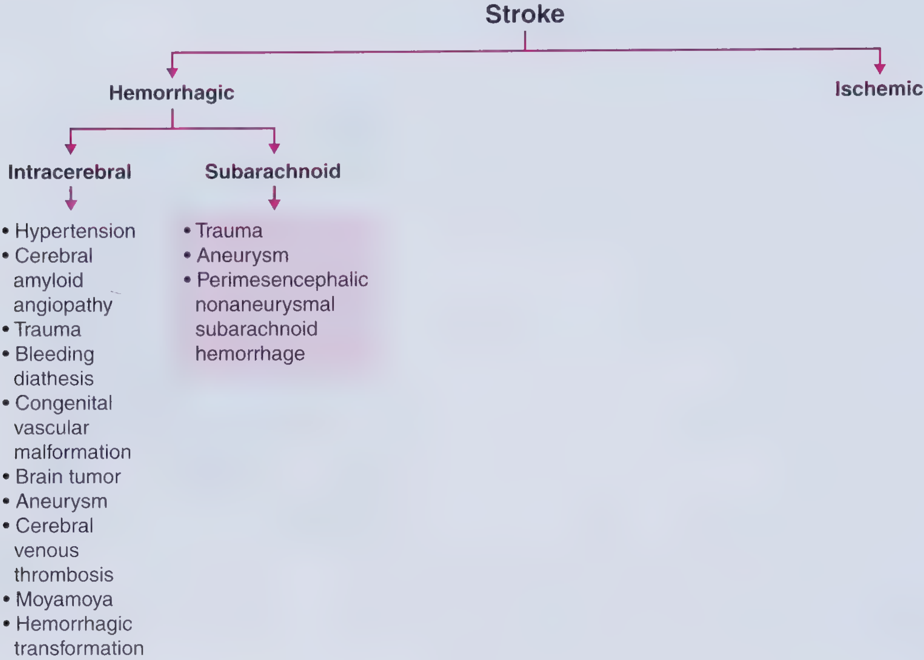
A. ISCHEMIC STROKE (~80% of all strokes)
TOAST Classification (Trial of ORG 10172 in Acute Stroke Treatment)
1. Large Artery Atherosclerosis (Large Vessel Disease)
- Mechanism: In situ thrombosis at an atherosclerotic plaque OR artery-to-artery embolism from a proximal stenosis (carotid, vertebral, basilar, MCA).
- Diagnostic criteria: >50% stenosis or occlusion of a major cerebral or supplying artery; clinical and imaging findings consistent with cortical or cerebellar infarct.
- Clinical features: Often stuttering/stepwise onset; may be preceded by TIAs; cortical signs (aphasia, neglect, hemianopia); MCA territory most commonly involved.
- Imaging: Large territorial infarcts; MCA territory, posterior circulation infarcts.
- Important note (Grainger & Allison): MCA territory infarcts can arise from emboli from the heart or carotid artery, or from in situ thrombosis in the MCA itself.
2. Cardioembolism
- Mechanism: Embolus originating from the heart travels to occlude a cerebral artery. Accounts for approximately 20% of ischemic strokes.
- Sources:
- Atrial fibrillation (thrombus in left atrial appendage) - most common
- Myocardial infarction (mural thrombus on hypokinetic segment)
- Valvular disease / prosthetic valves
- Cardiomyopathy (low ejection fraction)
- Cardiac tumors (myxoma)
- Paradoxical emboli through patent foramen ovale (PFO)
- Clinical features: Sudden onset, maximal deficits at onset; cortical infarcts; multiple vascular territory involvement suggests cardiac source; peripheral infarcts involving multiple vascular territories must come from a proximal (cardiac) source.
- Imaging: Typically large cortical infarcts; can be hemorrhagic; bilateral or multiple territory involvement.
3. Small Vessel Occlusion (Lacunar Stroke)
- Mechanism: Occlusion of small, deep penetrating arteries (lenticulostriate, thalamoperforating, pontine perforators) - typically due to lipohyalinosis or microatheroma from chronic hypertension.
- Territories: Basal ganglia, thalamus, internal capsule, pons, corona radiata.
- Clinical syndromes (classic lacunar syndromes):
- Pure motor hemiparesis (posterior limb of internal capsule or pons)
- Pure sensory stroke (thalamus - VPL nucleus)
- Sensorimotor stroke
- Ataxic hemiparesis (pons or internal capsule)
- Dysarthria-clumsy hand syndrome
- Diagnostic criteria: Infarct <1.5 cm on MRI; no cortical involvement; no cardioembolic or large vessel source identified.
- Note (Blumenfeld): "Small-vessel infarcts are sometimes called lacunar infarcts because they resemble small lakes or cavities when the brain is examined on pathologic section."
- Important caveat (Grainger & Allison): Deep white matter infarcts are typically small vessel in nature but can result from emboli originating from large vessel atheroma or cardiac source - always rule these out.
4. Stroke of Other Determined Etiology
- Arterial dissection (carotid or vertebral) - traumatic or spontaneous; thrombus forms at dissection site and embolizes
- Hypercoagulable states: Protein C/S deficiency, Factor V Leiden, antiphospholipid syndrome, prothrombin gene mutation
- Vasculitis: Primary CNS vasculitis, SLE, temporal arteritis, polyarteritis nodosa, Wegener's granulomatosis
- Hematological: Sickle cell disease, polycythemia, hyperviscosity
- Non-inflammatory vessel wall disease: CADASIL, fibromuscular dysplasia, Moyamoya disease
- Infections: HIV, syphilis, TB, aspergillosis, trichinosis
- Migraine-associated stroke (migrainous infarction)
- Drug-induced: Cocaine, amphetamines
5. Cryptogenic / Undetermined Source
- All investigations are negative OR two or more possible causes are identified, OR workup is incomplete.
- ESUS (Embolic Stroke of Undetermined Source): A newer subcategory - non-lacunar infarct with no atrial fibrillation, no high-risk cardiac source, no significant ipsilateral large-artery stenosis. Implies presumed occult embolism as the mechanism.
6. Hypoperfusion Stroke (Watershed / Hemodynamic)
- Mechanism: Global or regional cerebral hypoperfusion (severe hypotension, cardiac failure, carotid stenosis with hypotensive episode) causes ischemia in arterial border zones.
- Territories: ACA-MCA watershed; MCA-PCA watershed.
- Clinical features: "Man in the barrel" syndrome (proximal arm/leg weakness with relative sparing of face/hands); transcortical aphasia; higher-order visual processing disturbances.
- Key concept (Blumenfeld): "When the blood supply to two adjacent cerebral arteries is compromised, the regions between the two vessels are most susceptible to ischemia - these are called watershed zones."
B. HEMORRHAGIC STROKE (~20% of all strokes)
1. Intracerebral Hemorrhage (ICH) (~15%)
- Mechanism: Rupture of small intraparenchymal arteries/arterioles, most often at sites of chronic hypertensive damage (Charcot-Bouchard microaneurysms) or amyloid angiopathy.
- Common sites:
- Putamen (most common) - 35%
- Thalamus - 20%
- Cerebellum - 15%
- Pons - 10%
- Subcortical white matter (lobar) - often amyloid angiopathy in elderly
- Causes:
- Hypertension (primary; most common overall)
- Cerebral amyloid angiopathy (lobar, elderly patients)
- Vascular malformations: AVM, cavernoma
- Iatrogenic anticoagulation (warfarin, DOACs)
- Cocaine use
- Brain tumor (hemorrhage into tumor)
- Cerebral venous thrombosis with hemorrhagic venous infarction
- Hemorrhagic transformation of ischemic stroke
- Pathophysiology: ICP rises causing local neuronal damage; secondary vasoconstriction mediated by blood breakdown products can cause remote perfusion changes (diaschisis).
- Clinical: Sudden onset; headache common; neurological deficit progresses over minutes to hours (hematoma expansion); vomiting, hypertension; decreased consciousness.
- Risk factors: Advanced age, prior stroke, hypertension, tobacco, alcohol, Asian/African descent.
2. Subarachnoid Hemorrhage (SAH) (~5%)
- Mechanism: Hemorrhage into the subarachnoid space (between pia and arachnoid).
- Primary causes:
- Berry (saccular) aneurysm rupture - most common (~85%); typically at bifurcation points in the circle of Willis
- Vascular malformation rupture (AVM, dural AV fistula)
- Perimesencephalic nonaneurysmal SAH (PM-NASAH) - venous origin; limited to cisterns around midbrain; benign course
- Trauma (traumatic SAH)
- Classic presentation: Sudden-onset "thunderclap" headache - "worst headache of life" (sentinel headache may precede rupture by days); neck stiffness, photophobia, loss of consciousness.
- Common aneurysm sites (circle of Willis):
- Anterior communicating artery (AComm) - most common (~30%)
- Posterior communicating artery (PComm) - presents with CN III palsy
- MCA bifurcation
- ICA bifurcation
- Basilar tip ("top of the basilar")
TEMPORAL CLASSIFICATION
| Type | Duration | Definition |
|---|---|---|
| TIA (Transient Ischemic Attack) | <24 hours (typically <1 hour) | Transient neurological deficit, no infarction on MRI |
| RIND (Reversible Ischemic Neurological Deficit) | <72 hours | Now largely abandoned term |
| Completed Stroke | Permanent deficit | Established infarction |
| Stroke-in-evolution | Progressing | Worsening deficits over hours |
VASCULAR TERRITORY CLASSIFICATION
| Territory | Vessel | Key Deficits |
|---|---|---|
| Anterior | ACA | Contralateral leg > arm weakness (medial homunculus); abulia, alien hand |
| Middle | MCA (dominant) | Contralateral face/arm > leg weakness; Broca's or Wernicke's aphasia; hemianopia; hemisensory loss |
| Middle | MCA (non-dominant) | Contralateral face/arm > leg weakness; hemispatial neglect; anosognosia |
| Posterior | PCA | Contralateral hemianopia; cortical blindness (bilateral); thalamic pain; alexia without agraphia |
| Posterior fossa | Basilar / vertebral | Diplopia, dysarthria, dysphagia, ataxia, crossed sensorimotor deficits, locked-in syndrome |
| Brainstem | Perforators | Various alternating/crossed syndromes (Wallenberg, Weber, etc.) |
PART II: CIRCLE OF WILLIS
Anatomy and Structure
Anterior and Posterior Circulations
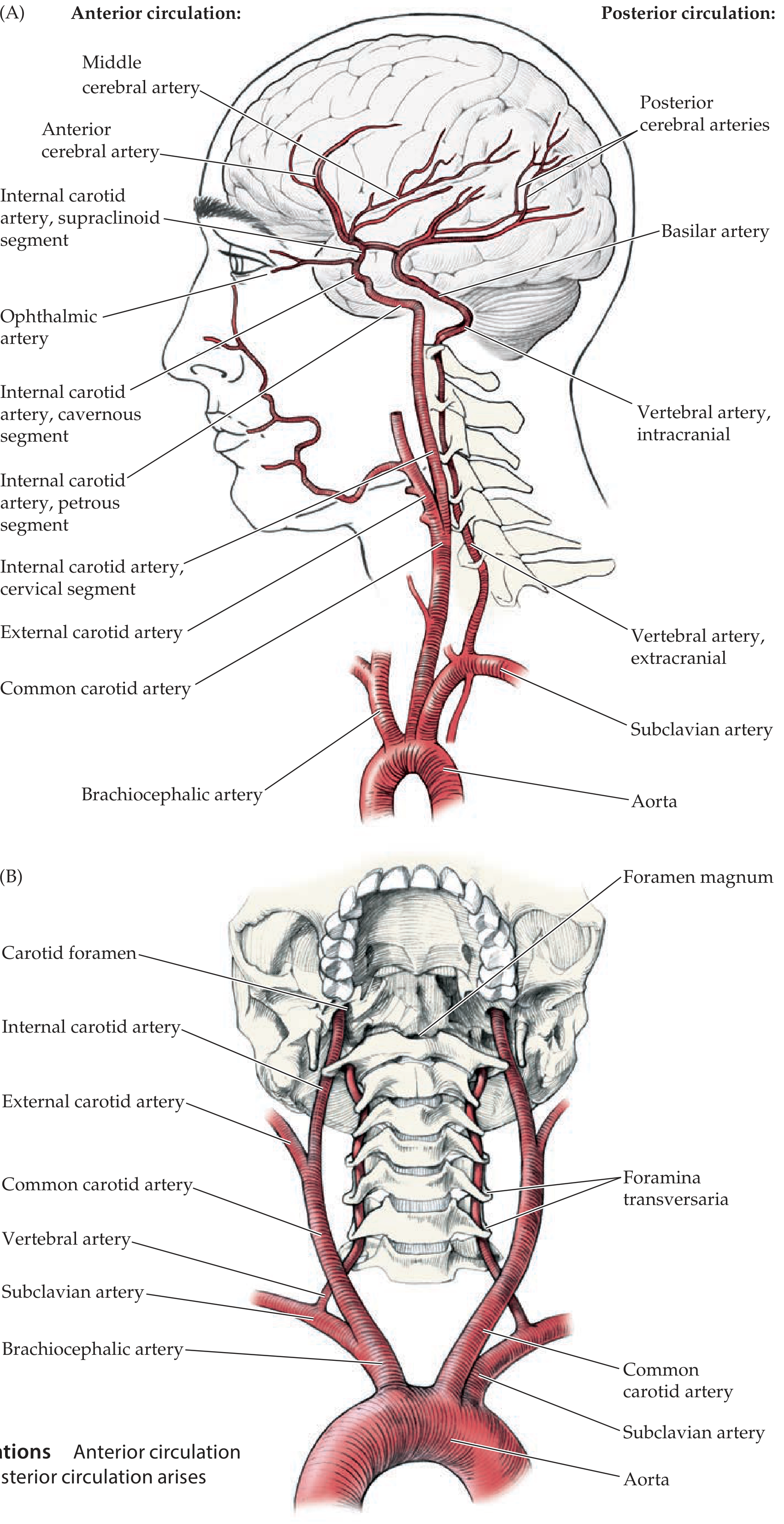
Anterior Circulation (Carotid System)
- Cervical segment - relatively vertical course in the neck; no branches
- Petrous segment - enters carotid canal of temporal bone; sharp horizontal bend
- Cavernous segment - S-shaped turn (carotid siphon) within the cavernous sinus
- Supraclinoid (intracranial) segment - passes anterior clinoid process, pierces dura, enters subarachnoid space
- O - Ophthalmic artery (enters optic foramen with optic nerve; main retinal blood supply)
- P - Posterior communicating artery (PComm)
- A - Anterior choroidal artery
- A - Anterior cerebral artery (ACA)
- M - Middle cerebral artery (MCA)
Posterior Circulation (Vertebrobasilar System)
- Vertebral artery branches: PICA (posterior inferior cerebellar artery), anterior spinal artery, posterior spinal artery
- Basilar artery branches: AICA (anterior inferior cerebellar artery), pontine perforators, SCA (superior cerebellar artery)
- Terminal branches of basilar artery: Two PCAs (posterior cerebral arteries) at the basilar tip
The Circle of Willis - Complete Anatomy

- Anterior communicating artery (AComm): Connects the two ACAs anteriorly
- Posterior communicating arteries (PComm) - bilateral: Connect the ICA (supraclinoid segment) to the ipsilateral PCA, linking anterior and posterior circulations
| Component | Origin |
|---|---|
| ACA (A1 segments - bilateral) | Terminal ICA |
| Anterior communicating artery (AComm) | Joins two ACAs |
| ICA (bilateral, supraclinoid) | Carotid system |
| PComm (bilateral) | ICA to PCA |
| PCA (P1 segments - bilateral) | Basilar tip |
| Basilar artery (top) | Vertebral junction |
Segment Nomenclature
- ACA: A1 (precommunicating) → A2 (postcommunicating, into interhemispheric fissure)
- MCA: M1 (sphenoidal/horizontal) → M2 (insular, within Sylvian fissure) → M3/M4 (opercular/cortical)
- PCA: P1 (precommunicating/interpeduncular) → P2 (ambient cistern) → P3/P4 (quadrigeminal/calcarine)
Functional Significance of the Circle of Willis
- A patient with complete occlusion of the ICA proximal to the circle may be entirely asymptomatic if collateral flow through AComm (from contralateral ICA) and PComm (from basilar) is sufficient.
- Conversely, patients with hypoplastic communicating arteries (common anatomical variant) have poor collateral capacity and suffer larger infarcts.
Vascular Territories of the Three Main Cerebral Arteries
- Superficial (cortical) branches - travel in subarachnoid space into sulci; supply cortex and subcortical white matter
- Deep (perforating) branches - arise from proximal segments near the circle; supply basal ganglia, thalamus, internal capsule
Anterior Cerebral Artery (ACA)
- Passes forward into the interhemispheric fissure, sweeps back over the corpus callosum
- Main branches: pericallosal and callosomarginal arteries
- Territory: Medial surface of frontal and anterior parietal lobes; medial sensorimotor cortex (leg area of homunculus)
- Deep perforators (Heubner's recurrent artery): Head of caudate, anterior limb of internal capsule
- Infarct deficits: Contralateral leg > arm weakness (medial homunculus); abulia; alien hand syndrome; urinary incontinence (parasagittal cortex)
Middle Cerebral Artery (MCA)
- Turns laterally into the Sylvian fissure; bifurcates into superior and inferior divisions (sometimes three or four branches)
- Territory: Most of the lateral convexity of the hemisphere (frontal, parietal, temporal lobes)
- Deep perforators (lenticulostriate arteries): Putamen, caudate, globus pallidus, posterior limb internal capsule
- Infarct deficits: Contralateral face and arm > leg weakness; hemisensory loss; hemianopia; aphasia (dominant) or hemispatial neglect (non-dominant)
- Note (Grainger & Allison): MCA territory infarcts can arise from cardiac emboli, carotid embolism, or in situ MCA thrombosis.
Posterior Cerebral Artery (PCA)
- Arises from the basilar tip; courses around the midbrain through the ambient and quadrigeminal cisterns
- Territory: Occipital lobe (primary visual cortex), medial temporal lobe (including hippocampus), inferior temporal lobe
- Deep perforators (thalamoperforators, posterior choroidal): Thalamus, midbrain, posterior limb of internal capsule, choroid plexus
- Infarct deficits: Contralateral homonymous hemianopia (macula spared due to dual MCA/PCA supply); thalamic pain; alexia without agraphia (dominant occipital + splenium); prosopagnosia; cortical blindness (bilateral PCA occlusion)
Common Aneurysm Sites in the Circle of Willis
| Site | Frequency | Clinical Pearl |
|---|---|---|
| AComm | ~30% | Most common; rupture causes SAH; may compress ACA |
| PComm (ICA-PComm junction) | ~25% | Presents with CN III palsy (ptosis, mydriasis, ophthalmoplegia) from compression of CN III |
| MCA bifurcation (M1-M2) | ~20% | Often presents with hemispheric hematoma |
| ICA bifurcation | ~10% | |
| Basilar tip | ~5-10% | "Top of the basilar" syndrome; bilateral PCA territory infarcts + thalamic infarcts |
| PICA origin | ~5% | Lateral medullary (Wallenberg) syndrome |
"Top of the Basilar" Syndrome
- Bilateral PCA territory infarcts (thalami + occipital lobes)
- Superior cerebellar artery territory infarcts
- Midbrain infarcts
- Clinical: Behavioral changes, memory loss, visual disturbances, oculomotor abnormalities, altered consciousness
Watershed (Border Zone) Zones
- ACA-MCA watershed: Cortex along the superior convexity parasagittally; causes proximal arm/shoulder > leg weakness ("man in the barrel")
- MCA-PCA watershed: Posterior parieto-occipital region; causes higher-order visual processing disturbances, Balint syndrome
- Internal (subcortical) watershed: Between superficial and deep MCA territories
Summary Comparison: TOAST Subtypes
| Subtype | % of Ischemic Strokes | Key Investigation | Secondary Prevention |
|---|---|---|---|
| Large vessel atherosclerosis | ~25% | Carotid duplex, CTA/MRA | Antiplatelet + statin + revascularization if indicated |
| Cardioembolism | ~20% | Echocardiogram, Holter/telemetry | Anticoagulation (AF → warfarin/DOAC) |
| Small vessel (lacunar) | ~25% | MRI (DWI <1.5cm); BP control | Antiplatelet + aggressive BP/DM management |
| Other determined | ~5% | Hypercoag panel, vessel imaging | Depends on etiology |
| Cryptogenic/ESUS | ~25% | Prolonged cardiac monitoring | Antiplatelet; consider DOAC if high embolic risk |
| Hemorrhagic | ~20% total | CT head (gold standard acute) | BP control; reverse anticoagulation |
- Neuroanatomy through Clinical Cases, 3rd Ed. (Blumenfeld) - KCC 10.1, 10.2, 10.4; Figures 10.2, 10.3
- Tintinalli's Emergency Medicine - Table 167-2 Stroke Classification
- Grainger & Allison's Diagnostic Radiology - TOAST classification and imaging correlates
- Fuster & Hurst's The Heart, 15th Ed. - Chapter 167: Stroke and Cerebrovascular Disease
- Miller's Anesthesia, 10th Ed. - Cerebral vascular anatomy and circle of Willis variants
- Frameworks for Internal Medicine - Stroke types flowchart
great now write it in notes formate flowchart easy to read & remember but coverd everything
🧠 STROKE — MASTER NOTES
PG-Level | Classification + Circle of Willis
━━━━━━━━━━━━━━━━━━━━━━━━
FLOWCHART 1 — STROKE MASTER TREE
━━━━━━━━━━━━━━━━━━━━━━━━
                          STROKE
                    (Focal neuro deficit
                     of vascular origin)
                            │
          ┌─────────────────┴──────────────────┐
          │                                     │
    ISCHEMIC (80%)                     HEMORRHAGIC (20%)
          │                                     │
    ┌─────┴──────┐                    ┌─────────┴──────────┐
    │            │                    │                     │
THROMBOTIC   EMBOLIC             INTRACEREBRAL         SUBARACHNOID
  (in situ)  (distant            HEMORRHAGE (ICH)       HEMORRHAGE
             source)              ~15%                    (SAH) ~5%
    │            │
    ↓            ↓
Large         Cardiogenic
vessel    OR  Artery-to-
thrombosis    artery
━━━━━━━━━━━━━━━━━━━━━━━━
FLOWCHART 2 — ISCHEMIC STROKE: TOAST CLASSIFICATION
━━━━━━━━━━━━━━━━━━━━━━━━
            ISCHEMIC STROKE
                  │
    ┌─────────────┼──────────────┐──────────────┬────────────────┐
    │             │              │              │                │
  LARGE         SMALL          CARDIO-       OTHER           CRYPTO-
  VESSEL        VESSEL         EMBOLIC       CAUSE           GENIC
  ATHERO-       (LACUNAR)       │            │              (ESUS)
  SCLEROSIS       │             │            │
    │             │           (see          (see
  ~25%           ~25%         Box A)        Box B)         ~25%
                              ~20%          ~5%
━━━━━━━━━━━━━━━━━━━━━━━━
SECTION 1 — ISCHEMIC STROKE SUBTYPES (TOAST)
━━━━━━━━━━━━━━━━━━━━━━━━
1️⃣ LARGE VESSEL ATHEROSCLEROSIS
┌─────────────────────────────────────────────────────────┐
│  MECHANISM:                                              │
│  ► In-situ thrombosis on atherosclerotic plaque         │
│  ► OR artery-to-artery embolism from proximal stenosis  │
│                                                         │
│  VESSELS: ICA > MCA > Vertebral > Basilar               │
│                                                         │
│  CLINICAL CLUES:                                        │
│  ✦ Stuttering/stepwise onset                           │
│  ✦ Preceded by TIAs                                    │
│  ✦ Cortical signs: aphasia, neglect, hemianopia        │
│                                                         │
│  CRITERIA:                                              │
│  ► >50% stenosis on imaging                            │
│  ► Cortical or cerebellar infarct on MRI               │
│                                                         │
│  Rx: Antiplatelet + Statin + Revascularization         │
└─────────────────────────────────────────────────────────┘
2️⃣ SMALL VESSEL (LACUNAR) STROKE
┌─────────────────────────────────────────────────────────┐
│  MECHANISM:                                              │
│  ► Lipohyalinosis / microatheroma of deep perforators   │
│  ► Chronic hypertension → Charcot-Bouchard aneurysms   │
│                                                         │
│  SITES: Putamen > Thalamus > Pons > Internal capsule    │
│         > Corona radiata                                │
│                                                         │
│  CLASSIC SYNDROMES (must memorize):                     │
│  ✦ Pure Motor Hemiparesis      → Post. limb IC / pons  │
│  ✦ Pure Sensory Stroke         → Thalamus (VPL)        │
│  ✦ Sensorimotor Stroke         → Thalamo-capsular      │
│  ✦ Ataxic Hemiparesis          → Pons / IC             │
│  ✦ Dysarthria-Clumsy Hand      → Pons / IC             │
│                                                         │
│  IMAGING: DWI infarct < 1.5 cm; no cortical involvement│
│                                                         │
│  ⚠ CAVEAT: Deep WM infarcts can be embolic — always    │
│    exclude large vessel + cardiac source first!         │
│                                                         │
│  Rx: Antiplatelet + strict BP/DM/lipid control         │
└─────────────────────────────────────────────────────────┘
3️⃣ CARDIOEMBOLIC STROKE
┌─────────────────────────────────────────────────────────┐
│  MECHANISM: Embolus from heart → cerebral artery        │
│  ~20% of all ischemic strokes                           │
│                                                         │
│  BOX A — CARDIAC SOURCES                               │
│  ┌────────────────────────────────────────┐            │
│  │  HIGH RISK:                            │            │
│  │  ► Atrial fibrillation (LAA thrombus)  │            │
│  │  ► Recent MI (mural thrombus)          │            │
│  │  ► Prosthetic valve                    │            │
│  │  ► Dilated cardiomyopathy (low EF)     │            │
│  │  ► Cardiac myxoma                      │            │
│  │  ► Infective endocarditis              │            │
│  │                                        │            │
│  │  MEDIUM RISK:                          │            │
│  │  ► Patent foramen ovale (PFO)          │            │
│  │  ► Atrial septal aneurysm              │            │
│  │  ► Mitral valve prolapse               │            │
│  └────────────────────────────────────────┘            │
│                                                         │
│  CLINICAL CLUES:                                        │
│  ✦ SUDDEN onset, maximal at onset                      │
│  ✦ Multiple territory infarcts → always cardiac        │
│  ✦ Hemorrhagic transformation common                   │
│  ✦ May have AF on ECG/Holter                           │
│                                                         │
│  Rx: Anticoagulation (warfarin / DOAC)                 │
└─────────────────────────────────────────────────────────┘
4️⃣ OTHER DETERMINED CAUSE
┌─────────────────────────────────────────────────────────┐
│  BOX B — THINK "YOUNG STROKE"                           │
│                                                         │
│  VASCULAR:                                              │
│  ► Arterial dissection (carotid/vertebral)             │
│  ► Fibromuscular dysplasia                             │
│  ► Moyamoya disease                                    │
│  ► CADASIL (hereditary WM disease)                     │
│                                                         │
│  INFLAMMATORY / VASCULITIS:                             │
│  ► Primary CNS vasculitis                              │
│  ► SLE, antiphospholipid syndrome                      │
│  ► Temporal arteritis, PAN, Wegener's                  │
│                                                         │
│  HYPERCOAGULABLE:                                       │
│  ► Protein C/S deficiency, Factor V Leiden             │
│  ► Antithrombin III deficiency                         │
│  ► Prothrombin gene mutation                           │
│  ► Antiphospholipid antibodies                         │
│  ► Sickle cell, polycythemia, hyperviscosity           │
│                                                         │
│  INFECTIONS: HIV, syphilis, TB meningitis              │
│  DRUGS: Cocaine, amphetamines                          │
│  OTHER: Migraine-associated infarction                 │
└─────────────────────────────────────────────────────────┘
5️⃣ CRYPTOGENIC / ESUS
┌─────────────────────────────────────────────────────────┐
│  CRYPTOGENIC: No cause found after full workup          │
│                                                         │
│  ESUS (Embolic Stroke of Undetermined Source):          │
│  ► Non-lacunar infarct on neuroimaging                 │
│  ► No AF detected                                      │
│  ► No >50% ipsilateral large-artery stenosis           │
│  ► No high-risk cardiac source                         │
│  ► Implies occult embolism                             │
│                                                         │
│  Rx: Antiplatelet; prolonged cardiac monitoring        │
└─────────────────────────────────────────────────────────┘
6️⃣ WATERSHED / HEMODYNAMIC STROKE
┌─────────────────────────────────────────────────────────┐
│  MECHANISM: Global/regional hypoperfusion →             │
│  ischemia at border zones between 2 territories        │
│                                                         │
│  ZONES & DEFICITS:                                      │
│  ┌─────────────────────────────────────────┐           │
│  │  ACA-MCA watershed                      │           │
│  │  → Proximal arm > leg weakness          │           │
│  │  → "MAN IN THE BARREL" syndrome        │           │
│  │                                         │           │
│  │  MCA-PCA watershed                      │           │
│  │  → Higher-order visual disturbance      │           │
│  │  → Transcortical aphasia               │           │
│  │  → Balint syndrome (bilateral)         │           │
│  └─────────────────────────────────────────┘           │
│                                                         │
│  TRIGGERS: Cardiac arrest, severe hypotension,         │
│            tight carotid stenosis + hypotensive        │
│            episode                                      │
└─────────────────────────────────────────────────────────┘
━━━━━━━━━━━━━━━━━━━━━━━━
SECTION 2 — HEMORRHAGIC STROKE
━━━━━━━━━━━━━━━━━━━━━━━━
FLOWCHART 3 — ICH vs SAH
          HEMORRHAGIC STROKE
                  │
    ┌─────────────┴───────────────┐
    │                             │
   ICH                          SAH
(Intraparenchymal)          (Subarachnoid)
~15%                         ~5%
    │                             │
    ├── HTN → basal ganglia       ├── Berry aneurysm (85%)
    │   putamen (MC)              │   at CoW bifurcations
    ├── Amyloid angiopathy        │
    │   → lobar (elderly)         ├── AVM rupture
    ├── AVM / cavernoma           │
    ├── Anticoagulants            └── Perimesencephalic
    ├── Cocaine                       nonaneurysmal SAH
    ├── Hemorrhagic transformation    (benign; venous)
    └── Cerebral venous thrombosis
🔴 ICH — LOCATION GUIDE
┌──────────────────────────────────────────────────────────┐
│  SITE           │  CAUSE           │  KEY DEFICIT        │
├──────────────────────────────────────────────────────────┤
│  Putamen (35%)  │  HTN             │  Contralat. motor + │
│                 │                  │  hemisensory         │
├──────────────────────────────────────────────────────────┤
│  Thalamus (20%) │  HTN             │  Sensory > motor;   │
│                 │                  │  eye deviation DOWN  │
├──────────────────────────────────────────────────────────┤
│  Pons (10%)     │  HTN             │  Quadriplegia;      │
│                 │                  │  pinpoint pupils     │
├──────────────────────────────────────────────────────────┤
│  Cerebellum     │  HTN / AVM       │  Ataxia; NO motor   │
│  (15%)          │                  │  deficit initially   │
├──────────────────────────────────────────────────────────┤
│  Lobar (WM)     │  Amyloid         │  Variable; elderly; │
│                 │  angiopathy      │  recurrent bleeds    │
└──────────────────────────────────────────────────────────┘
🔴 SAH — KEY POINTS
┌─────────────────────────────────────────────────────────┐
│  PRESENTATION:                                          │
│  ✦ "THUNDERCLAP" headache — worst in life              │
│  ✦ Sentinel headache days before (warning leak)        │
│  ✦ Neck stiffness, photophobia, vomiting               │
│  ✦ CN III palsy → PComm aneurysm                      │
│                                                         │
│  ANEURYSM SITES AT CIRCLE OF WILLIS:                   │
│  AComm         ~30%  (MC overall)                      │
│  PComm         ~25%  (CN III palsy)                    │
│  MCA bifurc.   ~20%                                    │
│  ICA bifurc.   ~10%                                    │
│  Basilar tip   ~5-10% (top of basilar syndrome)        │
└─────────────────────────────────────────────────────────┘
━━━━━━━━━━━━━━━━━━━━━━━━
SECTION 3 — TEMPORAL CLASSIFICATION
━━━━━━━━━━━━━━━━━━━━━━━━
DURATION OF DEFICIT
        │
        ├── <24 hrs, no infarct on MRI ─── TIA
        │   (typically <1 hour in practice)
        │
        ├── Progressing over hours ──────── STROKE-IN-EVOLUTION
        │
        └── Permanent, established ──────── COMPLETED STROKE
━━━━━━━━━━━━━━━━━━━━━━━━
SECTION 4 — CIRCLE OF WILLIS
━━━━━━━━━━━━━━━━━━━━━━━━
FLOWCHART 4 — BLOOD SUPPLY TO BRAIN
          AORTA
            │
    ┌───────┴────────┐
    │                │
BRACHIOCEPHALIC    L. COMMON
  ARTERY           CAROTID
    │
  ┌─┴──────────────┐
  │                │
R. COMMON       R. SUBCLAVIAN
CAROTID             │
    │            VERTEBRAL A.
    │                │
CAROTID          ascends through
BIFURCATION      foramina
    │            transversaria
    │                │
    ├── ECA          │
    │            enters foramen
    └── ICA      magnum
          │          │
      (4 SEGMENTS)   │
          │          │
    CERVICAL         │
    PETROUS      ┌───┘
    CAVERNOUS    │
    SUPRACLINOID ├── joins at
                 │   pontomedullary
                 │   junction
                 │
              BASILAR
              ARTERY
                 │
                 └── BASILAR TIP
                           │
                      ┌────┴────┐
                      │         │
                    L.PCA     R.PCA
FLOWCHART 5 — CIRCLE OF WILLIS ANATOMY
          ANTERIOR COMMUNICATING A. (AComm)
                    │
          ┌─────────┴────────┐
          │                  │
        L.ACA              R.ACA
          │                  │
        L.ICA              R.ICA
    (supraclinoid)      (supraclinoid)
          │                  │
       L.PComm           R.PComm
          │                  │
        L.PCA              R.PCA
          └─────────┬────────┘
                    │
              BASILAR TIP
              (basilar artery)

COMPLETE RING present in only ~34% of individuals!
ICA BRANCHES MNEMONIC — "OPAAM"
┌─────────────────────────────────────────────────────────┐
│  O — Ophthalmic artery                                  │
│      (→ optic foramen; retinal supply)                 │
│  P — Posterior communicating artery (PComm)             │
│      (→ links ICA to PCA; aneurysm → CN III palsy)    │
│  A — Anterior choroidal artery                          │
│      (→ posterior limb IC, optic tract, hippocampus)  │
│  A — Anterior cerebral artery (ACA)                     │
│  M — Middle cerebral artery (MCA)                       │
└─────────────────────────────────────────────────────────┘
ICA SEGMENTS — QUICK TABLE
┌──────────────┬──────────────────────────────────────────┐
│  SEGMENT     │  COURSE / NOTE                           │
├──────────────┼──────────────────────────────────────────┤
│  Cervical    │  Vertical in neck; NO branches           │
├──────────────┼──────────────────────────────────────────┤
│  Petrous     │  Carotid canal; horizontal bend           │
├──────────────┼──────────────────────────────────────────┤
│  Cavernous   │  S-curve = carotid siphon; within        │
│              │  cavernous sinus                          │
├──────────────┼──────────────────────────────────────────┤
│  Supraclinoid│  Pierces dura; enters subarachnoid space │
│  (intracranial)  gives OPAAM branches                   │
└──────────────┴──────────────────────────────────────────┘
VERTEBROBASILAR BRANCHES — FLOWCHART
VERTEBRAL ARTERY
    ├── Posterior inferior cerebellar a. (PICA)
    │   → lateral medulla + inferior cerebellum
    │   → PICA infarct = Wallenberg syndrome
    ├── Anterior spinal artery
    └── Posterior spinal artery

BASILAR ARTERY
    ├── AICA (anterior inferior cerebellar a.)
    │   → lateral pons + anterior inferior cerebellum
    ├── Pontine perforators
    │   → medial pons
    ├── SCA (superior cerebellar a.)
    │   → superior cerebellum + midbrain
    └── PCA (terminal) × 2
        → occipital lobe + medial temporal + thalamus
VASCULAR TERRITORIES AT A GLANCE
┌──────────────────────────────────────────────────────────────┐
│  ARTERY   │  TERRITORY              │  INFARCT DEFICITS      │
├──────────────────────────────────────────────────────────────┤
│  ACA      │  Medial frontal +        │  Leg > arm weakness    │
│           │  anterior parietal       │  Abulia, alien hand    │
│           │  (medial homunculus)     │  Urinary incontinence  │
├──────────────────────────────────────────────────────────────┤
│  MCA      │  Lateral convexity       │  Face + arm > leg      │
│  dominant │  frontal/parietal/temp.  │  Broca's / Wernicke's  │
│           │                          │  Hemianopia            │
├──────────────────────────────────────────────────────────────┤
│  MCA      │  Same territory          │  Face + arm > leg      │
│  non-dom  │                          │  Hemispatial NEGLECT   │
│           │                          │  Anosognosia           │
├──────────────────────────────────────────────────────────────┤
│  PCA      │  Occipital, medial       │  Homonymous hemianopia │
│           │  temporal, thalamus      │  (macula SPARED)       │
│           │                          │  Thalamic pain         │
│           │                          │  Alexia w/o agraphia   │
├──────────────────────────────────────────────────────────────┤
│  Basilar  │  Pons, midbrain,         │  Diplopia, dysarthria  │
│           │  cerebellum              │  Dysphagia, ataxia     │
│           │                          │  Crossed syndromes     │
│           │                          │  Locked-in (total occ) │
└──────────────────────────────────────────────────────────────┘
DEEP PERFORATORS — HIGH-YIELD TABLE
┌──────────────────────┬─────────────────────────────────────┐
│  ARTERY              │  DEEP STRUCTURES SUPPLIED           │
├──────────────────────┼─────────────────────────────────────┤
│  Lenticulostriate    │  Putamen, caudate, globus pallidus, │
│  (from MCA M1)       │  posterior limb internal capsule    │
├──────────────────────┼─────────────────────────────────────┤
│  Heubner's recurrent │  Head of caudate,                   │
│  (from ACA A1-A2)    │  anterior limb internal capsule     │
├──────────────────────┼─────────────────────────────────────┤
│  Thalamoperforators  │  Thalamus (medial, intralaminar)    │
│  (from P1 / PComm)   │                                     │
├──────────────────────┼─────────────────────────────────────┤
│  Ant. choroidal      │  Post. limb IC, optic tract,        │
│  (from ICA)          │  hippocampus, lateral geniculate    │
├──────────────────────┼─────────────────────────────────────┤
│  Pontine perforators │  Medial pons (paramedian)           │
│  (from basilar)      │  → lacunar strokes in pons          │
└──────────────────────┴─────────────────────────────────────┘
WATERSHED ZONES — VISUAL SUMMARY
CORONAL VIEW (simplified):

    Medial surface          Lateral surface
         │                        │
    ACA territory           MCA territory
         │                        │
    ←──── ACA-MCA WATERSHED ────→
         (parasagittal strip)
         DEFICIT: proximal arm/shoulder weakness
         "MAN IN THE BARREL"

    MCA territory           PCA territory
         │                        │
    ←── MCA-PCA WATERSHED ──────→
         (posterior parieto-occipital)
         DEFICIT: visual processing, transcortical aphasia
━━━━━━━━━━━━━━━━━━━━━━━━
SECTION 5 — HIGH-YIELD QUICK RECALL BOX
━━━━━━━━━━━━━━━━━━━━━━━━
┌─────────────────────────────────────────────────────────────┐
│            STROKE PEARLS — DON'T MISS                       │
├─────────────────────────────────────────────────────────────┤
│  ► Stroke ~80% ischemic / ~20% hemorrhagic                  │
│  ► TOAST: Large vessel 25% | Lacunar 25% |                  │
│    Cardioembolic 20% | Other 5% | Cryptogenic 25%           │
│  ► Multiple territories = CARDIAC source until proven       │
│    otherwise                                                │
│  ► Deep WM infarct ≠ always lacunar; rule out emboli       │
│  ► Complete CoW in only ~34% of people                      │
│  ► PComm aneurysm → CN III palsy (surgical emergency)       │
│  ► AComm aneurysm = MC SAH site (~30%)                      │
│  ► Basilar tip occlusion = bilateral thalami + occipital    │
│    lobes + cerebellar infarcts                              │
│  ► "Man in barrel" = ACA-MCA watershed                      │
│  ► Thunderclap headache = SAH until proven otherwise        │
│  ► tPA window: 4.5 hours from onset                         │
│  ► Thrombectomy: up to 24 hours (with penumbral imaging)    │
│  ► PICA infarct = Wallenberg syndrome                       │
│    (ipsilat. face numbness + contralat. body numbness)      │
│  ► Putamen = MC site of HTN-related ICH                     │
└─────────────────────────────────────────────────────────────┘
━━━━━━━━━━━━━━━━━━━━━━━━
SECTION 6 — MNEMONIC BANK
━━━━━━━━━━━━━━━━━━━━━━━━
ICA branches (supraclinoid):  O P A A M
                              Ophthalmic
                              Posterior communicating
                              Anterior choroidal
                              Anterior cerebral
                              Middle cerebral

LACUNAR syndromes:  "Pure Motor, Pure Sensory, Senso-Motor,
                     Ataxic Hemi, Dysarthria-Clumsy Hand"

HTN ICH sites (frequency):  "Put That Pons Carefully Lobar"
                              Putamen > Thalamus > Pons >
                              Cerebellum > Lobar

SAH aneurysm sites:  A-COMM is the MOST COMMON
                     P-COMM gives CN 3 compression
                     BASILAR TIP = bilateral PCA devastation

TOAST order:  Large, Small, Cardio, Other, Cryptogenic
              "Loud Students Create Outstanding Chaos"
Create a printable PDF quick reference card for stroke classification & CoW anatomy
ls -la /tmp/workspace/ 2>/dev/null || mkdir -p /tmp/workspace && echo "workspace ready"
mkdir -p /tmp/workspace/stroke-refcard && python3 -c "from reportlab.lib import colors; print('reportlab OK')"
~/stroke-refcard/build_card.py
"""
Stroke & Circle of Willis - Printable Quick Reference Card
A4 landscape, 2 pages — built with ReportLab
"""
from reportlab.lib.pagesizes import A4, landscape
from reportlab.lib import colors
from reportlab.lib.units import mm, cm
from reportlab.platypus import (
SimpleDocTemplate, Table, TableStyle, Paragraph, Spacer,
HRFlowable, KeepTogether
)
from reportlab.lib.styles import getSampleStyleSheet, ParagraphStyle
from reportlab.lib.enums import TA_CENTER, TA_LEFT, TA_RIGHT
from reportlab.platypus.flowables import Flowable
from reportlab.graphics.shapes import Drawing, Rect, String, Line, Polygon
from reportlab.graphics import renderPDF
# ── Colour Palette ────────────────────────────────────────────────────────────
C_NAVY = colors.HexColor("#1a2e4a")
C_CRIMSON = colors.HexColor("#c0392b")
C_TEAL = colors.HexColor("#1abc9c")
C_AMBER = colors.HexColor("#f39c12")
C_PURPLE = colors.HexColor("#8e44ad")
C_BLUE = colors.HexColor("#2980b9")
C_GREEN = colors.HexColor("#27ae60")
C_ORANGE = colors.HexColor("#e67e22")
C_LIGHT = colors.HexColor("#ecf0f1")
C_DARKGRAY = colors.HexColor("#2c3e50")
C_MIDGRAY = colors.HexColor("#7f8c8d")
C_WHITE = colors.white
C_PALE_RED = colors.HexColor("#fdecea")
C_PALE_BLU = colors.HexColor("#e8f4fd")
C_PALE_GRN = colors.HexColor("#eafaf1")
C_PALE_YLW = colors.HexColor("#fef9e7")
C_PALE_PUR = colors.HexColor("#f5eef8")
C_PALE_TEA = colors.HexColor("#e8f8f5")
PAGE_W, PAGE_H = landscape(A4) # 297 × 210 mm
MARGIN = 8 * mm
# ── Style helpers ─────────────────────────────────────────────────────────────
def sty(name, **kw):
base = getSampleStyleSheet()["Normal"]
kw.setdefault("fontName", "Helvetica")
kw.setdefault("fontSize", 7.5)
kw.setdefault("leading", 10)
kw.setdefault("textColor", C_DARKGRAY)
return ParagraphStyle(name, parent=base, **kw)
H1 = sty("H1", fontName="Helvetica-Bold", fontSize=16, leading=20,
textColor=C_WHITE, alignment=TA_CENTER)
H2 = sty("H2", fontName="Helvetica-Bold", fontSize=9, leading=12,
textColor=C_WHITE, alignment=TA_CENTER)
H3 = sty("H3", fontName="Helvetica-Bold", fontSize=8, leading=11,
textColor=C_NAVY)
BODY= sty("BODY",fontSize=7, leading=9.5)
BODW= sty("BODW",fontSize=7, leading=9.5, textColor=C_WHITE)
BOLD= sty("BOLD",fontName="Helvetica-Bold", fontSize=7, leading=9.5)
BOLW= sty("BOLW",fontName="Helvetica-Bold", fontSize=7, leading=9.5, textColor=C_WHITE)
SML = sty("SML", fontSize=6.2, leading=8.5, textColor=C_MIDGRAY)
CTR = sty("CTR", fontSize=7, leading=9.5, alignment=TA_CENTER)
CTRB= sty("CTRB",fontName="Helvetica-Bold", fontSize=7.5, leading=10,
alignment=TA_CENTER, textColor=C_NAVY)
def P(txt, style=BODY): return Paragraph(txt, style)
def SP(h=2): return Spacer(1, h*mm)
def col_header(txt, bg=C_NAVY, fg=C_WHITE, size=8):
return Paragraph(f"<b>{txt}</b>",
sty("ch", fontName="Helvetica-Bold", fontSize=size,
leading=11, textColor=fg, alignment=TA_CENTER))
# ── Section title bar ─────────────────────────────────────────────────────────
def section_bar(txt, bg=C_NAVY, width=None):
w = width or (PAGE_W - 2*MARGIN)
d = Drawing(w, 14)
d.add(Rect(0, 0, w, 14, fillColor=bg, strokeColor=None))
d.add(String(6, 3.5, txt, fontName="Helvetica-Bold",
fontSize=8.5, fillColor=colors.white))
return d
# ── Flowable box ──────────────────────────────────────────────────────────────
class ColorBox(Flowable):
"""A filled rounded rectangle containing text lines."""
def __init__(self, lines, bg, w, h, text_color=C_DARKGRAY,
font="Helvetica", fsize=6.8, bold_first=False):
super().__init__()
self.lines = lines
self.bg = bg
self.w, self.h = w, h
self.text_color = text_color
self.font = font
self.fsize = fsize
self.bold_first = bold_first
def wrap(self, *args): return self.w, self.h
def draw(self):
c = self.canv
c.setFillColor(self.bg)
c.roundRect(0, 0, self.w, self.h, 3, fill=1, stroke=0)
c.setFillColor(self.text_color)
y = self.h - self.fsize - 3
for i, ln in enumerate(self.lines):
fn = ("Helvetica-Bold" if (i == 0 and self.bold_first)
else self.font)
c.setFont(fn, self.fsize)
c.drawString(5, y, ln)
y -= self.fsize + 2
if y < 3: break
# ══════════════════════════════════════════════════════════════════════════════
# PAGE 1
# ══════════════════════════════════════════════════════════════════════════════
def page1_elements():
elems = []
W = PAGE_W - 2*MARGIN # usable width
# ── MASTER HEADER ─────────────────────────────────────────────────────────
hdr = Drawing(W, 26)
hdr.add(Rect(0, 0, W, 26, fillColor=C_NAVY, strokeColor=None, rx=4, ry=4))
hdr.add(String(W/2, 15, "STROKE — QUICK REFERENCE CARD",
fontName="Helvetica-Bold", fontSize=13,
fillColor=colors.white, textAnchor="middle"))
hdr.add(String(W/2, 4, "Classification • Pathophysiology • TOAST • Circle of Willis",
fontName="Helvetica", fontSize=7,
fillColor=colors.HexColor("#bdc3c7"), textAnchor="middle"))
elems.append(hdr); elems.append(SP(2))
# ── ROW 1: Master tree + TOAST table ──────────────────────────────────────
# Left: Master stroke tree
tree_w = 88*mm
tree = Drawing(tree_w, 62)
tree.add(Rect(0, 0, tree_w, 62, fillColor=C_PALE_BLU, strokeColor=C_BLUE, strokeWidth=0.5, rx=3))
tree.add(String(tree_w/2, 52, "STROKE MASTER TREE",
fontName="Helvetica-Bold", fontSize=7.5,
fillColor=C_NAVY, textAnchor="middle"))
# stroke box
bx = 34; by = 40; bw = 38; bh = 10
tree.add(Rect(bx, by, bw, bh, fillColor=C_NAVY, rx=2))
tree.add(String(bx+bw/2, by+2.5, "STROKE",
fontName="Helvetica-Bold", fontSize=7.5,
fillColor=colors.white, textAnchor="middle"))
# branches
cx = bx + bw/2
tree.add(Line(cx-16, by, 28, 29, strokeColor=C_NAVY, strokeWidth=1))
tree.add(Line(cx+16, by, tree_w-28, 29, strokeColor=C_NAVY, strokeWidth=1))
# ischemic box
tree.add(Rect(tree_w-72, 18, 44, 12, fillColor=C_BLUE, rx=2))
tree.add(String(tree_w-72+22, 20.5, "ISCHEMIC 80%",
fontName="Helvetica-Bold", fontSize=7,
fillColor=colors.white, textAnchor="middle"))
# haem box
tree.add(Rect(4, 18, 44, 12, fillColor=C_CRIMSON, rx=2))
tree.add(String(26, 20.5, "HEMORRHAGIC 20%",
fontName="Helvetica-Bold", fontSize=7,
fillColor=colors.white, textAnchor="middle"))
# sub-branches ischemic
ix = tree_w-72+22
tree.add(Line(ix-10, 18, ix-16, 10, strokeColor=C_BLUE, strokeWidth=0.8))
tree.add(Line(ix+10, 18, ix+16, 10, strokeColor=C_BLUE, strokeWidth=0.8))
tree.add(Rect(tree_w-82, 1, 28, 10, fillColor=colors.HexColor("#d6eaf8"), rx=2))
tree.add(String(tree_w-82+14, 3, "Ischemic",
fontName="Helvetica", fontSize=5.5,
fillColor=C_BLUE, textAnchor="middle"))
tree.add(String(tree_w-82+14, -3.5, "(thrombotic/",
fontName="Helvetica", fontSize=5,
fillColor=C_BLUE, textAnchor="middle"))
tree.add(Rect(tree_w-50, 1, 28, 10, fillColor=colors.HexColor("#d6eaf8"), rx=2))
tree.add(String(tree_w-50+14, 3, "Embolic",
fontName="Helvetica", fontSize=5.5,
fillColor=C_BLUE, textAnchor="middle"))
# sub-branches haem
hx = 26
tree.add(Line(hx-10, 18, hx-8, 10, strokeColor=C_CRIMSON, strokeWidth=0.8))
tree.add(Line(hx+10, 18, hx+10, 10, strokeColor=C_CRIMSON, strokeWidth=0.8))
tree.add(Rect(2, 1, 22, 9, fillColor=colors.HexColor("#fadbd8"), rx=2))
tree.add(String(13, 3, "ICH ~15%",
fontName="Helvetica-Bold", fontSize=5.8,
fillColor=C_CRIMSON, textAnchor="middle"))
tree.add(Rect(26, 1, 22, 9, fillColor=colors.HexColor("#fadbd8"), rx=2))
tree.add(String(37, 3, "SAH ~5%",
fontName="Helvetica-Bold", fontSize=5.8,
fillColor=C_CRIMSON, textAnchor="middle"))
# Right: TOAST classification table
toast_w = W - tree_w - 4*mm
toast_data = [
[col_header("TOAST CLASSIFICATION OF ISCHEMIC STROKE", bg=C_BLUE, size=7.5)],
[Table(
[
[P("<b>SUBTYPE</b>", BOLD), P("<b>%</b>", BOLD),
P("<b>MECHANISM</b>", BOLD), P("<b>KEY FEATURE</b>", BOLD),
P("<b>Rx</b>", BOLD)],
[P("Large Vessel\nAtherosclerosis", BOLD),
P("~25%", CTR),
P("Thrombosis / A-to-A\nembolism at plaque"),
P("Stuttering onset\nTIAs; cortical signs\n>50% stenosis"),
P("Antiplatelet\n+ Statin\n+ Revasc.")],
[P("Small Vessel\n(Lacunar)", BOLD),
P("~25%", CTR),
P("Lipohyalinosis of\ndeep perforators\n(HTN)"),
P("DWI <1.5 cm; deep\nno cortical signs\n5 classic syndromes"),
P("Antiplatelet\nBP/DM/\nlipid control")],
[P("Cardioembolism", BOLD),
P("~20%", CTR),
P("Thrombus from\nheart → brain"),
P("Sudden onset;\nmulti-territory;\nHemorrhagic Tx"),
P("Anticoag.\n(DOAC/warfarin)")],
[P("Other Cause", BOLD),
P("~5%", CTR),
P("Dissection, vasculitis,\nhypercoag, Moyamoya\nCADASIL, sickle cell"),
P("Young stroke\nworkup; rare\netiologies"),
P("Cause-\nspecific")],
[P("Cryptogenic\n(ESUS)", BOLD),
P("~25%", CTR),
P("No cause found;\nESUS = non-lacunar\nno AF, no stenosis"),
P("Prolonged cardiac\nmonitoring;\noccult embolism"),
P("Antiplatelet;\nconsider\nDOAC")],
],
colWidths=[toast_w*0.18, toast_w*0.07, toast_w*0.26,
toast_w*0.28, toast_w*0.19],
style=TableStyle([
("BACKGROUND", (0,0), (-1,0), C_NAVY),
("TEXTCOLOR", (0,0), (-1,0), C_WHITE),
("FONTNAME", (0,0), (-1,0), "Helvetica-Bold"),
("FONTSIZE", (0,0), (-1,0), 6.5),
("ROWBACKGROUNDS", (0,1), (-1,-1),
[C_PALE_BLU, C_WHITE, C_PALE_BLU, C_WHITE, C_PALE_BLU]),
("GRID", (0,0), (-1,-1), 0.3, colors.HexColor("#bdc3c7")),
("VALIGN", (0,0), (-1,-1), "TOP"),
("TOPPADDING", (0,0), (-1,-1), 2),
("BOTTOMPADDING", (0,0), (-1,-1), 2),
("LEFTPADDING", (0,0), (-1,-1), 2),
])
)]
]
row1 = Table(
[[tree, Table(toast_data,
colWidths=[toast_w],
style=TableStyle([
("GRID", (0,0), (-1,-1), 0, colors.white),
("TOPPADDING", (0,0), (-1,-1), 0),
("BOTTOMPADDING", (0,0), (-1,-1), 0),
("LEFTPADDING", (0,0), (-1,-1), 0),
("RIGHTPADDING", (0,0), (-1,-1), 0),
]))]],
colWidths=[tree_w + 4*mm, toast_w],
style=TableStyle([
("VALIGN", (0,0), (-1,-1), "TOP"),
("LEFTPADDING", (0,0), (-1,-1), 0),
("RIGHTPADDING", (0,0), (-1,-1), 0),
("TOPPADDING", (0,0), (-1,-1), 0),
("BOTTOMPADDING", (0,0), (-1,-1), 0),
])
)
elems.append(row1); elems.append(SP(2))
# ── ROW 2: Lacunar syndromes | ICH locations | SAH pearls | Temporal ─────
# Lacunar syndromes
lac_data = [
[col_header("LACUNAR SYNDROMES", bg=C_TEAL)],
[Table([
[P("<b>SYNDROME</b>", BOLD), P("<b>SITE</b>", BOLD)],
[P("Pure Motor Hemiplegia"), P("Post. limb IC / Pons")],
[P("Pure Sensory Stroke"), P("Thalamus (VPL)")],
[P("Sensorimotor Stroke"), P("Thalamo-capsular")],
[P("Ataxic Hemiparesis"), P("Pons / IC")],
[P("Dysarthria-Clumsy Hand"), P("Pons / IC")],
], colWidths=[36*mm, 30*mm],
style=TableStyle([
("BACKGROUND", (0,0), (-1,0), C_TEAL),
("TEXTCOLOR", (0,0), (-1,0), C_WHITE),
("ROWBACKGROUNDS", (0,1),(-1,-1),[C_PALE_TEA, C_WHITE]*5),
("GRID", (0,0),(-1,-1), 0.3, colors.HexColor("#a9cce3")),
("FONTSIZE", (0,0),(-1,-1), 6.5),
("TOPPADDING", (0,0),(-1,-1), 1.5),
("BOTTOMPADDING", (0,0),(-1,-1), 1.5),
("LEFTPADDING", (0,0),(-1,-1), 2),
]))]
]
# ICH locations
ich_data = [
[col_header("ICH LOCATIONS (HTN)", bg=C_CRIMSON)],
[Table([
[P("<b>SITE</b>", BOLD), P("<b>%</b>", BOLD), P("<b>KEY SIGN</b>", BOLD)],
[P("Putamen"), P("35%",CTR), P("Contralat. motor+sensory")],
[P("Thalamus"), P("20%",CTR), P("Sensory>motor; downgaze")],
[P("Pons"), P("10%",CTR), P("Quadriplegia; pinpoint pupils")],
[P("Cerebellum"), P("15%",CTR), P("Ataxia; no early motor ↓")],
[P("Lobar (WM)"), P("20%",CTR), P("Amyloid angiopathy; elderly")],
], colWidths=[22*mm, 10*mm, 36*mm],
style=TableStyle([
("BACKGROUND", (0,0), (-1,0), C_CRIMSON),
("TEXTCOLOR", (0,0), (-1,0), C_WHITE),
("ROWBACKGROUNDS", (0,1),(-1,-1),[C_PALE_RED, C_WHITE]*5),
("GRID", (0,0),(-1,-1), 0.3, colors.HexColor("#f1948a")),
("FONTSIZE", (0,0),(-1,-1), 6.5),
("TOPPADDING", (0,0),(-1,-1), 1.5),
("BOTTOMPADDING", (0,0),(-1,-1), 1.5),
("LEFTPADDING", (0,0),(-1,-1), 2),
]))]
]
# SAH pearls
sah_data = [
[col_header("SAH ANEURYSM SITES", bg=C_PURPLE)],
[Table([
[P("<b>SITE</b>", BOLD), P("<b>%</b>", BOLD), P("<b>PEARL</b>", BOLD)],
[P("AComm"), P("30%",CTR), P("MC; behavioural Δ")],
[P("PComm"), P("25%",CTR), P("CN III palsy →surgical emerg.")],
[P("MCA bifurc."), P("20%",CTR), P("Hemispheric haematoma")],
[P("ICA bifurc."), P("10%",CTR), P("—")],
[P("Basilar tip"), P("8%",CTR), P("Bilateral PCA + thalami")],
], colWidths=[20*mm, 9*mm, 38*mm],
style=TableStyle([
("BACKGROUND", (0,0), (-1,0), C_PURPLE),
("TEXTCOLOR", (0,0), (-1,0), C_WHITE),
("ROWBACKGROUNDS", (0,1),(-1,-1),[C_PALE_PUR, C_WHITE]*5),
("GRID", (0,0),(-1,-1), 0.3, colors.HexColor("#c39bd3")),
("FONTSIZE", (0,0),(-1,-1), 6.5),
("TOPPADDING", (0,0),(-1,-1), 1.5),
("BOTTOMPADDING", (0,0),(-1,-1), 1.5),
("LEFTPADDING", (0,0),(-1,-1), 2),
]))]
]
# Temporal + treatment
temp_data = [
[col_header("TEMPORAL + TREATMENT", bg=C_AMBER)],
[Table([
[P("<b>TYPE</b>", BOLD), P("<b>DURATION</b>", BOLD)],
[P("TIA"), P("<24 h; no MRI infarct")],
[P("Stroke-in-evolution"), P("Progressing over hours")],
[P("Completed stroke"), P("Permanent deficit")],
[P("<b>tPA window</b>", BOLD), P("<b>≤4.5 hours</b>", BOLD)],
[P("<b>Thrombectomy</b>", BOLD), P("<b>Up to 24 h (LVO)</b>", BOLD)],
], colWidths=[28*mm, 36*mm],
style=TableStyle([
("BACKGROUND", (0,0), (-1,0), C_AMBER),
("TEXTCOLOR", (0,0), (-1,0), C_WHITE),
("ROWBACKGROUNDS", (0,1),(-1,-1),[C_PALE_YLW, C_WHITE]*5),
("BACKGROUND", (0,4), (-1,-1), colors.HexColor("#fdebd0")),
("GRID", (0,0),(-1,-1), 0.3, colors.HexColor("#f0b27a")),
("FONTSIZE", (0,0),(-1,-1), 6.5),
("TOPPADDING", (0,0),(-1,-1), 1.5),
("BOTTOMPADDING", (0,0),(-1,-1), 1.5),
("LEFTPADDING", (0,0),(-1,-1), 2),
]))]
]
def make_box(data, w):
return Table(data, colWidths=[w],
style=TableStyle([
("GRID", (0,0),(-1,-1), 0, colors.white),
("TOPPADDING", (0,0),(-1,-1), 0),
("BOTTOMPADDING", (0,0),(-1,-1), 0),
("LEFTPADDING", (0,0),(-1,-1), 0),
("RIGHTPADDING", (0,0),(-1,-1), 0),
]))
lw = 68*mm; iw = 70*mm; sw = 69*mm; tw = 66*mm
gap = (W - lw - iw - sw - tw) / 3
row2 = Table(
[[make_box(lac_data, lw),
make_box(ich_data, iw),
make_box(sah_data, sw),
make_box(temp_data, tw)]],
colWidths=[lw+gap, iw+gap, sw+gap, tw],
style=TableStyle([
("VALIGN", (0,0), (-1,-1), "TOP"),
("LEFTPADDING", (0,0), (-1,-1), 0),
("RIGHTPADDING", (0,0), (-1,-1), 0),
("TOPPADDING", (0,0), (-1,-1), 0),
("BOTTOMPADDING", (0,0), (-1,-1), 0),
])
)
elems.append(row2); elems.append(SP(2))
# ── BOTTOM PEARLS STRIP ───────────────────────────────────────────────────
pearls = [
"⚡ Multiple territories → CARDIAC source",
"⚡ Deep WM infarct ≠ always lacunar — exclude emboli",
"⚡ Thunderclap headache → SAH until proven otherwise",
"⚡ PComm aneurysm → CN III palsy (surgical emergency)",
"⚡ 'Man in barrel' → ACA-MCA watershed",
"⚡ ESUS = non-lacunar + no AF + no stenosis",
]
pearl_cells = [[P(f"<b>{p}</b>",
sty("pp", fontName="Helvetica-Bold", fontSize=6.2,
leading=8, textColor=C_NAVY))]
for p in pearls]
strip_tbl = Table(
[pearl_cells[i] for i in range(len(pearl_cells))],
colWidths=[W],
style=TableStyle([
("BACKGROUND", (0,0), (-1,-1), colors.HexColor("#fef9e7")),
("BOX", (0,0), (-1,-1), 0.5, C_AMBER),
("TOPPADDING", (0,0), (-1,-1), 1.5),
("BOTTOMPADDING", (0,0), (-1,-1), 1.5),
("LEFTPADDING", (0,0), (-1,-1), 4),
])
)
# Arrange pearls in 3 columns
pc = [P(f"<b>{p}</b>",
sty("pp2", fontName="Helvetica-Bold", fontSize=6,
leading=8.5, textColor=C_NAVY))
for p in pearls]
strip = Table(
[[pc[0], pc[1], pc[2]], [pc[3], pc[4], pc[5]]],
colWidths=[W/3, W/3, W/3],
style=TableStyle([
("BACKGROUND", (0,0), (-1,-1), colors.HexColor("#fef9e7")),
("BOX", (0,0), (-1,-1), 0.8, C_AMBER),
("INNERGRID", (0,0), (-1,-1), 0.3, colors.HexColor("#f9e79f")),
("TOPPADDING", (0,0), (-1,-1), 2),
("BOTTOMPADDING", (0,0), (-1,-1), 2),
("LEFTPADDING", (0,0), (-1,-1), 4),
])
)
elems.append(strip)
return elems
# ══════════════════════════════════════════════════════════════════════════════
# PAGE 2
# ══════════════════════════════════════════════════════════════════════════════
def page2_elements():
elems = []
W = PAGE_W - 2*MARGIN
# ── HEADER ────────────────────────────────────────────────────────────────
hdr = Drawing(W, 26)
hdr.add(Rect(0, 0, W, 26, fillColor=C_TEAL, strokeColor=None, rx=4, ry=4))
hdr.add(String(W/2, 15, "CIRCLE OF WILLIS — ANATOMY & CLINICAL CORRELATES",
fontName="Helvetica-Bold", fontSize=13,
fillColor=colors.white, textAnchor="middle"))
hdr.add(String(W/2, 4, "Neuroanatomy through Clinical Cases (Blumenfeld 3e) | Miller's Anesthesia 10e",
fontName="Helvetica", fontSize=7,
fillColor=colors.HexColor("#d5f5f0"), textAnchor="middle"))
elems.append(hdr); elems.append(SP(2))
# ── ROW 1: CoW diagram + ICA segments + Vertebrobasilar ───────────────────
# -- CoW schematic diagram (drawn) --
cow_w = 82*mm
cow_h = 95*mm
dg = Drawing(cow_w, cow_h)
dg.add(Rect(0, 0, cow_w, cow_h,
fillColor=colors.HexColor("#f0fafa"),
strokeColor=C_TEAL, strokeWidth=0.5, rx=4))
# Title
dg.add(String(cow_w/2, cow_h-8, "CIRCLE OF WILLIS — Inferior View",
fontName="Helvetica-Bold", fontSize=6.5,
fillColor=C_NAVY, textAnchor="middle"))
cx = cow_w / 2 # centre x
cy = 46 # centre y of the ring
def artery(x1,y1,x2,y2, col=C_CRIMSON, w=2):
dg.add(Line(x1,y1,x2,y2, strokeColor=col, strokeWidth=w))
def label(x,y,txt, col=C_NAVY, sz=5.5, anchor="start"):
dg.add(String(x,y,txt, fontName="Helvetica", fontSize=sz,
fillColor=col, textAnchor=anchor))
def blabel(x,y,txt, col=C_NAVY, sz=5.8, anchor="start"):
dg.add(String(x,y,txt, fontName="Helvetica-Bold", fontSize=sz,
fillColor=col, textAnchor=anchor))
# === DRAW THE RING ===
# Basilar artery (vertical centre-bottom)
artery(cx, 4, cx, cy-12, col=C_ORANGE, w=2.5)
blabel(cx+2, 8, "Basilar A.", col=C_ORANGE)
# Vertebrals
artery(cx-8, 4, cx, 10, col=C_ORANGE, w=1.5)
artery(cx+8, 4, cx, 10, col=C_ORANGE, w=1.5)
label(2, 2, "L. Vertebral", col=C_ORANGE, sz=5)
label(cx+10, 2, "R. Vertebral", col=C_ORANGE, sz=5)
# PCA left & right (from basilar tip)
artery(cx, cy-12, cx-22, cy-2, col=C_PURPLE, w=1.8)
artery(cx, cy-12, cx+22, cy-2, col=C_PURPLE, w=1.8)
label(4, cy-2, "L. PCA", col=C_PURPLE, sz=5.5)
label(cx+24, cy-2, "R. PCA", col=C_PURPLE, sz=5.5)
# PComm left & right (PCA to ICA)
artery(cx-22, cy-2, cx-22, cy+12, col=C_GREEN, w=1.5)
artery(cx+22, cy-2, cx+22, cy+12, col=C_GREEN, w=1.5)
label(2, cy+5, "PComm", col=C_GREEN, sz=5.2)
label(cx+24, cy+5, "PComm", col=C_GREEN, sz=5.2)
# ICA left & right (vertical)
artery(cx-22, cy+12, cx-22, cy+28, col=C_BLUE, w=2.5)
artery(cx+22, cy+12, cx+22, cy+28, col=C_BLUE, w=2.5)
label(2, cy+25, "L. ICA", col=C_BLUE, sz=5.5)
label(cx+24, cy+25, "R. ICA", col=C_BLUE, sz=5.5)
# ACA left & right (from ICA upward, then medial)
artery(cx-22, cy+28, cx-6, cy+38, col=colors.HexColor("#1a5276"), w=2)
artery(cx+22, cy+28, cx+6, cy+38, col=colors.HexColor("#1a5276"), w=2)
# AComm (horizontal top)
artery(cx-6, cy+38, cx+6, cy+38, col=C_GREEN, w=2)
blabel(cx-14, cy+41, "AComm", col=C_GREEN, sz=5.5, anchor="middle")
# ACA labels
label(2, cy+36, "L.ACA", col=colors.HexColor("#1a5276"), sz=5.5)
label(cx+10, cy+36, "R.ACA", col=colors.HexColor("#1a5276"), sz=5.5)
# MCA left & right (from ICA, lateral)
artery(cx-22, cy+20, cx-46, cy+20, col=colors.HexColor("#c0392b"), w=2)
artery(cx+22, cy+20, cx+46, cy+20, col=colors.HexColor("#c0392b"), w=2)
label(2, cy+22, "L.MCA", col=colors.HexColor("#c0392b"), sz=5.5)
label(cx+28, cy+22, "R.MCA", col=colors.HexColor("#c0392b"), sz=5.5)
# SCA
artery(cx, cy-6, cx-16, cy-14, col=C_ORANGE, w=1.2)
artery(cx, cy-6, cx+16, cy-14, col=C_ORANGE, w=1.2)
label(2, cy-16, "SCA", col=C_ORANGE, sz=5)
label(cx+17, cy-16, "SCA", col=C_ORANGE, sz=5)
# Anterior choroidal
artery(cx-22, cy+14, cx-36, cy+8, col=C_MIDGRAY, w=1)
label(2, cy+7, "Ant.Choroidal", col=C_MIDGRAY, sz=4.8)
# Ophthalmic
artery(cx-22, cy+26, cx-38, cy+32, col=C_MIDGRAY, w=1)
label(2, cy+30, "Ophthalmic A.", col=C_MIDGRAY, sz=4.8)
# Complete ring note
dg.add(Rect(10, cow_h-24, cow_w-20, 13,
fillColor=colors.HexColor("#fef9e7"),
strokeColor=C_AMBER, strokeWidth=0.7, rx=2))
dg.add(String(cow_w/2, cow_h-15,
"Complete ring: only ~34% of individuals",
fontName="Helvetica-Bold", fontSize=5.5,
fillColor=C_CRIMSON, textAnchor="middle"))
dg.add(String(cow_w/2, cow_h-22,
"Collateral flow when a branch is occluded",
fontName="Helvetica", fontSize=5.2,
fillColor=C_NAVY, textAnchor="middle"))
# ── ICA segments ──────────────────────────────────────────────────────────
ica_w = 62*mm
ica_data = [
[col_header("ICA SEGMENTS + OPAAM BRANCHES", bg=C_BLUE)],
[Table([
[P("<b>SEGMENT</b>", BOLD), P("<b>COURSE / NOTES</b>", BOLD)],
[P("1. Cervical"), P("Vertical in neck; NO branches")],
[P("2. Petrous"), P("Carotid canal; sharp horizontal bend")],
[P("3. Cavernous"), P("S-curve = carotid siphon within cavernous sinus")],
[P("4. Supraclinoid"),P("Pierces dura → subarachnoid space; gives OPAAM")],
[P("", BOLD), P("", BOLD)],
[P("<b>OPAAM MNEMONIC</b>", BOLD), P("<b>BRANCH</b>", BOLD)],
[P("O — Ophthalmic"), P("Retinal supply via optic foramen")],
[P("P — Post.Comm."), P("Links ICA → PCA; aneurysm → CN III palsy")],
[P("A — Ant.Choroidal"), P("Post. limb IC, optic tract, hippocampus")],
[P("A — Ant.Cerebral"), P("→ ACA (medial frontal/parietal)")],
[P("M — Mid.Cerebral"), P("→ MCA (lateral convexity; largest branch)")],
], colWidths=[28*mm, ica_w-28*mm],
style=TableStyle([
("BACKGROUND", (0,0), (-1,0), C_BLUE),
("TEXTCOLOR", (0,0), (-1,0), C_WHITE),
("BACKGROUND", (0,5), (-1,5), C_NAVY),
("TEXTCOLOR", (0,5), (-1,5), C_WHITE),
("ROWBACKGROUNDS", (0,1), (-1,4), [C_PALE_BLU, C_WHITE]*3),
("ROWBACKGROUNDS", (0,6), (-1,-1),[C_PALE_BLU, C_WHITE]*5),
("GRID", (0,0), (-1,-1), 0.3, colors.HexColor("#aed6f1")),
("FONTSIZE", (0,0), (-1,-1), 6.2),
("TOPPADDING", (0,0), (-1,-1), 1.5),
("BOTTOMPADDING", (0,0), (-1,-1), 1.5),
("LEFTPADDING", (0,0), (-1,-1), 2),
]))]
]
# ── Vertebrobasilar + territories ─────────────────────────────────────────
vb_w = W - cow_w - ica_w - 6*mm
vb_data = [
[col_header("VERTEBROBASILAR BRANCHES", bg=C_ORANGE)],
[Table([
[P("<b>ARTERY</b>", BOLD), P("<b>SUPPLIES</b>", BOLD)],
[P("PICA"), P("Lat.medulla + inf.cerebellum → Wallenberg")],
[P("Ant.Spinal"), P("Ant. 2/3 spinal cord")],
[P("AICA"), P("Lat.pons + ant.inf.cerebellum")],
[P("Pontine perf."), P("Medial pons (lacunar strokes)")],
[P("SCA"), P("Superior cerebellum + midbrain")],
[P("PCA"), P("Occipital lobe, med.temporal, thalamus")],
], colWidths=[18*mm, vb_w-18*mm],
style=TableStyle([
("BACKGROUND", (0,0), (-1,0), C_ORANGE),
("TEXTCOLOR", (0,0), (-1,0), C_WHITE),
("ROWBACKGROUNDS", (0,1),(-1,-1),[C_PALE_YLW, C_WHITE]*4),
("GRID", (0,0),(-1,-1), 0.3, colors.HexColor("#f0b27a")),
("FONTSIZE", (0,0),(-1,-1), 6.2),
("TOPPADDING", (0,0),(-1,-1), 1.5),
("BOTTOMPADDING", (0,0),(-1,-1), 1.5),
("LEFTPADDING", (0,0),(-1,-1), 2),
]))]
]
def wrap_box(data, w):
return Table(data, colWidths=[w],
style=TableStyle([
("GRID", (0,0),(-1,-1),0,colors.white),
("TOPPADDING",(0,0),(-1,-1),0),
("BOTTOMPADDING",(0,0),(-1,-1),0),
("LEFTPADDING",(0,0),(-1,-1),0),
("RIGHTPADDING",(0,0),(-1,-1),0),
]))
row1 = Table(
[[dg,
wrap_box(ica_data, ica_w),
wrap_box(vb_data, vb_w)]],
colWidths=[cow_w + 3*mm, ica_w + 3*mm, vb_w],
style=TableStyle([
("VALIGN",(0,0),(-1,-1),"TOP"),
("LEFTPADDING",(0,0),(-1,-1),0),
("RIGHTPADDING",(0,0),(-1,-1),0),
("TOPPADDING",(0,0),(-1,-1),0),
("BOTTOMPADDING",(0,0),(-1,-1),0),
])
)
elems.append(row1); elems.append(SP(2))
# ── ROW 2: Vascular territories + Deep perforators + Watershed ───────────
# Territories
ter_w = 130*mm
ter_data = [
[col_header("VASCULAR TERRITORIES — INFARCT DEFICITS", bg=C_NAVY, size=7.5)],
[Table([
[P("<b>ARTERY</b>",BOLD), P("<b>TERRITORY</b>",BOLD),
P("<b>DOMINANT HEMISPHERE</b>",BOLD), P("<b>NON-DOMINANT</b>",BOLD)],
[P("<b>ACA</b>",sty("b",fontName="Helvetica-Bold",fontSize=7,
textColor=colors.HexColor("#1a5276"))),
P("Medial frontal + ant.parietal (leg homunculus)"),
P("Leg>arm weakness; abulia; alien hand; incontinence"), P("Same ± akinetic mutism")],
[P("<b>MCA</b>",sty("b2",fontName="Helvetica-Bold",fontSize=7,
textColor=colors.HexColor("#c0392b"))),
P("Lateral convexity — frontal/parietal/temporal"),
P("Face+arm>leg; Broca's or Wernicke's aphasia; hemianopia"),
P("Face+arm>leg; hemispatial NEGLECT; anosognosia")],
[P("<b>PCA</b>",sty("b3",fontName="Helvetica-Bold",fontSize=7,
textColor=C_PURPLE)),
P("Occipital, med.temporal, thalamus"),
P("Hemianopia (macular sparing); alexia w/o agraphia; thalamic pain"),
P("Hemianopia; prosopagnosia; cortical blindness if bilateral")],
[P("<b>Basilar</b>",sty("b4",fontName="Helvetica-Bold",fontSize=7,
textColor=C_ORANGE)),
P("Pons, midbrain, cerebellum"),
P("Diplopia, dysarthria, dysphagia, ataxia, crossed signs"),
P("Locked-in syndrome (total occlusion)")],
], colWidths=[14*mm, 40*mm, 38*mm, ter_w-92*mm],
style=TableStyle([
("BACKGROUND", (0,0),(-1,0), C_NAVY),
("TEXTCOLOR", (0,0),(-1,0), C_WHITE),
("ROWBACKGROUNDS",(0,1),(-1,-1),[C_PALE_BLU,C_WHITE]*5),
("GRID",(0,0),(-1,-1),0.3,colors.HexColor("#bdc3c7")),
("FONTSIZE",(0,0),(-1,-1),6.2),
("VALIGN",(0,0),(-1,-1),"TOP"),
("TOPPADDING",(0,0),(-1,-1),2),
("BOTTOMPADDING",(0,0),(-1,-1),2),
("LEFTPADDING",(0,0),(-1,-1),2),
]))]
]
# Deep perforators
dp_w = 70*mm
dp_data = [
[col_header("DEEP PERFORATING ARTERIES", bg=C_TEAL)],
[Table([
[P("<b>ARTERY</b>",BOLD), P("<b>FROM</b>",BOLD), P("<b>SUPPLIES</b>",BOLD)],
[P("Lenticulostriate"), P("MCA M1"), P("Putamen, caudate, post.limb IC")],
[P("Heubner's recurrent"),P("ACA A1-A2"),P("Head caudate, ant.limb IC")],
[P("Thalamoperforators"),P("P1/PComm"), P("Thalamus (medial/intralaminar)")],
[P("Ant.Choroidal"), P("ICA"), P("Post.limb IC, optic tract, hippocampus")],
[P("Pontine perforators"),P("Basilar"), P("Medial pons → lacunar strokes")],
], colWidths=[28*mm, 16*mm, dp_w-44*mm],
style=TableStyle([
("BACKGROUND",(0,0),(-1,0),C_TEAL),
("TEXTCOLOR", (0,0),(-1,0),C_WHITE),
("ROWBACKGROUNDS",(0,1),(-1,-1),[C_PALE_TEA,C_WHITE]*4),
("GRID",(0,0),(-1,-1),0.3,colors.HexColor("#a2d9ce")),
("FONTSIZE",(0,0),(-1,-1),6.2),
("VALIGN",(0,0),(-1,-1),"TOP"),
("TOPPADDING",(0,0),(-1,-1),2),
("BOTTOMPADDING",(0,0),(-1,-1),2),
("LEFTPADDING",(0,0),(-1,-1),2),
]))]
]
# Watershed zones
ws_w = W - ter_w - dp_w - 4*mm
ws_data = [
[col_header("WATERSHED ZONES", bg=C_AMBER)],
[Table([
[P("<b>ZONE</b>",BOLD), P("<b>DEFICIT</b>",BOLD)],
[P("ACA-MCA\n(parasagittal)"),P("'Man in Barrel'\nProximal arm > shoulder weakness")],
[P("MCA-PCA\n(post.parieto-occipital)"), P("Higher-order visual Δ\nTranscortical aphasia")],
[P("Internal\n(subcortical)"),P("Deep MCA superficial\nvs.deep border zone")],
[P(""),P("")],
[P("<b>TRIGGER:</b>",BOLD), P("Cardiac arrest,\nsevere hypotension,\nICA occlusion")],
], colWidths=[ws_w*0.42, ws_w*0.58],
style=TableStyle([
("BACKGROUND",(0,0),(-1,0),C_AMBER),
("TEXTCOLOR", (0,0),(-1,0),C_WHITE),
("ROWBACKGROUNDS",(0,1),(-1,-1),[C_PALE_YLW,C_WHITE]*4),
("BACKGROUND",(0,4),(-1,4),colors.HexColor("#fdebd0")),
("GRID",(0,0),(-1,-1),0.3,colors.HexColor("#f0b27a")),
("FONTSIZE",(0,0),(-1,-1),6.2),
("VALIGN",(0,0),(-1,-1),"TOP"),
("TOPPADDING",(0,0),(-1,-1),2),
("BOTTOMPADDING",(0,0),(-1,-1),2),
("LEFTPADDING",(0,0),(-1,-1),2),
]))]
]
def wrap_box2(data, w):
return Table(data, colWidths=[w],
style=TableStyle([
("GRID",(0,0),(-1,-1),0,colors.white),
("TOPPADDING",(0,0),(-1,-1),0),
("BOTTOMPADDING",(0,0),(-1,-1),0),
("LEFTPADDING",(0,0),(-1,-1),0),
("RIGHTPADDING",(0,0),(-1,-1),0),
]))
g = (W - ter_w - dp_w - ws_w) / 2
row2 = Table(
[[wrap_box2(ter_data, ter_w),
wrap_box2(dp_data, dp_w),
wrap_box2(ws_data, ws_w)]],
colWidths=[ter_w+g, dp_w+g, ws_w],
style=TableStyle([
("VALIGN",(0,0),(-1,-1),"TOP"),
("LEFTPADDING",(0,0),(-1,-1),0),
("RIGHTPADDING",(0,0),(-1,-1),0),
("TOPPADDING",(0,0),(-1,-1),0),
("BOTTOMPADDING",(0,0),(-1,-1),0),
])
)
elems.append(row2); elems.append(SP(2))
# ── BOTTOM MNEMONICS STRIP ────────────────────────────────────────────────
mn = [
[P("<b>OPAAM</b> (ICA branches):", BOLD),
P("Ophthalmic · Post.Comm · Ant.Choroidal · ACA · MCA")],
[P("<b>LACUNAR</b> (5 syndromes):", BOLD),
P("Pure Motor · Pure Sensory · Sensorimotor · Ataxic Hemi · Dysarthria-Clumsy Hand")],
[P("<b>ICH SITES</b> (frequency):", BOLD),
P("Put That Pons Carefully Lobar → Putamen > Thalamus > Pons > Cerebellum > Lobar")],
[P("<b>TOAST</b> order:", BOLD),
P('"Loud Students Create Outstanding Chaos" → Large · Small · Cardio · Other · Cryptogenic')],
]
mnem = Table(mn, colWidths=[40*mm, W-40*mm],
style=TableStyle([
("BACKGROUND",(0,0),(-1,-1),colors.HexColor("#f0f3ff")),
("BOX",(0,0),(-1,-1),0.8,C_NAVY),
("INNERGRID",(0,0),(-1,-1),0.3,colors.HexColor("#d6dbf5")),
("FONTSIZE",(0,0),(-1,-1),6.5),
("TOPPADDING",(0,0),(-1,-1),2.5),
("BOTTOMPADDING",(0,0),(-1,-1),2.5),
("LEFTPADDING",(0,0),(-1,-1),4),
("VALIGN",(0,0),(-1,-1),"MIDDLE"),
]))
elems.append(mnem)
# ── FOOTER ────────────────────────────────────────────────────────────────
elems.append(SP(1.5))
footer_txt = (
"Sources: Neuroanatomy through Clinical Cases 3e (Blumenfeld) · Tintinalli's Emergency Medicine · "
"Grainger & Allison's Diagnostic Radiology · Fuster & Hurst's The Heart 15e · Miller's Anesthesia 10e"
)
elems.append(P(footer_txt,
sty("ft", fontSize=5.2, leading=7, textColor=C_MIDGRAY,
alignment=TA_CENTER)))
return elems
# ══════════════════════════════════════════════════════════════════════════════
# BUILD PDF
# ══════════════════════════════════════════════════════════════════════════════
OUT = "/tmp/workspace/stroke-refcard/Stroke_CoW_QuickRef.pdf"
doc = SimpleDocTemplate(
OUT,
pagesize=landscape(A4),
leftMargin=MARGIN, rightMargin=MARGIN,
topMargin=MARGIN, bottomMargin=MARGIN,
)
story = page1_elements()
from reportlab.platypus import PageBreak
story.append(PageBreak())
story += page2_elements()
doc.build(story)
print(f"PDF saved to: {OUT}")
cd /tmp/workspace/stroke-refcard && python3 build_card.py 2>&1
ls -lh /tmp/workspace/stroke-refcard/Stroke_CoW_QuickRef.pdf
Stroke CoW QuickRef
PDF Document · PDF
- Master stroke tree (Ischemic 80% vs Hemorrhagic 20%) as a visual flowchart
- Full TOAST classification table - all 5 subtypes with mechanism, %, key features, and treatment
- Lacunar syndromes table (all 5 syndromes + site)
- ICH location guide (Putamen → Thalamus → Pons → Cerebellum → Lobar with causes + key signs)
- SAH aneurysm sites table with CN III pearl
- Temporal classification + tPA/thrombectomy windows
- High-yield pearls strip across the bottom (6 must-know facts)
- Hand-drawn CoW schematic (inferior view) with all vessels labeled and colour-coded: ACA/MCA (blue/red), PCA (purple), PComm/AComm (green), vertebrobasilar (orange), with the "~34% complete" warning box
- ICA segments table (Cervical → Petrous → Cavernous → Supraclinoid) + full OPAAM mnemonic
- Vertebrobasilar branches table (PICA, AICA, SCA, PCA + what each supplies)
- Vascular territories table (ACA/MCA/PCA/Basilar with dominant vs non-dominant deficits)
- Deep perforating arteries table (Lenticulostriate, Heubner's, Thalamoperforators, Ant.Choroidal, Pontine)
- Watershed zones (ACA-MCA "man in barrel" + MCA-PCA)
- Mnemonics strip (OPAAM, TOAST mnemonic, ICH sites, Lacunar syndromes)
- Source attribution footer
vertebrobasilar artery syndrome comes under which classification?
Where VBS Falls in Stroke Classification
1. PRIMARY TYPE → Ischemic Stroke (~80%)
2. BY MECHANISM (TOAST) → Large Vessel Atherosclerosis (most commonly)
| TOAST Subtype | How VBS fits |
|---|---|
| Large Vessel Atherosclerosis | Most common - atherosclerosis of vertebral/basilar arteries causing in-situ thrombosis or A-to-A embolism |
| Cardioembolism | Embolus from heart lodges in basilar artery - "top of the basilar" syndrome |
| Small Vessel (Lacunar) | Pontine perforator occlusion - causes brainstem lacunar syndromes (part of the VBS spectrum) |
| Other Cause | Vertebral artery dissection (young patients), vasculitis |
3. BY VASCULAR TERRITORY → Posterior Circulation Stroke
POSTERIOR CIRCULATION STROKE
           │
    ┌──────┴───────┐
    │               │
VERTEBRAL A.    BASILAR A.
SYNDROME        SYNDROME
    │               │
    ├── PICA         ├── AICA syndrome
    │   (Wallenberg) ├── Pontine syndromes
    ├── Ant.Spinal   ├── SCA syndrome
    │   A. syndrome  └── "Top of Basilar"
    └── Medial           syndrome
        Medullary
        syndrome
4. BY ANATOMY → Infratentorial / Posterior Fossa Stroke
5. BY TEMPORAL PATTERN
- TIA: Vertebrobasilar TIA - transient episodes of vertigo, diplopia, drop attacks, dysarthria (<24 h, no infarct)
- Completed stroke: Fixed posterior fossa deficits
- Stroke-in-evolution: Basilar artery thrombosis - rapidly progressing, high mortality if untreated
Classic VBS Syndromes and Their Sub-Classification
| Syndrome | Artery Occluded | TOAST | Special Note |
|---|---|---|---|
| Wallenberg (lateral medullary) | PICA or vertebral | Large vessel / Other | Ipsilat. face + contralat. body numbness - "crossed" signs |
| Medial medullary | Ant. spinal / vertebral perforator | Small vessel / Large vessel | Contralat. hemiplegia + ipsilat. tongue palsy |
| AICA syndrome | AICA | Large vessel | Ipsilat. hearing loss + facial palsy + ataxia |
| "Locked-in" syndrome | Basilar (total) | Large vessel / Cardioembolic | Quadriplegia + anarthria; eye movement preserved |
| Top of basilar | Basilar tip | Cardioembolic (MC) | Bilateral PCA + thalami; behavioral Δ + visual loss |
| Pontine lacunar | Pontine perforators | Small vessel | Pure motor hemiplegia / ataxic hemiparesis |
| SCA syndrome | SCA | Large vessel | Ipsilat. cerebellar ataxia + contralat. pain/temp loss |
Summary Answer
VBS = Ischemic Stroke → Posterior Circulation → TOAST: Large Vessel Atherosclerosis (most common) OR Cardioembolism (basilar tip) OR Small Vessel (pontine perforators)