
Pancreatitis reason, causes, reports, diagnosis?
Reading File
Now let me search the medical library and PubMed in parallel for comprehensive pancreatitis content.
Finding Sources
Searching PubMed
"pancreatitis"[MeSH Terms] AND "diagnosis"[MeSH Terms]
Searching the Web
pancreatitis causes diagnosis laboratory findings 2025
Good results. Now let me read the most comprehensive textbook sections in parallel.
Reading File
Reading File
Finding Sources
Excellent data. Now let me get the full diagnosis and clinical features sections from Rosen's Emergency Medicine and Sleisenger & Fordtran's.
Reading File
Finding Sources
Now let me grab the diagnosis section from Rosen's and the severity scoring details in parallel.
Reading File
Finding Sources
Reading File
I now have comprehensive information to write a thorough answer. Let me also fetch the pancreas anatomy image that was referenced.
Loading Image
I now have all the information needed for a comprehensive answer.
Pancreatitis: Causes, Pathophysiology, Clinical Features, Lab Reports & Diagnosis
What Is Pancreatitis?
Pancreatitis is inflammation of the pancreas - a retroperitoneal organ with both endocrine (insulin, glucagon, somatostatin) and exocrine (digestive enzyme) functions. It is the most common pancreatic disease worldwide and one of the leading GI reasons for hospitalization in the United States. It ranges from mild self-limited disease to life-threatening multiorgan failure with mortality as high as 30% in severe cases.
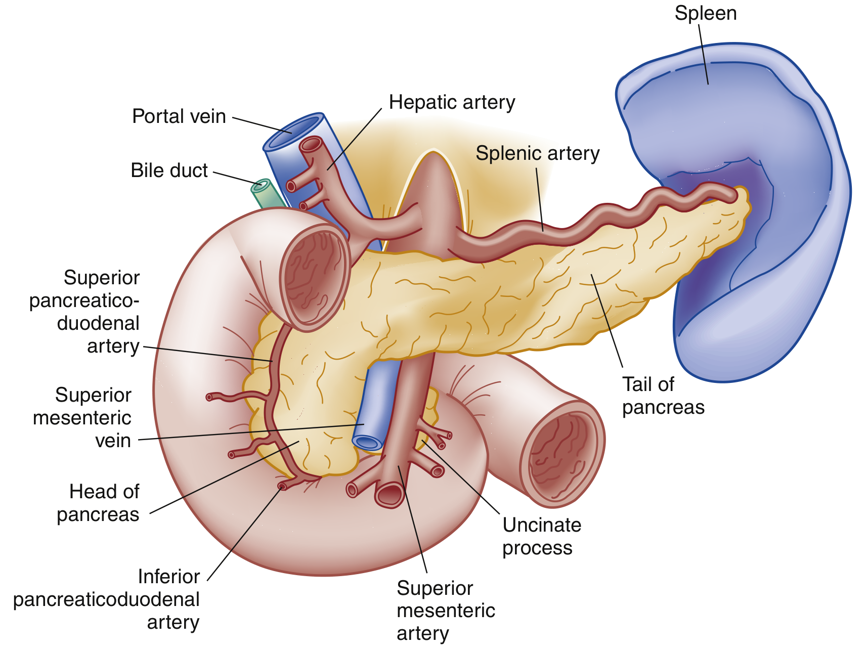
How It Starts: Pathophysiology
Injury begins with an inciting event - duct obstruction by a gallstone, exposure to alcohol, a drug, or another trigger. This:
- Disrupts normal membrane trafficking in pancreatic acinar cells
- Triggers inappropriate activation of trypsinogen → trypsin
- Activated trypsin activates other digestive enzymes, causing autodigestion of pancreatic tissue
- Recruits macrophages and neutrophils, triggering the inflammatory cascade
- Cytokine release increases vascular permeability → edema, hemorrhage, necrosis
- In severe cases: SIRS → sepsis → shock → multiorgan failure (ARDS, renal failure, DIC)
- ROSEN's Emergency Medicine, p. 1265
Causes (Etiology)
Most Common (accounts for ~65-100% of cases)
| Cause | Frequency |
|---|---|
| Gallstones (biliary pancreatitis) | 40-70% |
| Chronic alcohol use | 25-35% |
Other Causes (Organized by Category)
Toxic - Metabolic
- Hypertriglyceridemia (serum TG > 1000 mg/dL)
- Hypercalcemia
- Uremia
- Drugs (see below)
- Scorpion venom
Mechanical - Obstructive
- Biliary stones (most common)
- Post-ERCP (endoscopic retrograde cholangiopancreatography)
- Pancreas divisum, annular pancreas (congenital)
- Tumors (ampullary, neuroendocrine, pancreatic carcinoma)
- Ampullary dysfunction or stenosis
- Duodenal diverticulum
- Abdominal trauma
Infectious
- Viral: mumps, coxsackievirus, HIV, CMV, EBV, varicella
- Bacterial: TB, Salmonella, Campylobacter, Legionella, Mycoplasma
- Parasitic: Ascaris lumbricoides
Vascular
- Vasculitis
- Embolism
- Hypoperfusion / ischemia
- Hypercoagulability
Other
- Idiopathic (no cause found after workup)
- Hereditary / genetic (PRSS1, SPINK1, CFTR mutations)
- Autoimmune pancreatitis (IgG4-related)
- Diabetic ketoacidosis (DKA)
Note: Drug-induced pancreatitis tends to be mild and self-limited. Common implicated drugs include azathioprine, thiazide diuretics, tetracycline, sulfonamides, estrogens, valproic acid, and furosemide.
- ROSEN's Emergency Medicine, Box 77.1
Types of Pancreatitis
Acute Pancreatitis
Sudden-onset inflammation that is usually reversible. Two subtypes:
- Interstitial edematous (~90-95% of cases): diffuse gland enlargement with edema; typically resolves within the first week
- Necrotizing (~5-10% of cases): involves parenchymal and/or peripancreatic tissue necrosis; may remain sterile or become infected (infected necrosis dramatically increases morbidity)
Chronic Pancreatitis
Progressive, irreversible fibrosis and loss of exocrine/endocrine function. Typically caused by:
- Long-term alcohol abuse + smoking (classic presentation)
- Cardinal symptoms: recurrent abdominal pain, steatorrhea (fatty stools), weight loss, bloating, and eventually diabetes mellitus
Recurrent Acute Pancreatitis
Multiple acute attacks, often with an underlying cause (microlithiasis, sphincter of Oddi dysfunction, alcohol, hereditary factors).
Clinical Features (Symptoms & Signs)
Symptoms:
- Persistent epigastric or LUQ pain that radiates to the back, chest, or flanks
- Pain is usually moderate to severe; worsened by eating; relieved by leaning forward
- Nausea, vomiting, anorexia
Physical Examination Signs:
- Epigastric tenderness with or without guarding
- Distended abdomen, reduced or absent bowel sounds (ileus)
- Fever, tachycardia, tachypnea (from SIRS)
- Jaundice - suggests obstructive cause (gallstone, tumor)
- Cullen sign: bluish periumbilical discoloration (hemoperitoneum) - rare, poor prognosis
- Grey Turner sign: reddish-brown flank discoloration (retroperitoneal bleeding) - rare, poor prognosis
- Shallow respirations, basilar crackles, pleural effusion (present in up to 50% of patients, more common on left)
Systemic Complications (severe cases):
- ARDS, respiratory failure
- Cardiovascular collapse, shock
- Acute renal failure
- DIC, thrombocytopenia
- Hyperglycemia (decreased insulin), hypocalcemia
Diagnostic Criteria (Atlanta Criteria)
Acute pancreatitis is diagnosed when at least 2 of 3 criteria are met:
- Abdominal pain characteristic of acute pancreatitis (epigastric, radiating to back)
- Serum lipase or amylase ≥ 3× the upper limit of normal (ULN)
- Characteristic findings on imaging (CT, MRI, or ultrasound)
Laboratory Reports / Investigations
Primary Diagnostic Tests
| Test | Findings in Pancreatitis | Notes |
|---|---|---|
| Serum Lipase | ≥ 3× ULN = highly diagnostic | Preferred test - more sensitive AND specific than amylase; peaks quickly, stays elevated ~1-2 weeks |
| Serum Amylase | ≥ 3× ULN | Less specific; produced by salivary glands and other organs too; stays elevated 3-5 days |
Lipase is more sensitive and specific than amylase. In chronic pancreatitis (usually from alcohol), lipase may be elevated with a normal amylase.
False positives for amylase (elevated without pancreatitis): macroamylasemia, renal failure, salivary gland disease, liver disease, appendicitis, cholecystitis, intestinal obstruction, intestinal ischemia, peptic ulcer disease, DKA, burns, pregnancy.
Supporting Blood Tests
| Test | Purpose |
|---|---|
| CBC (WBC) | Leukocytosis indicates infection/SIRS; rising WBC = worsening disease |
| LFTs (ALT, AST, bilirubin) | ALT > 3× normal strongly suggests biliary (gallstone) etiology |
| Serum triglycerides | > 1000 mg/dL can be both a cause and finding |
| Serum calcium | Hypocalcemia in severe disease (bad prognostic sign) |
| Blood glucose | Hyperglycemia |
| Serum creatinine / BUN | Renal function, early predictor of severity |
| LDH | Elevated; used in Ranson's criteria |
| Arterial blood gases | Detect hypoxemia / ARDS |
| Hematocrit | Hemoconcentration (Hct > 44%) = poor outcome predictor |
| CRP (C-reactive protein) | > 150 mg/L at 48 hrs indicates severe disease |
| Coagulation profile (PT/PTT) | Assess for DIC in severe cases |
Imaging Studies
| Modality | Use |
|---|---|
| Abdominal Ultrasound | First-line imaging; best for detecting gallstones as the cause; limited for pancreas visualization due to bowel gas |
| CT Abdomen with IV Contrast (CECT) | Gold standard for confirming diagnosis, assessing severity, detecting necrosis (Balthazar grading / CT Severity Index); NOT routinely needed if diagnosis is clinically clear; indicated when diagnosis is uncertain, severe disease is present, or complications are suspected |
| MRI / MRCP | Excellent alternative to CT; superior for detecting bile duct stones, ductal anatomy; preferred in pregnancy and renal insufficiency |
| ERCP | Therapeutic (not diagnostic); used when urgent stone extraction is needed in biliary pancreatitis with cholangitis |
| Plain X-ray abdomen | Non-specific; may show "sentinel loop" (dilated bowel near pancreas) or "colon cut-off sign" |
Severity Assessment
Revised Atlanta Classification
- Mild: no organ failure, no local/systemic complications
- Moderately severe: transient organ failure (<48 hours) and/or local complications
- Severe: persistent organ failure (>48 hours), single or multiorgan
Ranson's Criteria (used at admission and at 48 hours)
Scores 0-2 = mild; 3-4 = severe; ≥5 = very severe (mortality >40%)
At Admission:
- Age > 55 years
- WBC > 16,000/mm³
- Glucose > 200 mg/dL
- LDH > 350 IU/L
- AST > 250 IU/L
At 48 Hours:
- Hematocrit decrease > 10%
- BUN increase > 5 mg/dL
- Calcium < 8 mg/dL
- PO₂ < 60 mmHg
- Base deficit > 4 mEq/L
- Fluid sequestration > 6 L
CT Severity Index (Balthazar Score)
Based on degree of pancreatic inflammation and presence of necrosis on CECT; scores 0-10.
Differential Diagnosis
Because epigastric pain can come from many sources, important conditions to rule out include:
- Peptic ulcer disease / perforated ulcer
- Acute cholecystitis / cholangitis
- Acute MI (especially inferior wall)
- Mesenteric ischemia
- Bowel obstruction
- Aortic dissection / AAA
- Ectopic pregnancy
Local Complications (by Atlanta 2012 Classification)
From Interstitial Edematous Pancreatitis:
- Acute peripancreatic fluid collection (within 4 weeks)
- Pancreatic pseudocyst (>4 weeks, well-defined wall)
From Necrotizing Pancreatitis:
- Acute necrotic collection (heterogeneous fluid + necrosis)
- Walled-off necrosis (>4 weeks, well-defined wall; intrapancreatic or extrapancreatic)
Sources:
- ROSEN's Emergency Medicine: Concepts and Clinical Practice, Chapter 77
- Sleisenger and Fordtran's Gastrointestinal and Liver Disease
- Fischer's Mastery of Surgery, 8th edition
- Yamada's Textbook of Gastroenterology, 7th edition
- 2026 iLATAM-AP Ibero-Latin American Guidelines on Acute Pancreatitis (PMID 42216617)
Depet line in anys
I want to make sure I understand your question correctly before proceeding.
Asking for Preferences
Could you clarify what you mean by 'Depet line in anys'? Did you mean one of these?
Reading File
Now let me search the medical library and web in parallel for the dentate line.
Finding Sources
Finding Sources
Searching the Web
dentate line anus anatomy clinical significance surgery
Great - found excellent sources. Now let me read the most comprehensive textbook sections.
Reading File
Reading File
Reading File
Excellent - comprehensive data collected. Let me also fetch the anal canal anatomy diagram.
Loading Image
I have everything needed for a thorough answer.
The Dentate Line (Pectinate Line) of the Anus
The dentate line - also called the pectinate line - is one of the most clinically important anatomical landmarks in the anal canal and in colorectal surgery.
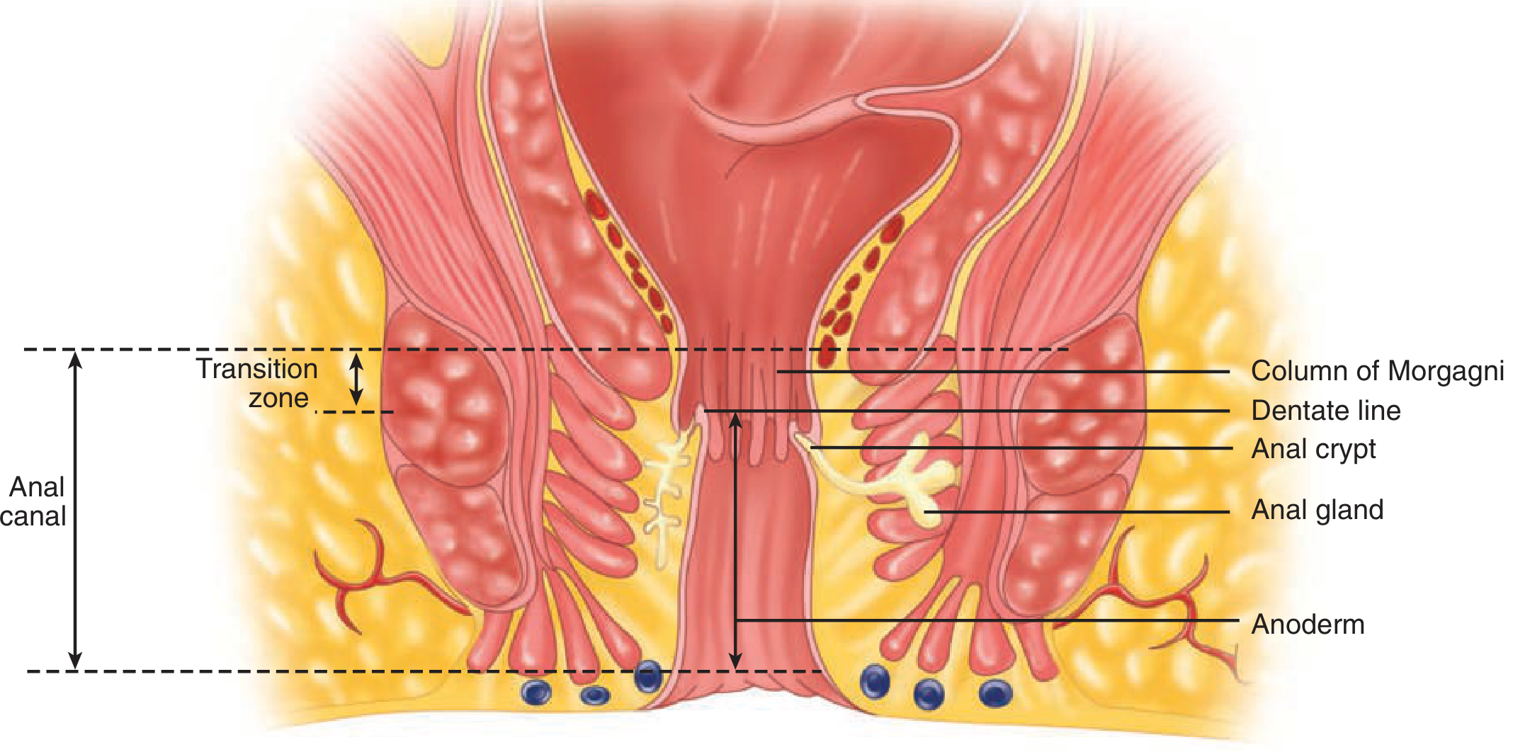
Location
-
Sits at the junction of the upper two-thirds and lower one-third of the anal canal
-
Located approximately 1.0-1.5 cm above the anal verge (the external opening of the anus)
-
The anorectal ring (upper border of the sphincter complex) lies about 1.0-1.5 cm above the dentate line
-
Maingot's Abdominal Operations, p. 1254-1256
What It Is - Embryological Significance
The dentate line is a true mucocutaneous junction - the meeting point of two embryological layers:
| Above Dentate Line | Below Dentate Line |
|---|---|
| Derived from hindgut (endoderm) | Derived from proctodeum (ectoderm) |
| Columnar/glandular epithelium | Squamous epithelium (anoderm) |
Just above the line, there is a transitional zone (6-12 mm) where the columnar epithelium of the rectum gradually changes to cuboidal epithelium before meeting the squamous epithelium at the dentate line. This transition zone can extend up to 15 cm proximal to the dentate line in some individuals.
- Schwartz's Principles of Surgery, 11th Edition
- Maingot's Abdominal Operations
Structures At and Around the Dentate Line
- Columns of Morgagni: 8-14 longitudinal folds of rectal mucosa just above the dentate line
- Anal crypts: small pockets/recesses between the bases of the columns of Morgagni, at the dentate line
- Anal glands: rudimentary glands that open into some anal crypts; they may extend through the internal sphincter into the intersphincteric plane (but NOT into the external sphincter). These are the source of cryptoglandular abscesses and fistula-in-ano
Why the Dentate Line Matters - Clinical Differences Above vs. Below
The dentate line divides the anal canal into two zones with completely different blood supply, venous drainage, lymphatics, and nerve supply. This has profound clinical consequences.
1. Nerve Supply (Pain Sensation)
| Above Dentate Line | Below Dentate Line |
|---|---|
| Visceral innervation (autonomic) | Somatic innervation (inferior rectal nerve branches) |
| Poorly defined dull sensation only (responds to stretch, not sharp pain) | Full cutaneous sensation: pain, touch, heat, cold |
| Internal hemorrhoids can be rubber-band ligated here without anaesthesia | Procedures here require local/general anaesthesia |
This is why internal hemorrhoids (above the dentate line) can be banded in the clinic without pain, while external hemorrhoids (below) are intensely painful.
2. Blood Supply (Arterial)
| Above Dentate Line | Below Dentate Line |
|---|---|
| Superior and middle rectal arteries (branches of inferior mesenteric artery and internal iliac artery) | Inferior rectal arteries (from internal pudendal artery) |
3. Venous Drainage
| Above Dentate Line | Below Dentate Line |
|---|---|
| Portal venous system (superior rectal vein → inferior mesenteric vein) | Systemic venous system (middle/inferior rectal veins → internal iliac vein) |
This is why portal hypertension can cause internal hemorrhoids (porto-systemic anastomosis at the anorectal junction).
4. Lymphatic Drainage
| Above Dentate Line | Below Dentate Line |
|---|---|
| Internal iliac lymph nodes (upward drainage with superior rectal vessels) | Superficial inguinal lymph nodes |
This distinction is critical for cancer staging and spread. Anal canal carcinomas above the dentate line metastasize to internal iliac nodes; those below spread to inguinal nodes. Inguinal lymphadenopathy in a patient with anal symptoms may indicate a distal anal canal tumor.
5. Hemorrhoid Classification
| Internal Hemorrhoids | External Hemorrhoids |
|---|---|
| Above the dentate line | Below the dentate line (at anal verge) |
| Covered by visceral mucosa - painless unless prolapsed/thrombosed | Covered by somatic skin - acutely painful if thrombosed |
| Treated by banding, sclerotherapy, or hemorrhoidectomy | Require excision under anaesthesia if thrombosed |
- Maingot's Abdominal Operations, pp. 1260-1267
- Mulholland and Greenfield's Surgery, 7th Edition
- Schwartz's Principles of Surgery, 11th Edition
Quick Summary Table
| Feature | Above Dentate Line | Below Dentate Line |
|---|---|---|
| Epithelium | Columnar (rectal mucosa) | Squamous (anoderm) |
| Embryology | Endoderm (hindgut) | Ectoderm (proctodeum) |
| Sensation | Visceral (dull/stretch) | Somatic (pain, temp, touch) |
| Arterial supply | Superior/middle rectal aa. | Inferior rectal aa. |
| Venous drainage | Portal system | Systemic (iliac) system |
| Lymphatics | Internal iliac nodes | Inguinal nodes |
| Hemorrhoids | Internal (painless) | External (painful) |
| Cancer spread | Internal iliac nodes | Inguinal nodes |
This is a shared conversation. Sign in to Orris to start your own chat.