
I had cough with stridor and wheeze for 1 week what is the probable diagnosis
croup laryngotracheobronchitis steeple sign X-ray
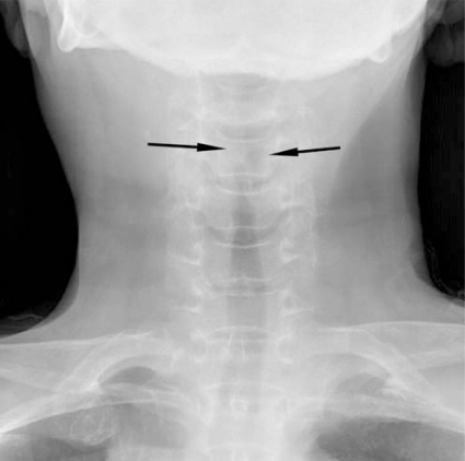
**Imaging Modality:** Anteroposterior (AP) plain film radiograph (X-ray). **Anatomical Region:** Soft tissue of the neck and upper respiratory tract. **Observed Pathology:** Subglottic narrowing of the airway column. **Characteristic Visual Features:** The image demonstrates a characteristic "steeple sign" (or "pencil point sign"), characterized by symmetrical, subglottic narrowing of the tracheal air column. This narrowing is caused by inflammatory soft tissue edema in the subglottic region. The proximal tracheal lumen is significantly constricted compared to the distal segment. **Key Diagnostic Features:** The tapering of the upper airway on an AP view is a classic radiologic hallmark of Croup (laryngotracheobronchitis). This feature helps differentiate the condition from epiglottitis, which typically presents with a "thumbprint sign" on a lateral neck radiograph. **Clinical Context:** The findings are highly suggestive of acute inflammatory airway obstruction in the subglottic space, typically seen in pediatric populations presenting with respiratory distress or stridor.
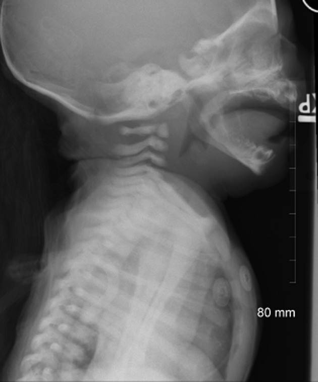
**Imaging Modality:** Lateral soft tissue X-ray of the neck and upper thorax. **Anatomical Region:** Pediatric cervical airway and upper respiratory tract. **Observed Pathology:** Findings are highly indicative of acute epiglottitis (supraglottitis). **Characteristic Visual Features:** * **"Thumb Sign":** Significant enlargement and rounding of the epiglottis, resulting in a thick, bulbous appearance resembling a thumb. * **Aryepiglottic Folds:** Notable thickening and convex bowing of the aryepiglottic folds. * **Arytenoid Enlargement:** Prominent swelling of the arytenoid soft tissues. * **Hypopharyngeal Distension:** Compensatory dilatation of the hypopharynx proximal to the site of supraglottic narrowing. * **Airway Caliber:** Reduction in the patency of the laryngeal inlet due to massive soft tissue edema. **Key Diagnostic Features:** The combination of the "thumb sign" and thickened aryepiglottic folds on a lateral view are pathognomonic for epiglottitis, distinguishing it from the "steeple sign" associated with subglottic narrowing in laryngotracheobronchitis (croup). Electrodes are visible on the chest wall, indicating clinical monitoring.
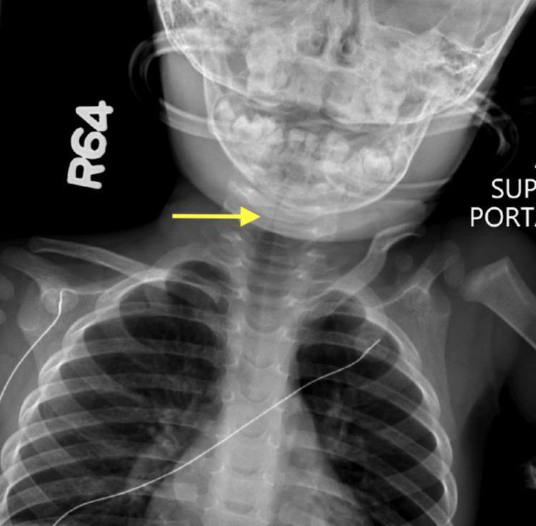
This diagnostic image is a posterior-anterior (PA) neck and upper chest radiograph of a pediatric patient. The central clinical finding is the classic 'steeple sign,' characterized by subglottic narrowing of the airway as it approaches the larynx, indicated by a yellow arrow. This tapering morphology represents edema in the subglottic region, commonly associated with viral laryngotracheobronchitis (croup). Beyond the focal subglottic stenosis, the radiograph demonstrates diffuse tracheal narrowing extending into the upper thoracic portion of the airway. Anatomical structures visible include the base of the skull, the cervical spine, bilateral clavicles, and the superior ribs. The soft tissues of the neck appear unremarkable except for the compromised airway lumen. This image serves as a key educational example of pediatric airway obstruction and the radiological manifestations of inflammatory subglottic stenosis in a clinical setting involving respiratory distress.
Probable Diagnosis: Croup (Acute Laryngotracheobronchitis)
What Is Croup?
Classic Clinical Triad
| Feature | Description |
|---|---|
| Barking/seal-like cough | Distinctive, harsh, "croupy" cough from subglottic swelling |
| Inspiratory stridor | High-pitched sound from narrowed larynx/subglottis |
| Wheeze | Lower airway component - tracheal/bronchial involvement |
- Usually preceded by 1-2 days of upper respiratory tract symptoms (runny nose, low-grade fever)
- Hoarse voice
- Symptoms often worse at night
- Child does NOT appear toxic (unlike epiglottitis)
- No drooling (key differentiating point from epiglottitis)
Who Gets It?
- Most common in children aged 6 months to 3 years (peak at age 2)
- Boys more affected than girls
- Seasonal peak in late autumn and winter
- Affects 3-6% of children under 6 years annually
Causative Organisms
- Influenza A and B
- RSV (Respiratory Syncytial Virus)
- Rhinovirus
- Adenovirus
- Metapneumovirus, Coronavirus (less common)
Diagnosis
Note: Classic steeple sign may be absent in up to 50% of croup cases, so a normal X-ray does not rule it out.
Important Differentials to Consider
| Condition | Key Distinguishing Features |
|---|---|
| Epiglottitis | Toxic appearance, drooling, high fever, NO barking cough, "tripod" position |
| Foreign body aspiration | Sudden onset, unilateral wheeze, no fever |
| Bacterial tracheitis | Toxic, high fever, does NOT respond to croup treatment |
| Spasmodic croup | Recurrent, no viral prodrome, resolves spontaneously |
| Asthma | Wheeze-predominant, usually no stridor, responds to bronchodilators |
Severity Assessment
| Severity | Features |
|---|---|
| Mild | Barking cough, no stridor at rest |
| Moderate | Stridor at rest, mild retractions |
| Severe | Biphasic stridor, significant retractions, O2 desaturation, altered consciousness |
Management (Brief)
- Mild: Supportive care at home, humidified air, reassurance
- Moderate-Severe: Dexamethasone (oral/IM, 0.15-0.6 mg/kg) - reduces oedema significantly
- Severe/life-threatening: Nebulised adrenaline (epinephrine) + dexamethasone + possible intubation