
Clinical anatomy to radial artery.
radial artery anatomy course forearm wrist diagram
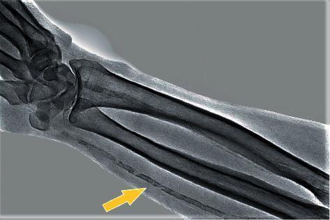
This diagnostic image is a plain radiograph of the distal forearm and wrist, demonstrating significant vascular pathology. The skeletal anatomy, including the radius, ulna, and carpal bones, serves as a reference for the prominent finding: diffuse calcification of the radial artery (indicated by a yellow arrow). The calcification presents as a continuous, linear, and somewhat beaded pattern of increased radiopacity tracing the anatomical course of the radial artery. This visual appearance is characteristic of Monckeberg medial calcific sclerosis (mediocalcinosis), where calcium deposits accumulate in the tunica media of the arterial wall. Unlike intimal atherosclerosis, which often presents with eccentric plaques, this medial calcification is typically non-obstructive but leads to increased arterial stiffness. This condition is frequently associated with advanced age, diabetes mellitus, and end-stage renal disease. The contrast between the dense cortical bone and the less dense, fragmented radiopacity of the calcified vessel highlights the extent of the vascular mineralization throughout the visible length of the forearm.
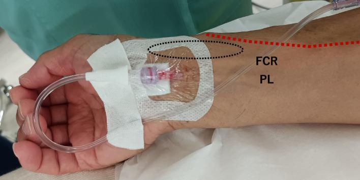
This clinical photograph illustrates an anatomical variation and its impact on arterial cannulation in the distal forearm. The image shows a right wrist with an arterial catheter secured in place. Annotations highlight the procedural anatomy: the catheter is positioned between the flexor carpi radialis (FCR) and palmaris longus (PL) tendons, a location corresponding to the course of the median nerve and median artery. A dashed black oval indicates the traditional site for radial artery palpation, where the pulse was difficult to identify. A red dashed line traces the path of a superficial radial artery, an anatomical variant that crosses over the anatomical snuffbox. This procedural image demonstrates incidental median artery cannulation, a clinical scenario encountered when standard radial access is complicated by vascular anomalies. It serves as an educational tool for anesthesiology and critical care training, emphasizing the importance of ultrasound guidance and awareness of forearm vascular variations to avoid unintentional cannulation of vessels associated with major nerves.
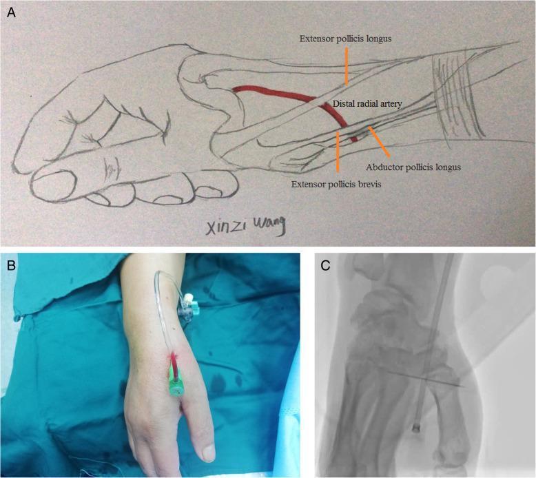
A multi-panel educational figure illustrating the anatomy and clinical application of distal radial artery (DRA) access. Panel A is an anatomical diagram of the hand and wrist highlighting the anatomical snuffbox. It depicts the distal radial artery in relation to key tendons: the extensor pollicis longus forms the medial border, while the abductor pollicis longus and extensor pollicis brevis form the lateral border. Panel B is a clinical photograph showing the procedural setup for distal transradial access (dTRA), featuring a 6Fr sheath successfully inserted into the snuffbox region with a connector tube attached. Panel C is a fluoroscopic diagnostic image (X-ray) providing a radiological view of the puncture site. It demonstrates the radiopaque sheath positioned within the DRA, localized near the radial styloid process and carpal bones. This figure is designed for medical professionals and interventionalists to understand the topographical and radiological landmarks necessary for arterial cannulation at the distal forearm/hand junction.
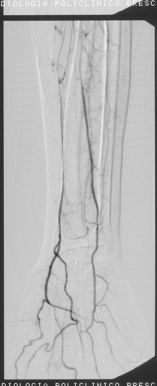
This diagnostic image is a digital subtraction angiogram (DSA) of the right forearm and wrist, captured following a thrombolysis procedure. The image depicts the arterial anatomy of the distal upper extremity, showing contrast opacification of the radial artery and its progression toward the hand. The radial artery exhibits continuous flow with a regular caliber, indicating a successful restoration of patency. In the wrist region, the image demonstrates clear vascularization with branching patterns and anastomoses forming the palmar arches. Smaller collateral vessels and tertiary branches are visible around the radio-carpal joint. The clinical significance of this imaging is to confirm the resolution of previous vasospasm or thromboembolic occlusion and to ensure adequate distal perfusion to the hand. This visual serves as an educational example of normal post-interventional vascular anatomy in the context of interventional radiology and vascular surgery.
Clinical Anatomy of the Radial Artery
Origin
Course in the Forearm (Anterior Compartment)
- Lies just deep to the brachioradialis muscle.
- The superficial branch of the radial nerve runs along its lateral side in the middle third of the forearm.
- Continues between brachioradialis laterally and the flexor carpi radialis medially.
- Emerges medial to the brachioradialis tendon and is covered only by deep fascia, superficial fascia, and skin - making it directly palpable.
- Lies immediately lateral to the flexor carpi radialis tendon and directly anterior to pronator quadratus and the distal radius.
- Clinical pearl: The flexor carpi radialis tendon is the reliable landmark for locating the radial pulse. The artery is gently compressed against the underlying bone to feel the pulse.
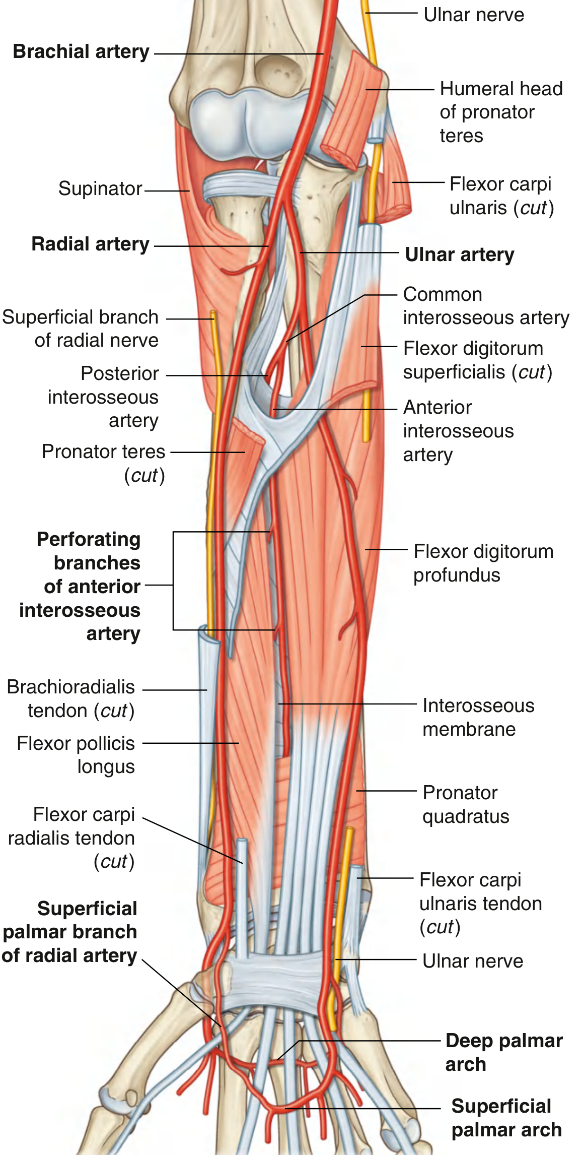
Branches in the Forearm
| Branch | Origin | Distribution |
|---|---|---|
| Radial recurrent artery | Proximal radial artery | Passes superiorly to anastomose around the elbow joint; also supplies lateral forearm muscles |
| Muscular branches | Throughout | Supply muscles on the radial/lateral side of the forearm |
| Palmar carpal branch | Distal forearm | Small; joins the palmar carpal arch - a vascular plexus supplying the carpal bones and joints |
| Superficial palmar branch | Distal forearm / wrist | Passes through or superficial to the thenar muscles; anastomoses with the superficial palmar arch formed by the ulnar artery |
| Posterior carpal branch | At the wrist | Passes to the posterior carpal arch on the dorsum of the wrist |
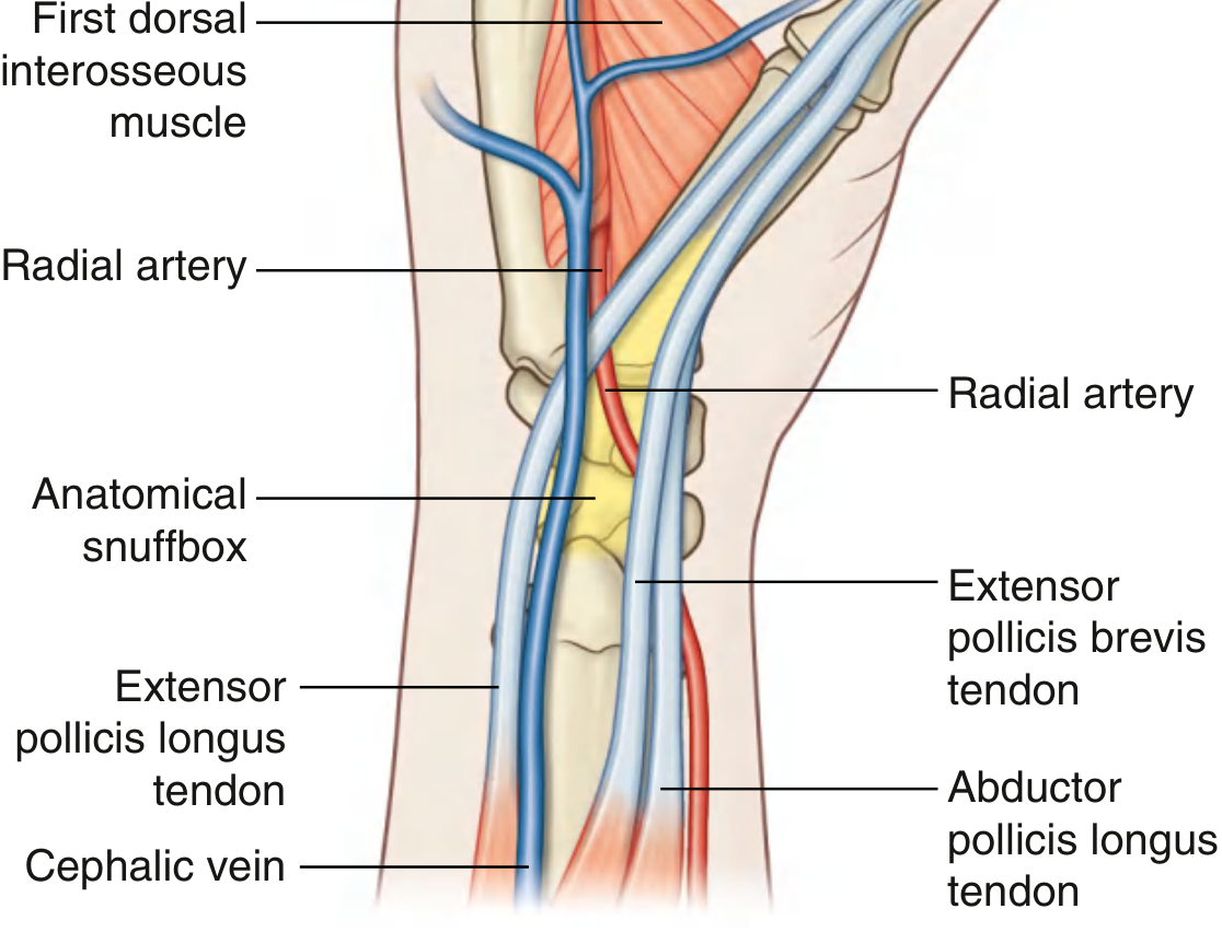
At the Wrist - Anatomical Snuffbox
- Lateral border: Tendons of abductor pollicis longus and extensor pollicis brevis
- Medial border: Tendon of extensor pollicis longus
- Floor: Scaphoid and trapezium bones, plus distal tendons of extensor carpi radialis longus and brevis
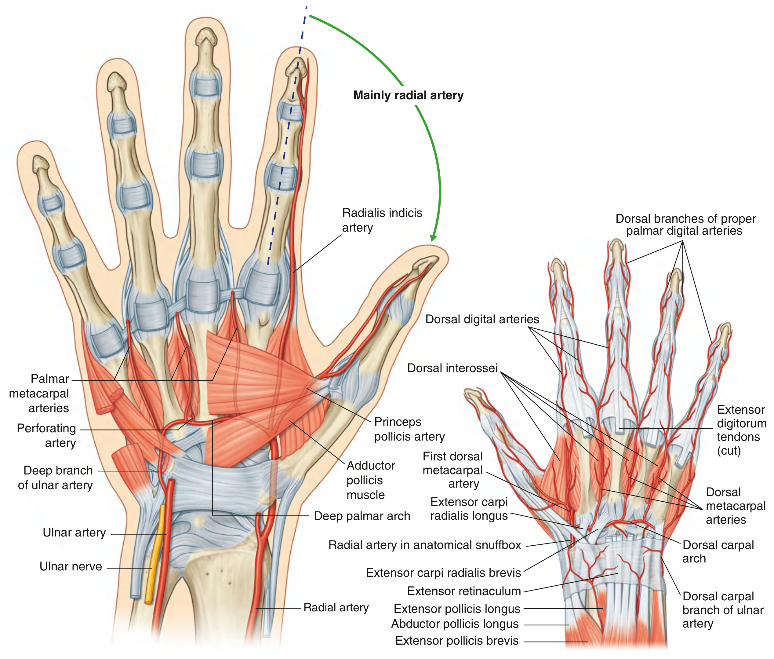
Course in the Hand and Terminal Branches
- Passes between the two heads of the first dorsal interosseous muscle
- Then passes between the two heads of the adductor pollicis to enter the deep plane of the palm
- Becomes the deep palmar arch
- Dorsal carpal branch - crosses medially to form the dorsal carpal arch, giving off three dorsal metacarpal arteries which divide into small dorsal digital arteries
- First dorsal metacarpal artery - supplies the adjacent sides of the index finger and thumb
- Princeps pollicis artery - major blood supply to the thumb
- Radialis indicis artery - supplies the lateral side of the index finger
- Three palmar metacarpal arteries - join the common palmar digital arteries from the superficial palmar arch
- Three perforating branches - pass posteriorly between the dorsal interossei to anastomose with the dorsal metacarpal arteries
Summary of the Complete Course
Brachial artery (cubital fossa)
    ↓  bifurcates at neck of radius
Radial artery (lateral)
    ↓  deep to brachioradialis (proximal forearm)
    ↓  lateral to superficial radial nerve (middle third)
    ↓  medial to brachioradialis tendon, lateral to FCR tendon (distal forearm)
    ↓  passes around lateral wrist
Anatomical snuffbox (floor = scaphoid + trapezium)
    ↓  passes between heads of 1st dorsal interosseous
    ↓  passes between heads of adductor pollicis
Deep palmar arch (deep to long flexors, on metacarpal bases)
    → Princeps pollicis artery (thumb)
    → Radialis indicis artery (lateral index finger)
    → Palmar metacarpal arteries
Clinical Anatomy Points
1. Radial Pulse
2. Radial Artery Cannulation (Arterial Lines)
- Technically easy to cannulate
- Superficial and accessible at the wrist
- Rarely causes ischemic complications due to collateral flow via the ulnar artery and palmar arches
- Slogoff et al. documented 1700 cardiovascular surgical patients with no ischemic complications despite arterial occlusion occurring post-decannulation in >25% of cases - Miller's Anesthesia, 10e
3. Modified Allen Test
- Occlude both radial and ulnar arteries with digital pressure; patient clenches fist to exsanguinate the hand
- Open the hand; release ulnar artery occlusion only
- Normal: rubor returns within 5-10 seconds
- Abnormal (positive): pallor persists >5-15 seconds after release - suggests inadequate ulnar collateral flow; avoid radial cannulation at that site
4. Radial Artery as a Surgical Conduit
5. Anatomical Variations
- The radial recurrent artery's origin is variable; it arises from the radial artery in only ~65% of the population, and may originate from the brachial artery or the ulnar-interosseous trunk
- A superficial radial artery variant (crossing over the anatomical snuffbox) exists and can complicate standard wrist cannulation
- The superficial palmar branch of the radial artery that contributes to the superficial palmar arch is highly variable - Imaging Anatomy Vol. 3; General Anatomy THIEME Atlas