
Follicular carcinoma of thyroid
"thyroid carcinoma"[MeSH Terms] AND follicular
follicular thyroid carcinoma diagnosis treatment ATA guideline
follicular thyroid carcinoma histology capsular and vascular invasion
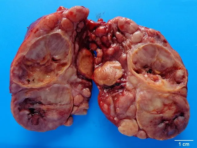
This gross pathology photograph documents a resected thyroid gland specimen, anterior view, with bilateral lobes separated for inspection. The background is a blue-friendly field and a 1 cm scale bar is present for size reference. The specimen displays multiple firm, tan to brown nodules replacing native thyroid parenchyma, with irregular contours and areas of marginal hemorrhage. The left and right lobes show expansion of the thyroid capsule, and prominent nodularity suggests follicular architecture disrupted by neoplastic growth. The caption indicates widely invasive follicular carcinoma of the thyroid, consistent with extrathyroidal extension and potential vascular invasion; gross features include invasion beyond the capsule and involvement of perithyroid soft tissue. No papillary nuclear features are assessable on gross inspection; histology would confirm follicular differentiation, capsular and vascular invasion, and tumor margins. Clinically, this specimen supports a diagnosis of aggressive differentiated thyroid carcinoma associated with higher risk of hematogenous metastasis. The imaging context is macroscopic pathology; the modality is gross photography used for documentation, education, and archival purposes. This image is relevant for surgical pathology education, thyroid cancer staging, case-based discussions, and radiologic-pathologic correlation exercises; useful for learners reviewing follicular thyroid carcinoma, aggressive variants, and the significance of extrathyroidal extension in prognosis.
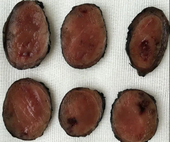
Gross image of six transverse sections from excised thyroid tissue representing Hurthle cell adenoma (oncocytic adenoma) of the thyroid gland. Modality: clinical gross pathology photography; specimen: thyroid nodules; orientation: macroscopic cross-sectional slices laid in a grid on a neutral background for documentation. The nodules exhibit well-defined, encapsulated margins with a uniform to slightly variegated tan-to-reddish cut surface. Capsular integrity appears preserved in most slices, consistent with a benign, noninvasive follicular lesion; occasional focal hemorrhagic or fibrous creases are present. The tissue demonstrates abundant eosinophilic cytoplasm in Hurthle cells (oncocytic change) at the gross level, though microarchitectural features cannot be resolved without histology. The image is intended to aid gross-pathology assessment and surgical pathology correlation, supporting the differential diagnosis of Hurthle cell adenoma versus Hurthle cell carcinoma and other thyroid follicular lesions such as follicular adenoma or nodular hyperplasia. Clinically, this finding is relevant to surgical planning, thyroid function assessment, and malignancy risk stratification; final classification relies on microscopic capsular and vascular invasion status. This depiction may be used in educational contexts to illustrate gross nodularity, encapsulation, and the appearance of Hurthle cell-containing thyroid adenomas. Educational use: suitable for medical students and residents encountering thyroid pathology. Correlates with histology, radiology reference.
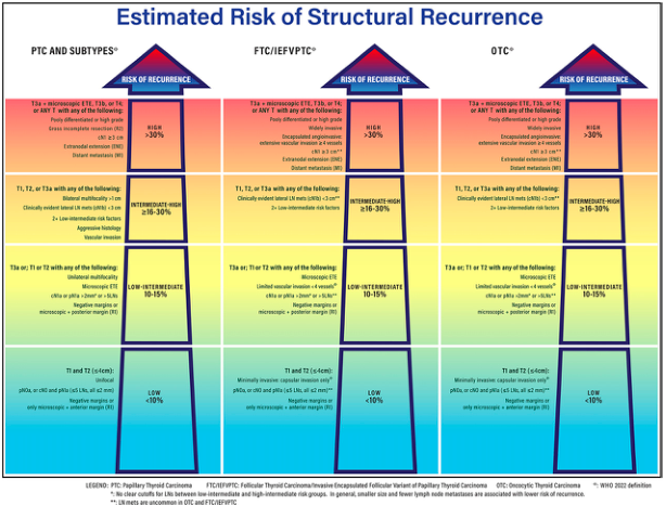
Summary : This figure presents a structured risk stratification chart for the estimated risk of structural recurrence in thyroid carcinoma, comparing three types: Papillary Thyroid Carcinoma (PTC) and subtypes, Follicular Thyroid Carcinoma/Invasive Encapsulated Follicular Variant of Papillary Thyroid Carcinoma (FTC/IEFVPTC), and Oncocytic Thyroid Carcinoma (OTC). The chart categorizes risk into four levels (High, Intermediate-High, Low-Intermediate, Low) based on specific clinical and pathological criteria.
https://www.thyroid.org/patient-thyroid-information/ct-for-p…
Follicular carcinoma of the thyroid (FTC)
Key pathological feature
- Capsular invasion: tumor penetrates through its capsule into surrounding thyroid tissue.
- Vascular invasion (angioinvasion): tumor cells invade blood vessels in or beyond the capsule.
Types
-
Minimally invasive FTC
- Limited capsular invasion, with or without limited vascular invasion.
- Generally has an excellent prognosis, particularly when vascular invasion is absent or minimal.
-
Widely invasive FTC
- Extensive infiltration of thyroid tissue and/or blood vessels.
- Has a higher risk of distant metastasis and recurrence.
Spread
- Common distant sites: bone, lungs, and less commonly brain or liver.
- Cervical lymph-node involvement is less common than in papillary carcinoma.
- Distant metastases may occasionally be the initial presentation. Scott-Brown's Otorhinolaryngology Head & Neck Surgery, Vol. 1, p. 990.
Clinical presentation
- Solitary thyroid nodule, usually painless
- Possible enlarging neck mass
- Hoarseness, dysphagia, or dyspnea if locally advanced
- Bone pain, pathological fracture, or respiratory symptoms can suggest distant metastasis
Investigation
- Serum TSH and thyroid ultrasound
- Ultrasound-guided FNA for a thyroid nodule
- FNA may report “follicular neoplasm” or “suspicious for follicular neoplasm”, but cannot establish carcinoma
- Diagnostic thyroid lobectomy may be needed for final histologic diagnosis
- After cancer confirmation: assessment of tumor size, invasion, vascular invasion, lymph nodes, and distant disease; thyroglobulin is useful for postoperative surveillance
Management
- Low-risk, intrathyroidal disease: thyroid lobectomy may be sufficient in selected patients.
- Invasive disease, substantial vascular invasion, large tumors, extrathyroidal extension, or metastases: total thyroidectomy is commonly considered.
- Radioactive iodine (RAI): used selectively after total thyroidectomy, particularly for higher-risk disease or iodine-avid metastatic disease.
- TSH suppression with levothyroxine: individualized to recurrence risk and response to therapy.
- External-beam radiotherapy or systemic treatment: reserved for selected unresectable, recurrent, or RAI-refractory disease.
Prognosis
- Older age
- Large primary tumor
- Extensive vascular invasion
- Widely invasive histology
- Extrathyroidal extension
- Distant metastases
- Incomplete surgical resection
Follicular adenoma and FTC may look alike on FNA. FTC is diagnosed only when capsular and/or vascular invasion is demonstrated on histopathology.
Follicular carcinoma of thyroid in detail
"follicular thyroid carcinoma"
2025 American Thyroid Association differentiated thyroid cancer guidelines follicular thyroid carcinoma PDF
follicular thyroid carcinoma microscopic histology capsular vascular invasion
Summary : This figure presents a structured risk stratification chart for the estimated risk of structural recurrence in thyroid carcinoma, comparing three types: Papillary Thyroid Carcinoma (PTC) and subtypes, Follicular Thyroid Carcinoma/Invasive Encapsulated Follicular Variant of Papillary Thyroid Carcinoma (FTC/IEFVPTC), and Oncocytic Thyroid Carcinoma (OTC). The chart categorizes risk into four levels (High, Intermediate-High, Low-Intermediate, Low) based on specific clinical and pathological criteria.
Gross image of six transverse sections from excised thyroid tissue representing Hurthle cell adenoma (oncocytic adenoma) of the thyroid gland. Modality: clinical gross pathology photography; specimen: thyroid nodules; orientation: macroscopic cross-sectional slices laid in a grid on a neutral background for documentation. The nodules exhibit well-defined, encapsulated margins with a uniform to slightly variegated tan-to-reddish cut surface. Capsular integrity appears preserved in most slices, consistent with a benign, noninvasive follicular lesion; occasional focal hemorrhagic or fibrous creases are present. The tissue demonstrates abundant eosinophilic cytoplasm in Hurthle cells (oncocytic change) at the gross level, though microarchitectural features cannot be resolved without histology. The image is intended to aid gross-pathology assessment and surgical pathology correlation, supporting the differential diagnosis of Hurthle cell adenoma versus Hurthle cell carcinoma and other thyroid follicular lesions such as follicular adenoma or nodular hyperplasia. Clinically, this finding is relevant to surgical planning, thyroid function assessment, and malignancy risk stratification; final classification relies on microscopic capsular and vascular invasion status. This depiction may be used in educational contexts to illustrate gross nodularity, encapsulation, and the appearance of Hurthle cell-containing thyroid adenomas. Educational use: suitable for medical students and residents encountering thyroid pathology. Correlates with histology, radiology reference.
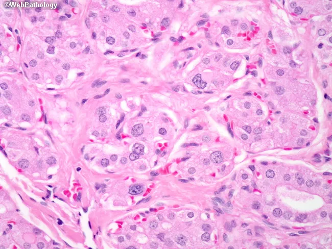
Imaging modality and specimen context: This is a high power light microscopic histology slide of thyroid gland tissue stained with Hematoxylin and Eosin, prepared from a surgically excised or biopsy specimen. Location: cervical thyroid region, thyroid parenchyma comprising follicular epithelium. The slide captures a dyshormonogenetic goiter pattern: hypercellular nodules formed by small, compact follicles, with lumina that are diminutive or nearly occluded and containing little to no colloid. Cytologic atypia is mild to minimal, lacking prominent nuclear features of papillary carcinoma. The background stroma may be edematous or fibrous, with scattered colloid-poor follicular structures. The morphological phenotype reflects defective thyroid hormone synthesis (dyshormonogenesis) with compensatory follicular hyperplasia, and may resemble multinodular goiter but with reduced colloid and more crowded epithelium. Diagnostic significance: histology supports dyshormonogenetic goiter as etiologic basis of congenital or acquired goiter, helps distinguish from autoimmune thyroiditis (lymphocytic infiltrate, germinal centers), Graves disease (diffuse hyperplasia with scalloped colloid), or follicular neoplasm (capsular/vascular invasion absent). Clinical correlation: management typically favors lifelong thyroid hormone replacement; surgical thyroidectomy is reserved for compression symptoms or diagnostic uncertainty. Relevance to clinical practice and research includes understanding thyroid biosynthesis defects, pediatric goiters, and surgical decision-making. Use cases include educational pathology, differential diagnosis practice, and radiologic-pathologic correlation in thyroid disease.
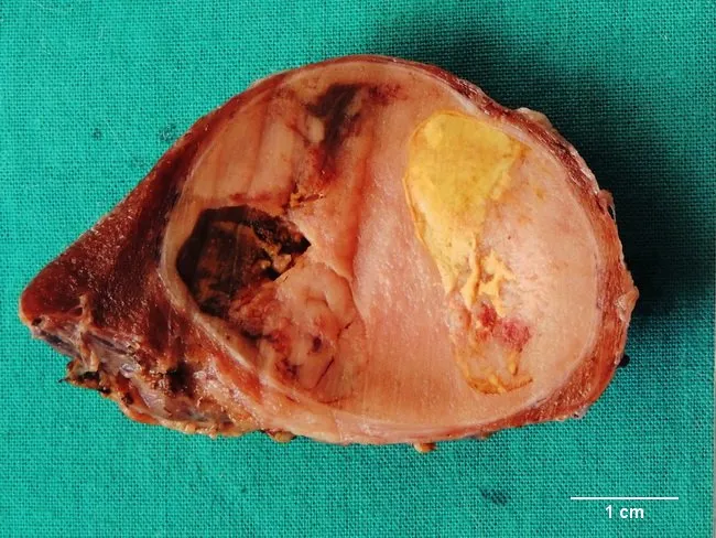
Grossly, this is a macroscopic photograph of a partial thyroidectomy specimen showing a well-circumscribed, tan to light brown nodule with a distinct fibrous capsule. The cut surface reveals a solid lesion with encapsulation, containing areas of hemorrhagic transformation and cystic degeneration, and surrounding scar tissue. These features are typical of a long-standing follicular adenoma of the thyroid, a benign neoplasm composed of uniform follicular epithelium forming colloid-filled follicles separated from adjacent thyroid parenchyma by a capsule. The degenerative changes reflect chronicity; hemorrhagic and cystic changes may obscure small follicles on microscopic examination. The presence of a capsule and lack of gross invasiveness support benignity, though definitive distinction from follicular carcinoma requires microscopic assessment for capsular and vascular invasion. Clinically, the patient is euthyroid; management typically involves lobectomy or partial thyroidectomy with histologic confirmation. This image is valuable for education and research by illustrating gross-pathology correlations with histology in thyroid nodules and the spectrum of degenerative changes in benign thyroid neoplasms. The 1 cm scale bar is visible, enabling rough size estimation. Knowledge of such gross features helps in differential diagnosis, correlation with capsule integrity, and understanding how long-standing nodules evolve over time.
- Definition, epidemiology, and risk factors
- Pathogenesis and molecular alterations
- Pathology and classification
- Clinical features and routes of spread
- Diagnostic work-up and differential diagnosis
- Staging and prognostic factors
- Treatment and follow-up
- Comparison with papillary carcinoma and follicular adenoma
Which section should I cover in full first?
Follicular carcinoma of the thyroid (FTC)
Key pathological feature
- Capsular invasion: tumor penetrates through its capsule into surrounding thyroid tissue.
- Vascular invasion (angioinvasion): tumor cells invade blood vessels in or beyond the capsule.
Types
-
Minimally invasive FTC
- Limited capsular invasion, with or without limited vascular invasion.
- Generally has an excellent prognosis, particularly when vascular invasion is absent or minimal.
-
Widely invasive FTC
- Extensive infiltration of thyroid tissue and/or blood vessels.
- Has a higher risk of distant metastasis and recurrence.
Spread
- Common distant sites: bone, lungs, and less commonly brain or liver.
- Cervical lymph-node involvement is less common than in papillary carcinoma.
- Distant metastases may occasionally be the initial presentation. Scott-Brown's Otorhinolaryngology Head & Neck Surgery, Vol. 1, p. 990.
Clinical presentation
- Solitary thyroid nodule, usually painless
- Possible enlarging neck mass
- Hoarseness, dysphagia, or dyspnea if locally advanced
- Bone pain, pathological fracture, or respiratory symptoms can suggest distant metastasis
Investigation
- Serum TSH and thyroid ultrasound
- Ultrasound-guided FNA for a thyroid nodule
- FNA may report “follicular neoplasm” or “suspicious for follicular neoplasm”, but cannot establish carcinoma
- Diagnostic thyroid lobectomy may be needed for final histologic diagnosis
- After cancer confirmation: assessment of tumor size, invasion, vascular invasion, lymph nodes, and distant disease; thyroglobulin is useful for postoperative surveillance
Management
- Low-risk, intrathyroidal disease: thyroid lobectomy may be sufficient in selected patients.
- Invasive disease, substantial vascular invasion, large tumors, extrathyroidal extension, or metastases: total thyroidectomy is commonly considered.
- Radioactive iodine (RAI): used selectively after total thyroidectomy, particularly for higher-risk disease or iodine-avid metastatic disease.
- TSH suppression with levothyroxine: individualized to recurrence risk and response to therapy.
- External-beam radiotherapy or systemic treatment: reserved for selected unresectable, recurrent, or RAI-refractory disease.
Prognosis
- Older age
- Large primary tumor
- Extensive vascular invasion
- Widely invasive histology
- Extrathyroidal extension
- Distant metastases
- Incomplete surgical resection
**Follicular adenoma and FTC may look alike on FNA. FTC is diagnosed only when capsular and/or vascular invasion is demonstrated on histopathology.**A detailed account can be organized as:
- Definition, epidemiology, and risk factors
- Pathogenesis and molecular alterations
- Pathology and classification
- Clinical features and routes of spread
- Diagnostic work-up and differential diagnosis
- Staging and prognostic factors
- Treatment and follow-up
- Comparison with papillary carcinoma and follicular adenoma
- Pathology: histology, capsular/vascular invasion, classification
- Diagnosis: clinical features, ultrasound, FNA, Bethesda, molecular tests
- Treatment: surgery, radioactive iodine, TSH suppression, follow-up
- Full exam note: all sections in a structured, detailed format