
Azygos vein
azygos vein anatomy posterior mediastinum diagram
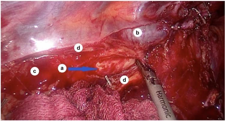
An intraoperative endoscopic photograph showing the surgical anatomy of the posterior mediastinum during a thoracic procedure. The operative field highlights the sulcus azygoaortalis (labeled 'c'), a physiological groove located between the descending thoracic aorta and the azygos vein. The adventitia of the thoracic aorta (labeled 'a') is visible through the dissection site. The azygos vein (labeled 'b') is positioned adjacent to the aortic wall. The mediastinal pleura has been incised and reflected, with its edges (labeled 'd') clearly demarcated at the periphery of the dissection. A harmonic scalpel or ultrasonic dissector is visible in the lower right, positioned near the operative site. This image demonstrates the critical anatomical relationships required for mobilization of the esophagus and identification of the thoracic duct, which typically resides within the loose connective tissue of this sulcus. The orientation reflects a sagittal surgical plane used during lymphadenectomy or esophageal surgery.
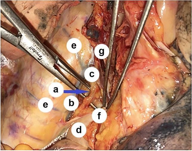
This clinical photograph provides an intraoperative view of the posterior mediastinum during a surgical procedure, specifically highlighting the anatomical relationships surrounding the azygos venous system and the thoracic duct. The image demonstrates the thoracic duct (a) being carefully isolated and elevated by a surgical dissector, appearing as a thin, translucent, yellowish vessel. Anatomical landmarks include the azygos vein (b) and the arch of the azygos vein (c) positioned laterally. The esophagus (d) is visible deep to the reflected mediastinal pleura (f). Posteriorly, intercostal veins (e) are seen draining into the azygos system. Also visible are branches of the vagal nerve (g) providing innervation in the periesophageal region. This image is educational for thoracic surgery and surgical anatomy, illustrating the critical proximity of the thoracic duct to the azygos vein and esophagus, which is essential for preventing intraoperative lymphatic injury during esophagectomy or mediastinal lymphadenectomy.
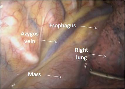
This endoscopic clinical photograph displays a intraoperative thoracoscopic view (VATS) of the right posterior mediastinum. The image highlights critical anatomical landmarks and their spatial relationships to a localized pathology. Centrally, the azygos vein is visible as a prominent bluish, tubular vascular structure. Parallel and adjacent to it lies the esophagus, appearing as a paler, vertically oriented muscular tube. A pathological mass is identified in the inferior aspect of the field, situated in the space between the azygos vein and the esophagus. The mass appears as a rounded, well-circumscribed, and highly vascularized structure with a smooth surface, identified in clinical context as hyaline-vascular Castleman disease. To the right of the surgical field, the parenchyma of the right lung is visible, demonstrating typical anthracotic pigment. This image serves as an educational reference for thoracic surgery and mediastinal anatomy, illustrating the typical presentation and surgical orientation for resecting posterior mediastinal tumors while avoiding injury to the azygos vein and esophagus.
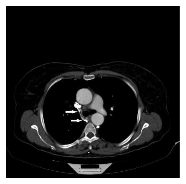
This axial thoracic CT angiography image demonstrates the anatomy of the mediastinum at the level of the aortic arch. The primary focus is the right azygos vein, marked with white arrows, as it arches anteriorly to discharge into the posterior aspect of the superior vena cava (SVC). The SVC appears slightly compressed or displaced, which context suggests is due to an aneurysmal dilatation of the adjacent ascending aorta. Other visible anatomical structures include the large diameter of the ascending aorta, the descending aorta situated posteriorly to the left, the trachea at the midline, and the esophagus. The pulmonary trunk and portions of the lungs are also visible. The image highlights the drainage pathway of the azygos system and illustrates how adjacent vascular pathologies, such as an aortic aneurysm, can exert mass effect on the low-pressure venous structures of the mediastinum. This content is valuable for students and radiologists studying thoracic vascular anatomy and the clinical presentation of aortic aneurysms.
Azygos Vein
Overview
Origin
- Right ascending lumbar vein, and
- Right subcostal vein
Course
- Enters the thorax through the aortic hiatus of the diaphragm (or posterior to / through the right crus)
- Ascends in the posterior mediastinum, to the right of the thoracic duct, close to the bodies of the inferior eight thoracic vertebrae
- At approximately vertebral level T4, it arches anteriorly over the root of the right lung
- Terminates by joining the superior vena cava just before the SVC enters the pericardial sac
- Gray's Anatomy for Students; Fischer's Mastery of Surgery
Diagram - Azygos System of Veins
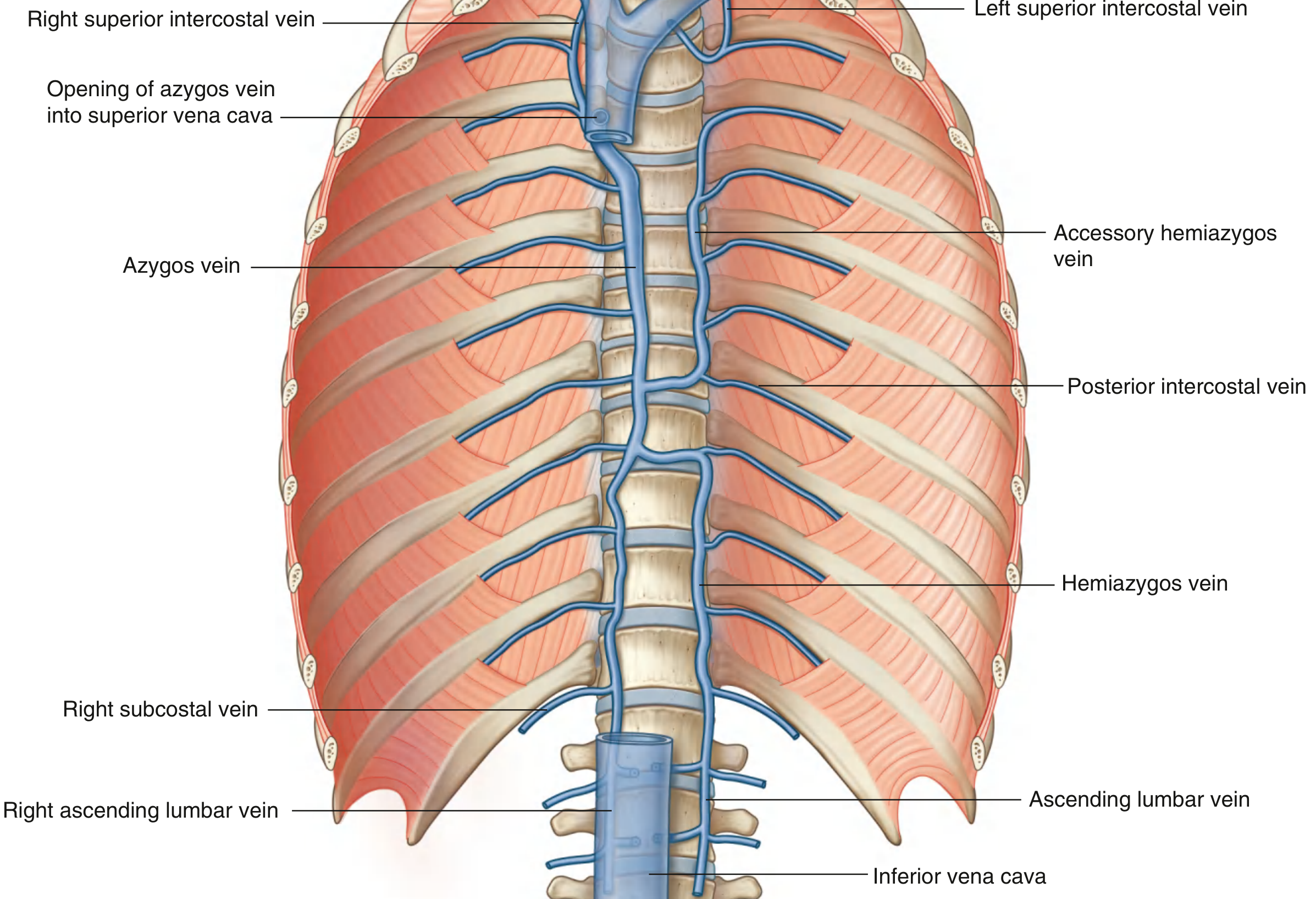
Tributaries of the Azygos Vein
| Tributary | Details |
|---|---|
| Right superior intercostal vein | Formed by 2nd, 3rd, and 4th intercostal veins (1st drains into brachiocephalic or vertebral vein) |
| 5th-11th right posterior intercostal veins | Direct segmental drainage |
| Hemiazygos vein | Joins at ~T9 after crossing midline |
| Accessory hemiazygos vein | Joins at ~T7/T8 after crossing midline |
| Esophageal veins | |
| Mediastinal veins | |
| Pericardial veins | |
| Right bronchial veins | |
| Vertebral venous plexuses | Draining the back, vertebrae, and structures in the vertebral canal |
- Gray's Anatomy for Students; Fischer's Mastery of Surgery
The Azygos System - Full Anatomical Diagram
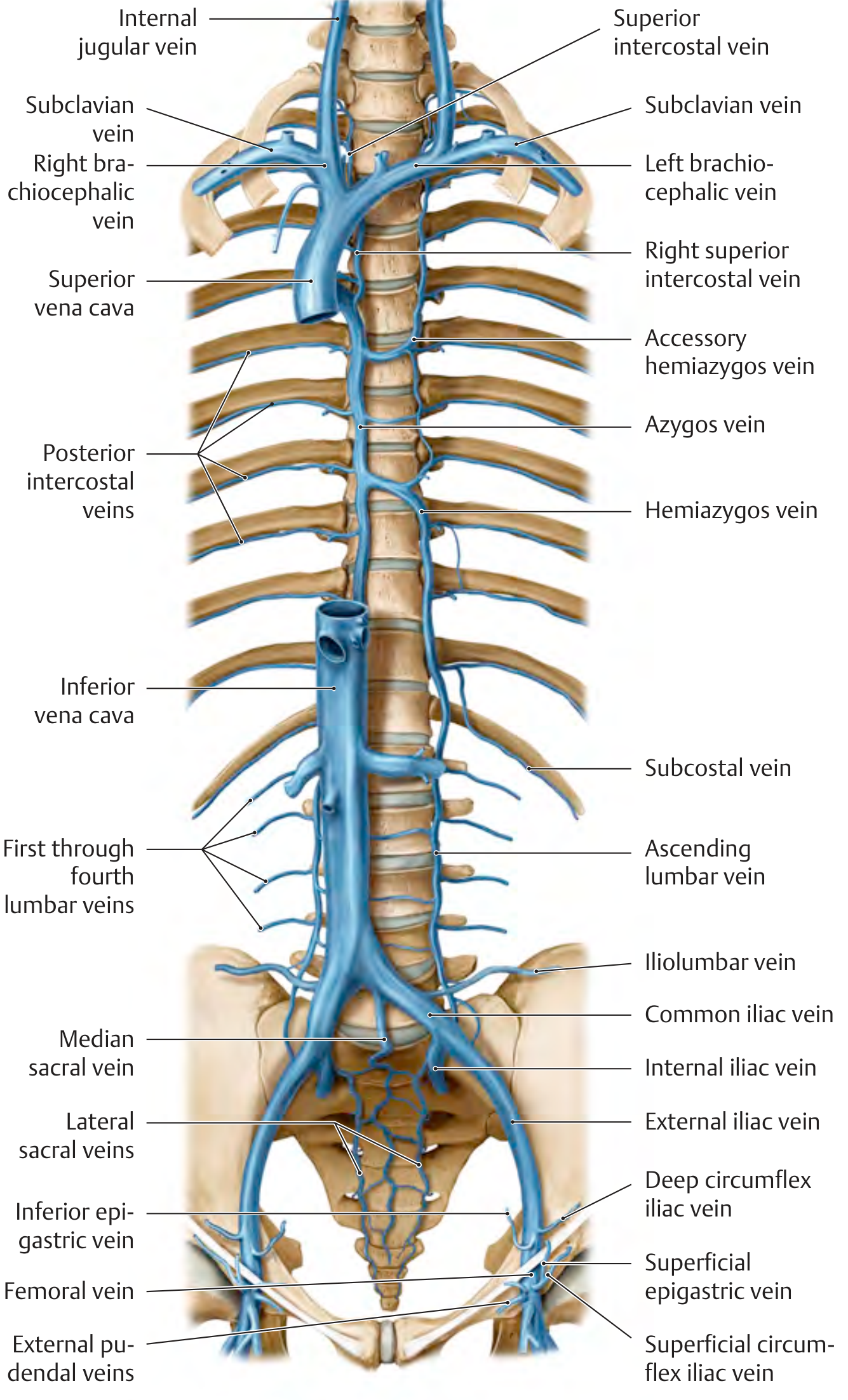
Companion Vessels
Hemiazygos Vein (Inferior Hemiazygos)
- Arises from the left ascending lumbar vein and left subcostal vein; may connect to the left renal vein
- Enters thorax through the left crus of the diaphragm
- Ascends on the left side of the posterior mediastinum to ~T9, then crosses the midline (posterior to thoracic aorta, esophagus, and thoracic duct) to drain into the azygos vein
- Tributaries: lowest 4-5 left posterior intercostal veins, esophageal and mediastinal veins
Accessory Hemiazygos Vein (Superior Hemiazygos)
-
Begins at the medial end of the 4th or 5th intercostal space
-
Descends on the left side from T5 to T8, then crosses to drain into the azygos vein at T7/T8
-
Tributaries: left 4th-8th posterior intercostal veins, sometimes the left bronchial vein
-
May join the hemiazygos vein before entering the azygos
-
Fischer's Mastery of Surgery
Clinical Significance
1. SVC-IVC Collateral Pathway
2. Portal Hypertension / Portosystemic Anastomosis
3. SVC Obstruction
4. Azygos Continuation of IVC
5. Lobe of the Azygos Vein (Azygos Lobe)
6. Thoracic Duct Relationship
Intraoperative View
Summary Table
| Feature | Detail |
|---|---|
| Origin | Junction of right ascending lumbar + right subcostal veins (L1-L2) |
| Entry into thorax | Aortic hiatus or right crus of diaphragm |
| Side | Right side of posterior mediastinum |
| Termination | SVC at ~T4, after arching over right lung root |
| Drains into | Superior vena cava |
| Receives from | Hemiazygos, accessory hemiazygos, right intercostal veins, bronchial, esophageal, pericardial, mediastinal veins |
| Key collateral | SVC ↔ IVC; Portal ↔ Caval (esophageal varices) |
| Surgical landmark | Lies lateral to thoracic duct; medial to right lung root |