
from dm vasudevan a textbook of biochemistry for Medical Students 11th edition write a 5 mark short note with proper content including fowchart, diagram, tables and clinical application for biochemistry theory university exam on Metabolism of Tyrosine
tyrosine metabolism pathway catecholamine melanin thyroid hormone diagram
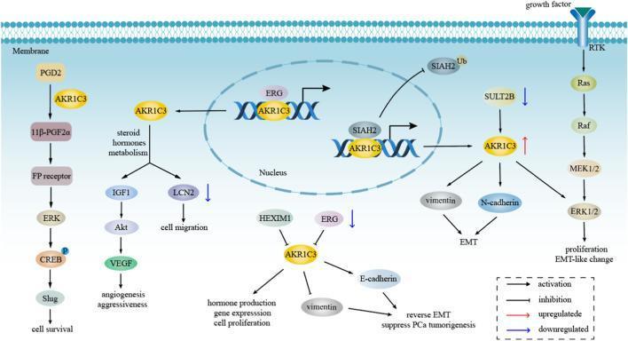
A pathophysiology diagram illustrating the intracellular signaling pathways of AKR1C3 in the context of cancer progression, specifically hormone-dependent tumors like prostate cancer. The diagram maps several concurrent pathways: 1) A growth factor-induced Receptor Tyrosine Kinase (RTK) pathway activating the Ras/Raf/MEK/ERK cascade leading to proliferation and EMT-like changes. 2) Prostaglandin metabolism where AKR1C3 converts PGD2 to 11β-PGF2α, signaling through the FP receptor/ERK/CREB/Slug axis to promote cell survival. 3) Steroid hormone metabolism where AKR1C3 influences IGF1/Akt/VEGF for angiogenesis and LCN2 for cell migration. 4) Nuclear interactions where AKR1C3 complexes with ERG and SIAH2 to modulate gene expression. 5) Cytoplasmic regulation where AKR1C3 upregulates vimentin and N-cadherin (promoting EMT) or downregulates E-cadherin to affect PCa tumorigenesis. The visual uses arrows to denote activation, T-bars for inhibition, and color-coded arrows for up/down-regulation, highlighting AKR1C3 as a central mediator of oncogenic signaling, epithelial-mesenchymal transition, and therapeutic resistance.
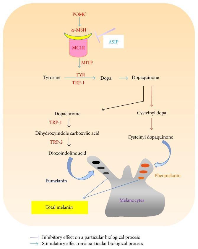
A pathophysiology diagram illustrating the molecular signaling cascade and biochemical pathways of melanogenesis in human hair follicles. The top section depicts the upstream signaling sequence: POMC stimulates ̑-MSH, which binds to the MC1R receptor on the melanocyte membrane, subsequently activating MITF. This stimulatory pathway is antagonized by ASIP, which exerts an inhibitory effect on the ̑-MSH/MC1R interaction. The central section details the enzymatic conversion of Tyrosine to Dopa and Dopaquinone, catalyzed by Tyrosinase (TYR) and Tyrosinase-Related Protein 1 (TRP-1). The pathway then bifurcates: the left branch involves the conversion of Dopaquinone to Dopachrome, Dihydroxyindole carboxylic acid, and Dioxoindoline acid (facilitated by TRP-1 and TRP-2) to produce Eumelanin (black pigment). The right branch shows the conversion of Dopaquinone to Cysteinyl dopa and Cysteinyl dopaquinone to produce Pheomelanin (orange/red pigment). Both pigment types are sequestered within melanocytes, contributing to 'Total melanin.' The diagram uses a legend to distinguish between stimulatory arrows and inhibitory flat-head connectors, representing the complex regulation of hair pigmentation.
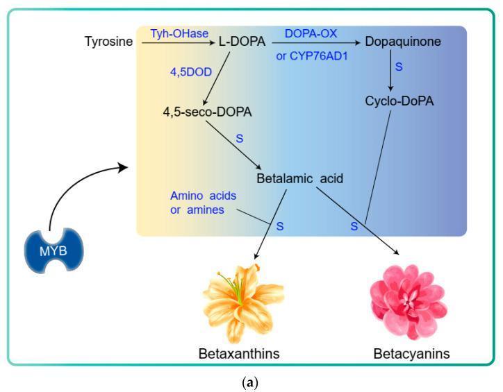
This biochemical flowchart illustrates the betalain biosynthesis pathway, a secondary metabolic process originating from the amino acid tyrosine. The diagram outlines the enzymatic and spontaneous steps leading to the formation of yellow-orange betaxanthins and red-violet betacyanins. The pathway begins with the conversion of tyrosine to L-DOPA via the enzyme Tyh-OHase (tyrosine hydroxylase). L-DOPA then branches into two routes: one mediated by 4,5DOD (DOPA-4,5-dioxygenase) to form 4,5-seco-DOPA, which spontaneously converts to betalamic acid; and another mediated by DOPA-OX (DOPA oxidase) or CYP76AD1 to form dopaquinone, which leads to cyclo-DOPA. Betalamic acid serves as a central intermediate, spontaneously conjugating with amino acids or amines to yield betaxanthins, or with cyclo-DOPA to produce betacyanins. A regulatory element, the MYB transcription factor, is shown influencing the initial tyrosine-to-L-DOPA conversion step. This illustration serves as an educational tool for understanding plant-based pigment metabolism, enzymatic regulation, and the shikimate-derived biosynthetic pathways relevant to pharmacognosy and nutritional biochemistry.
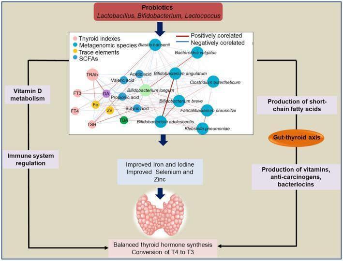
This pathophysiology diagram illustrates the 'gut-thyroid axis' and the influence of probiotics on thyroid function. At the top, probiotics including Lactobacillus, Bifidobacterium, and Lactococcus initiate a signaling cascade. A central network diagram shows correlations between metagenomic species (e.g., Bifidobacterium longum, Faecalibacterium prausnitzii), short-chain fatty acids (SCFAs like acetic and butyric acid), trace elements (Fe, Zn), and thyroid indexes (TRAb, FT3, FT4, TSH). Blue lines indicate negative correlations, while red lines indicate positive correlations. The pathway branches into two main regulatory loops: the left side focuses on Vitamin D metabolism and immune system regulation, while the right side emphasizes SCFA production and the secretion of vitamins, anti-carcinogens, and bacteriocins. These processes converge to improve the bioavailability of essential trace elements (iron, iodine, selenium, and zinc), ultimately leading to balanced thyroid hormone synthesis and the peripheral conversion of T4 to T3. The diagram serves as an educational tool for understanding how gut microbiota modulation supports endocrine homeostasis.
Metabolism of Tyrosine
Introduction
Sources of Tyrosine
- Dietary intake (proteins containing tyrosine)
- Hydroxylation of phenylalanine by phenylalanine hydroxylase (PAH), requiring tetrahydrobiopterin (BH4) as cofactor
FLOWCHART: Metabolic Fates of Tyrosine
                        PHENYLALANINE
                             │  phenylalanine hydroxylase (BH4)
                             ▼
                          TYROSINE  ←─── Diet
              ┌────────────┬──────────────┬───────────────┐
              │            │              │               │
              ▼            ▼              ▼               ▼
       CATABOLISM   CATECHOLAMINES    MELANIN       THYROID
       (degradation)  (dopamine,    (melanocytes)   HORMONES
                    epinephrine,                   (T3, T4)
                   norepinephrine)
Pathway 1: Catabolism / Degradation of Tyrosine
TYROSINE
   │  tyrosine aminotransferase (PLP)        ← Tyrosinemia Type II if blocked
   ▼
p-Hydroxyphenylpyruvate
   │  p-hydroxyphenylpyruvate dioxygenase (Vit C required)
   ▼
Homogentisate (Homogentisic acid)
   │  homogentisate oxidase                  ← Alkaptonuria if blocked
   ▼
Maleylacetoacetate
   │  maleylacetoacetate isomerase
   ▼
Fumarylacetoacetate
   │  fumarylacetoacetate hydrolase          ← Tyrosinemia Type I if blocked
   ▼
FUMARATE + ACETOACETATE
(enters TCA)   (ketone body → Acetyl-CoA)
Pathway 2: Synthesis of Catecholamines
TYROSINE
   │  tyrosine hydroxylase (rate-limiting; BH4)
   ▼
DOPA (3,4-dihydroxyphenylalanine)
   │  DOPA decarboxylase (PLP, Vit B6)
   ▼
DOPAMINE
   │  dopamine β-hydroxylase (Vit C)
   ▼
NOREPINEPHRINE (Noradrenaline)
   │  phenylethanolamine-N-methyltransferase (PNMT; SAM as methyl donor)
   ▼
EPINEPHRINE (Adrenaline)
Pathway 3: Melanin Synthesis
TYROSINE
   │  tyrosinase (copper-containing enzyme; in melanosomes)
   ▼
DOPA
   │  tyrosinase
   ▼
DOPAQUINONE
   │  spontaneous cyclization
   ▼
DOPACHROME → EUMELANIN (brown/black pigment)
                 or
   │  + cysteine
   ▼
CYSTEINYL DOPA → PHEOMELANIN (yellow/red pigment)
Pathway 4: Thyroid Hormone Synthesis
TYROSINE (in thyroglobulin) + Iodine (I⁻ oxidized by peroxidase)
   ▼
Monoiodotyrosine (MIT)  →  Diiodotyrosine (DIT)
MIT + DIT  →  T3 (Triiodothyronine)
DIT + DIT  →  T4 (Thyroxine)
DIAGRAM: Degradative Pathway with Disease Blocks
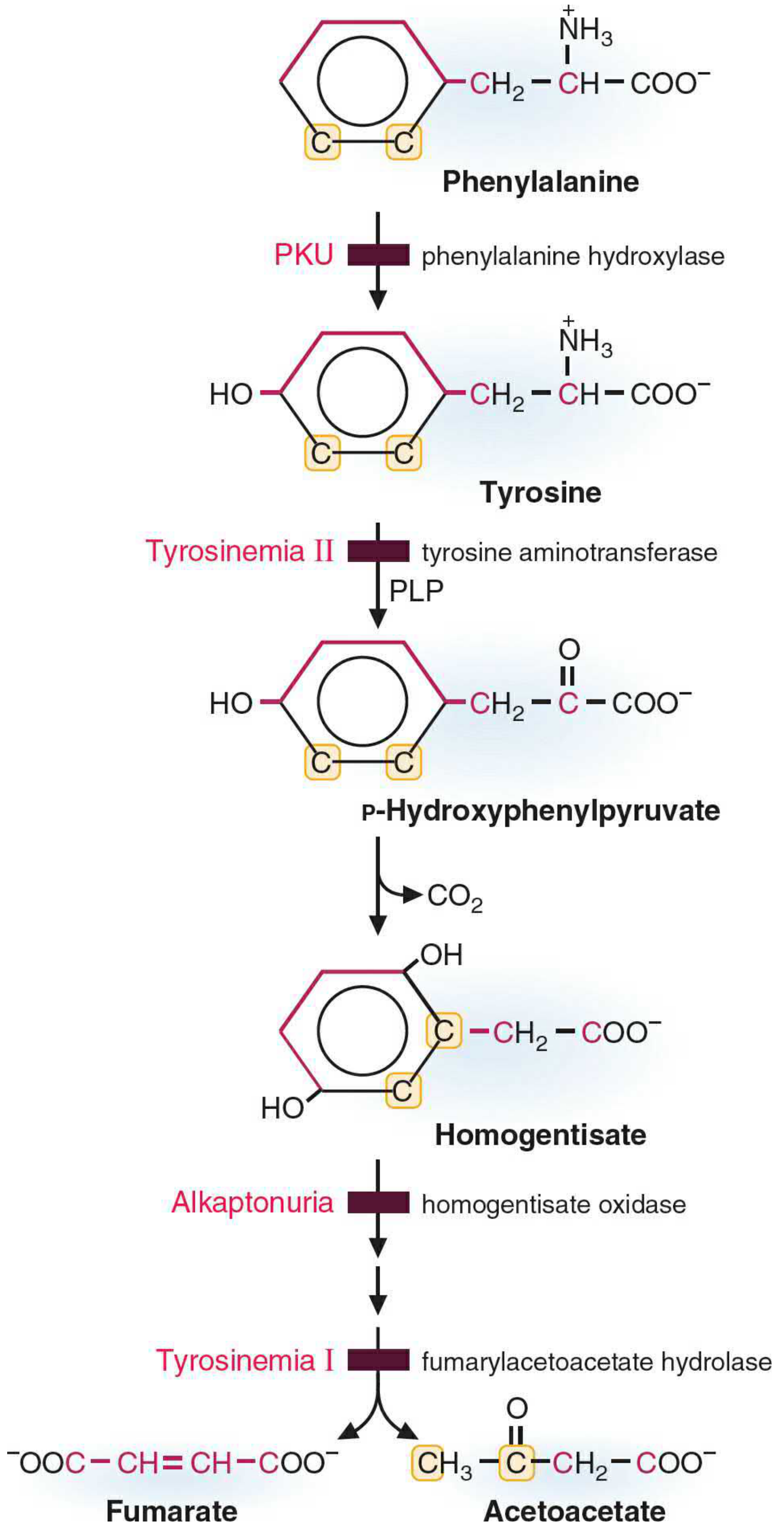
TABLE: Inborn Errors of Tyrosine Metabolism (Clinical Applications)
| Disease | Deficient Enzyme | Accumulated Metabolite | Clinical Features |
|---|---|---|---|
| PKU (Phenylketonuria) | Phenylalanine hydroxylase (PAH) | Phenylalanine | Intellectual disability, mousy odor, fair skin/hair |
| Tyrosinemia Type I (Tyrosinosis) | Fumarylacetoacetate hydrolase | Fumarylacetoacetate | Liver failure, early death; Fanconi syndrome |
| Tyrosinemia Type II (Richner-Hanhart syndrome) | Tyrosine aminotransferase | Tyrosine | Keratosis of palms/soles, corneal ulcers, neurological defects |
| Alkaptonuria | Homogentisate oxidase | Homogentisic acid | Black urine on standing, ochronosis (blue-black pigmentation of cartilage), arthritis |
| Albinism | Tyrosinase | No melanin produced | Lack of skin/hair/eye pigmentation; photosensitivity, nystagmus |
Melanin Synthesis - Additional Detail (Melanogenesis)
- Tyrosinase is the key enzyme - it is a copper-containing enzyme located in melanosomes of melanocytes
- Deficiency of tyrosinase → Albinism (oculocutaneous type)
- MSH (melanocyte-stimulating hormone) stimulates tyrosinase activity and melanin production
TABLE: Physiological Products of Tyrosine Metabolism
| Product | Pathway | Physiological Importance |
|---|---|---|
| Fumarate + Acetoacetate | Oxidative degradation | Energy production (TCA cycle) |
| Dopamine | Catecholamine synthesis | Neurotransmitter; Parkinson's disease when deficient |
| Norepinephrine | Catecholamine synthesis | Neurotransmitter, vasoconstriction |
| Epinephrine | Catecholamine synthesis | Stress response, glycogenolysis |
| Melanin | Melanogenesis | Skin/hair/eye pigmentation, UV protection |
| T3, T4 | Thyroid hormone synthesis | Metabolic rate regulation |
Key Enzymes and Cofactors Summary
| Enzyme | Reaction | Cofactor |
|---|---|---|
| Phenylalanine hydroxylase | Phe → Tyr | BH4 (tetrahydrobiopterin) |
| Tyrosine aminotransferase | Tyr → p-hydroxyphenylpyruvate | PLP (pyridoxal phosphate) |
| p-HPPD | p-HPP → Homogentisate | Vitamin C (ascorbate) |
| Tyrosine hydroxylase | Tyr → DOPA | BH4 (rate-limiting for catecholamine synthesis) |
| DOPA decarboxylase | DOPA → Dopamine | PLP (Vitamin B6) |
| Dopamine β-hydroxylase | Dopamine → Norepinephrine | Vitamin C |
| PNMT | NE → Epinephrine | SAM (S-adenosylmethionine) |
| Tyrosinase | Tyr → DOPA → Dopaquinone | Copper (Cu²⁺) |
Clinical Applications
-
Alkaptonuria - First described by Sir Archibald Garrod (early 20th century) as one of the original "inborn errors of metabolism." Urine turns dark/black on standing (oxidation of homogentisic acid). In late stages, homogentisic acid polymerizes to dark pigment that deposits in cartilage and connective tissue (ochronosis), causing disabling arthritis. Earliest known case detected in a 1500 BC Egyptian mummy.
-
Tyrosinemia Type I - Most severe form; presents with acute liver failure in infancy and is potentially fatal without treatment. Management: diet low in phenylalanine and tyrosine; NTBC (nitisinone) drug therapy.
-
Tyrosinemia Type II (Richner-Hanhart syndrome) - Presents with palmoplantar keratosis and painful corneal erosions. Managed with low-protein diet.
-
Albinism - Tyrosinase deficiency prevents melanin synthesis. Patients have photosensitivity, visual problems (nystagmus, reduced acuity), and increased risk of skin cancer.
-
Parkinson's Disease - Deficiency of dopamine in the substantia nigra (due to dopaminergic neuron degeneration). Treated with L-DOPA (levodopa), which crosses the blood-brain barrier and is converted to dopamine by DOPA decarboxylase.
-
Pheochromocytoma - Tumor of adrenal medulla causing excess epinephrine/norepinephrine secretion. Urinary VMA (vanillylmandelic acid) - catecholamine metabolite - is elevated and used as a diagnostic marker.
-
Hypothyroidism - Defective iodination of tyrosine residues in thyroglobulin leads to reduced T3/T4 synthesis.
-
Scurvy and Tyrosine Metabolism - Vitamin C is required by p-hydroxyphenylpyruvate dioxygenase; deficiency causes accumulation of p-hydroxyphenylpyruvate, contributing to tyrosinemia-like urinary excretion of tyrosine metabolites.
Memory Aid
"The DOCTOR Makes Every Patient Nicer"
- Dopamine
- Ochre (ochronosis - alkaptonuria)
- Catecholamines
- Thyroid hormones (T3/T4)
- Oxidative degradation → Fumarate + Acetoacetate
- Richner-Hanhart (Tyrosinemia II)
- Melanin
- Epinephrine
- PKU / Pheochromocytoma
- Norepinephrine