
Garm staining
Reading File
Gram staining is a foundational microbiology technique used to classify bacteria into two major groups based on their cell wall structure. Let me search the library for detailed content.
Searching sources...
Finding Sources
Finding Sources
Reading File
Reading File
Reading File
Loading Image
Reading File
Here is a thorough overview of Gram staining based on authoritative medical microbiology textbooks:
Gram Staining
History
The Gram stain was described in 1884 by the Danish physician Hans Christian Gram. It remains one of the most widely used and clinically useful procedures in microbiology and medicine. - Sherris & Ryan's Medical Microbiology, 8th Ed.
Principle
The Gram stain is a differential staining technique that separates bacteria into two fundamental groups - Gram-positive and Gram-negative - based on differences in their cell wall structure.
The key chemical event is the formation of a crystal violet-iodine complex inside the bacterial cell. The outcome then depends on how the cell wall responds to a decolorizing agent (acetone/alcohol):
- Gram-positive bacteria: have a thick peptidoglycan cell wall that retains the dye complex after decolorization -> appear purple
- Gram-negative bacteria: have a thin peptidoglycan layer covered by an outer lipid membrane; the decolorizer extracts the dye complex -> appear pink/red after counterstaining
The differential Gram reaction is fundamentally a property of cell wall structure and is correlated with many other morphologic and phylogenetic properties. - Jawetz, Melnick & Adelberg's Medical Microbiology, 28th Ed.
Steps of the Procedure
(From Pye's Surgical Handicraft & Henry's Clinical Diagnosis and Management)
| Step | Action | Effect |
|---|---|---|
| 1 | Prepare smear, air-dry, heat-fix (or methanol-fix) | Adherence to slide |
| 2 | Apply crystal violet (primary stain) | All bacteria stain purple |
| 3 | Apply Gram's iodine (mordant, ~10 seconds) | Forms insoluble crystal violet-iodine complex inside cells |
| 4 | Decolorize with acetone-alcohol (2-5 seconds) | Gram-negative cells lose the complex; Gram-positive retain it |
| 5 | Wash immediately with water | Stops decolorization |
| 6 | Apply safranin (or dilute carbol fuchsin) counterstain (10-20 s) | Gram-negative cells take up red/pink color |
| 7 | Wash, dry, examine under oil immersion (1000x) | Final interpretation |
Visual Overview
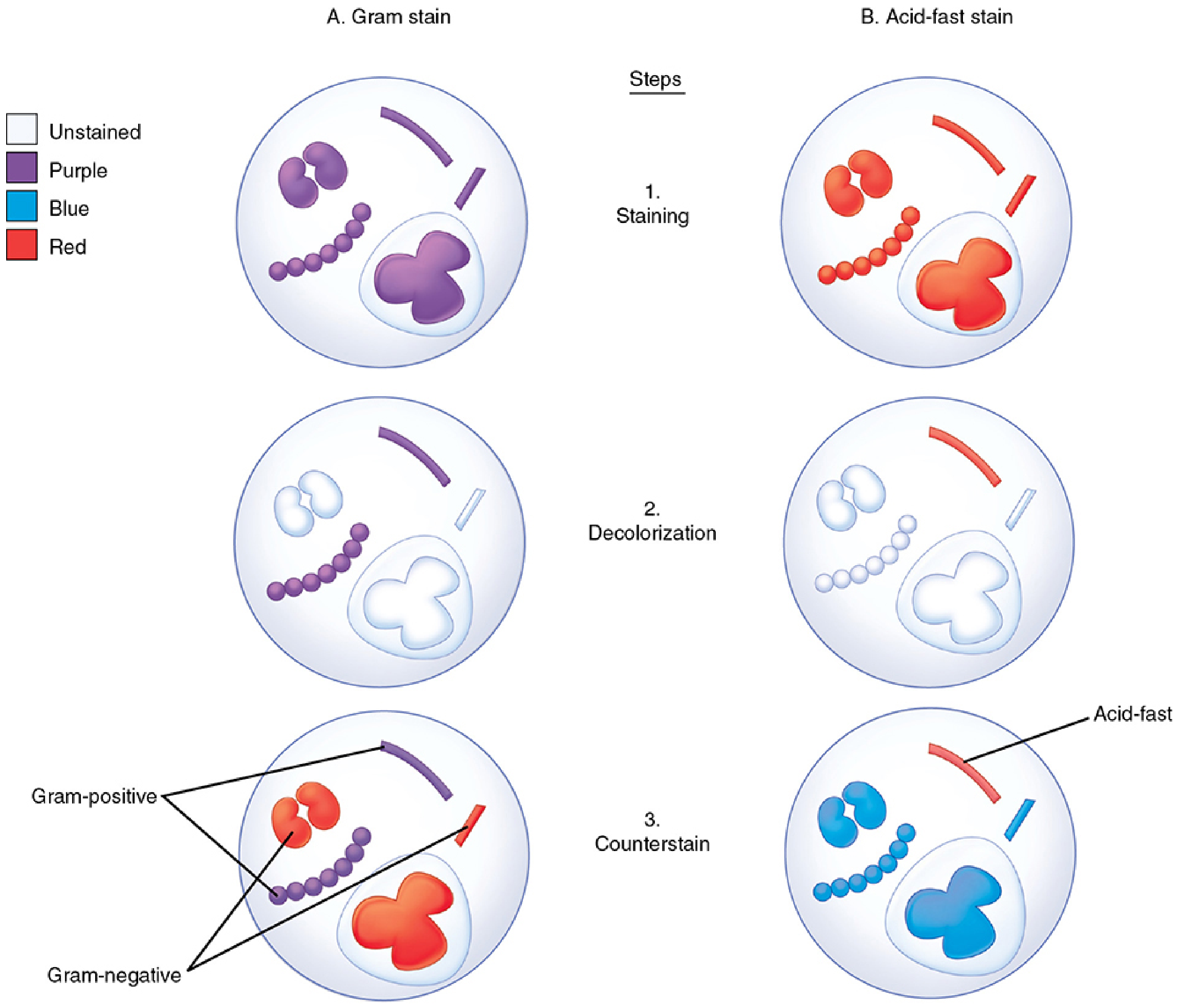
Gram stain (A) and Acid-fast stain (B) compared step by step. After crystal violet staining (Step 1), all bacteria are purple. After decolorization (Step 2), Gram-positive cells remain purple; Gram-negative lose color. After safranin counterstain (Step 3), Gram-negatives stain red/orange while Gram-positives retain purple. - Sherris & Ryan's Medical Microbiology
Reagents Summary
| Reagent | Role | Color Imparted |
|---|---|---|
| Crystal violet | Primary stain | Purple (all bacteria) |
| Gram's iodine | Mordant (fixes dye) | - |
| Acetone-alcohol | Decolorizer | - |
| Safranin (or carbol fuchsin) | Counterstain | Pink/red |
Interpretation
- Gram-positive (+) = Purple/violet (thick peptidoglycan wall retains the complex)
- Gram-negative (-) = Pink/red (outer lipid membrane is dissolved by decolorizer, releasing the complex)
When examining the stained smear, organisms are evaluated for:
- Size
- Shape (cocci, bacilli, coccobacilli, filaments)
- Arrangement (clusters, chains, pairs, single)
- Gram reaction
- Intracellular vs. extracellular location
Reporting should be as descriptive as possible - for example, "Gram-positive cocci in pairs" is more clinically useful than just "Gram-positive cocci." - Henry's Clinical Diagnosis and Management by Laboratory Methods
Specimen Concentration Requirement
At least 10^5 organisms/mL must be present to see one organism per oil immersion field (1000x). Sterile body fluids such as CSF should be concentrated 10-100x by centrifugation before smear preparation. - Henry's Clinical Diagnosis
Limitations and Causes of False Results
- Old, dead, or antimicrobial-damaged Gram-positive bacteria may fail to retain the stain and appear falsely Gram-negative
- Overly prolonged decolorization can falsely decolorize Gram-positive organisms
- Contaminated reagents can produce false-positive results
- Some organisms are weakly Gram-staining (e.g., Nocardia, Rhodococcus equi) and may require special stains
- Mycobacteria are poorly/not stained by Gram stain - they require the Acid-Fast stain instead
Clinical Significance
The Gram stain result provides rapid, preliminary guidance for antibiotic selection while awaiting culture results. It helps the laboratory:
- Assess specimen quality (squamous epithelial cells indicate oral contamination)
- Quantify organisms relative to white cells (more WBCs + more organisms = greater significance)
- Indicate if additional specialized media are needed (e.g., Gram-negative coccobacilli in respiratory specimen -> add chocolate agar for Haemophilus)
Common Gram-Positive and Gram-Negative Pathogens
| Gram-Positive | Gram-Negative |
|---|---|
| Staphylococcus (clusters) | Escherichia coli |
| Streptococcus (chains/pairs) | Klebsiella pneumoniae |
| Bacillus spp. | Pseudomonas aeruginosa |
| Clostridium spp. | Neisseria spp. (diplococci) |
| Enterococcus | Haemophilus influenzae |
| Listeria | Bacteroides spp. |
Sources: Jawetz, Melnick & Adelberg's Medical Microbiology 28th Ed.; Sherris & Ryan's Medical Microbiology 8th Ed.; Henry's Clinical Diagnosis and Management by Laboratory Methods; Pye's Surgical Handicraft 22nd Ed.
Procedure
The Gram staining procedure in detail is already covered above. Let me pull more granular procedural content from the textbooks.
Searching sources...
Finding Sources
Finding Sources
Finding Sources
Finding Sources
Reading File
Reading File
Here is the detailed Gram Staining Procedure from authoritative textbooks:
Gram Staining - Detailed Procedure
Reagents Required
| Reagent | Purpose |
|---|---|
| Crystal violet | Primary stain |
| Gram's iodine (iodine in potassium iodide) | Mordant |
| 30% Acetone-alcohol | Decolorizer |
| Safranin (or dilute carbol fuchsin) | Counterstain |
Step-by-Step Procedure
Step 1 - Smear Preparation
- Place a small amount of specimen (pus, sputum, CSF sediment, colony, etc.) on a clean glass slide
- Spread into a thin, even smear
- For sterile body fluids like CSF: centrifuge first to concentrate 10-100x, then smear the sediment
- Allow to air-dry completely
Step 2 - Fixation
- Fix the smear using either:
- Heat fixation: pass the slide briefly through a flame 2-3 times (underside of slide)
- Methanol fixation: flood with methanol for 1 minute (preferred - less distortion)
- Fixation kills organisms and adheres the smear to the slide
Step 3 - Primary Stain (Crystal Violet)
- Flood the smear with crystal violet solution
- Leave for 10-30 seconds
- Rinse with water - do not blot
- All bacteria stain purple at this point
Step 4 - Mordant (Gram's Iodine)
- Apply Gram's iodine solution
- Leave for 10-30 seconds
- Rinse with water - do not blot
- Iodine cross-links crystal violet to peptidoglycan in the cell wall
- All bacteria remain purple
Step 5 - Decolorization (Critical Step)
- Apply 30% acetone-alcohol with gentle agitation
- Duration: 10-30 seconds - until stain no longer flows off the slide
- Immediately rinse with water to stop decolorization
- This is the most critical step - over-decolorization gives false Gram-negatives; under-decolorization gives false Gram-positives
- After this step:
- Gram-positive cells: remain purple
- Gram-negative cells: colorless (decolorized)
Step 6 - Counterstain (Safranin)
- Flood with safranin solution
- Leave for 10-30 seconds
- Rinse with water, air-dry or blot dry gently
Step 7 - Microscopic Examination
- Examine under oil immersion (1000x)
- First scan under low power to find areas of interest, then switch to oil immersion
- Evaluate organisms for: size, shape, arrangement, and Gram reaction
(Source: Jawetz, Melnick & Adelberg's Medical Microbiology, 28th Ed., Table 47-1; Tietz Textbook of Laboratory Medicine, 7th Ed., Table 84.1)
Summary Flowchart
Smear → Air-dry → Fix
         ↓
    Crystal violet (10-30 s) → All bacteria PURPLE
         ↓
    Gram's iodine (10-30 s) → CV-iodine complex forms
         ↓
    Acetone-alcohol (10-30 s) → Gram-negatives DECOLORIZED
         ↓
    Safranin (10-30 s) → Gram-negatives turn PINK/RED
         ↓
    Examine under oil immersion (1000x)
Final Results
| Result | Appearance | Basis |
|---|---|---|
| Gram-positive | Purple/violet | Thick peptidoglycan wall retains CV-iodine complex |
| Gram-negative | Pink/red | Thin peptidoglycan + outer lipid membrane; complex washed out |
Key Tips & Common Errors
| Error | Consequence |
|---|---|
| Over-decolorization | Gram-positive appears falsely Gram-negative |
| Under-decolorization | Gram-negative appears falsely Gram-positive |
| Old/dead organisms | Gram-positives may lose staining ability |
| Antibiotic-damaged organisms | Cell wall disrupted; stain unreliable |
| Too thick smear | Difficult to decolorize properly |
| Insufficient heat fixation | Cells wash off slide |
What to Report
When examining, report:
- Gram reaction (positive or negative)
- Morphology (cocci, bacilli, coccobacilli, filaments)
- Arrangement (clusters, chains, pairs, singly)
- Relative quantity (rare, few, moderate, many)
- Relationship to WBCs (intracellular or extracellular)
- WBC and squamous epithelial cell counts (to assess specimen quality)
Example: "Gram-positive cocci in pairs and short chains, with many neutrophils" is more useful than just "Gram-positive cocci."
Sources: Jawetz, Melnick & Adelberg's Medical Microbiology 28th Ed.; Tietz Textbook of Laboratory Medicine 7th Ed.; Henry's Clinical Diagnosis and Management by Laboratory Methods; Pye's Surgical Handicraft 22nd Ed.
This is a shared conversation. Sign in to Orris to start your own chat.