
Discuss the role of Opioid free anaesthesia in present day practice of anaesthesia. List its potential benefits and limitations in perioperative pain management. (Jan 2025) Recent Advances in anesthesia for intrauterine fetal Surgery. (Jan 2025) Use of ultrasound for airway assessment and management. (Jan 2025) What is hospital acquired infections? Describe the causes, prevention and management of ventilator associated pneumonia. (Sept 2025) Give answer for this question from standard anaesthesia text book on exam pov of view and how to write
HOW TO WRITE ANAESTHESIA EXAM ANSWERS - GENERAL TIPS
- 10-mark question: Introduction (2-3 lines) + Body with headings (7-8 marks) + Conclusion (1-2 lines)
- Always use headings, subheadings, and bullet points - examiners scan, not read
- Start every answer with a definition or brief introduction
- Use tables where comparison or classification is asked
- End with a brief conclusion showing clinical perspective
- For "discuss" questions, cover: definition, mechanism, clinical use, benefits, limitations, complications
QUESTION 1: Opioid Free Anaesthesia (OFA) - Role, Benefits and Limitations in Perioperative Pain Management
INTRODUCTION
BACKGROUND AND RATIONALE
- The opioid crisis - widespread prescription opioid abuse
- Increased awareness of postoperative delirium in ICU/elderly patients
- Opioid-induced hyperalgesia (OIH)
- Postoperative nausea and vomiting (PONV)
- Respiratory depression
- Immunosuppressive effects of opioids
- Opioid tolerance and dependence
PHARMACOLOGICAL COMPONENTS OF OFA
| Category | Drugs Used |
|---|---|
| Alpha-2 agonists | Dexmedetomidine, Clonidine |
| NMDA antagonists | Ketamine (S-ketamine), Magnesium sulphate |
| Local anaesthetic infusions | Systemic Lidocaine (IV infusion) |
| NSAIDs / COX-2 inhibitors | Ketorolac, Parecoxib, Celecoxib |
| Alpha-2-delta ligands | Gabapentin, Pregabalin |
| Glucocorticoids | Dexamethasone |
| Paracetamol | Regular IV/oral acetaminophen |
| Regional anaesthesia | Epidural, TAP block, PEC block, peripheral nerve blocks |
| Volatile anaesthetics | High-dose inhalational agents |
| Beta-blockers | Esmolol (to suppress surgical stress response) |
MECHANISMS OF NON-OPIOID ANALGESIA
- Dexmedetomidine: Acts on presynaptic alpha-2 receptors in locus coeruleus, reduces norepinephrine release, provides sedation, analgesia, and anxiolysis
- Ketamine: NMDA receptor antagonism prevents central sensitisation and wind-up; reduces opioid requirements by 30-50%
- IV Lidocaine: Blocks sodium channels, reduces cytokine release, anti-hyperalgesic
- Gabapentin/Pregabalin: Blocks voltage-gated calcium channels, reduces presynaptic glutamate release
- Dexamethasone: Anti-inflammatory, reduces prostaglandin synthesis, prolongs regional block duration
POTENTIAL BENEFITS OF OFA
1. Reduction of PONV
- Studies show OFA reduces postoperative nausea and vomiting, particularly in bariatric surgery, beyond triple antiemetic prophylaxis (Morgan & Mikhail 7e)
2. Avoidance of Respiratory Depression
- Particularly important in obese patients, obstructive sleep apnoea, and paediatric tonsillectomy
3. Prevention of Opioid-Induced Hyperalgesia (OIH)
- High-dose intraoperative opioids activate anti-nociceptive pathways paradoxically, worsening postoperative pain
4. Reduced Postoperative Delirium
- Relevant in elderly ICU patients where opioids are a recognised modifiable risk factor
5. Better Surgical Conditions
- Reduced ileus, earlier return of bowel function (especially important in colorectal/bariatric surgery)
- Faster extubation and discharge (key in ERAS protocols)
6. Reduced Risk of Opioid Dependence
- Especially relevant in opioid-naive patients and those with substance use disorders
7. Improved Immune Function
- Opioids suppress NK cell activity; avoiding them may preserve perioperative immune function
8. Benefit in Chronic Pain Patients
- Avoids exacerbating existing opioid tolerance
LIMITATIONS OF OFA
1. Haemodynamic Instability
- Large proof-of-concept studies have been terminated prematurely due to high incidences of hypoxia and bradycardia
- Some studies showed increased demand for vasopressors, hypotension, and bradycardia (Miller's Anesthesia 10e)
- Dexmedetomidine causes bradycardia and hypotension
2. Postoperative Sedation and Falls
- Residual effects of dexmedetomidine can cause sedation extending into the postoperative period
3. Inadequate Analgesia for Major Surgery
- For highly nociceptive procedures (thoracotomy, open abdominal surgery), complete opioid avoidance may be insufficient
4. Dependence on Regional Anaesthesia
- OFA works best when combined with regional techniques, which carry their own risks (block failure, nerve injury, haematoma)
5. Drug Interactions and Side Effects of Adjuvants
- Ketamine: dysphoria, hallucinations, raised ICP
- NSAIDs: renal impairment, GI bleeding, platelet dysfunction
- Pregabalin: dizziness, sedation
6. Evidence Gaps
- Most evidence comes from specific populations (bariatric, gynaecology); evidence for major cardiac/thoracic surgery is limited
- No large RCTs conclusively demonstrating superiority over opioid-sparing analgesia
7. Not a Universal Solution
- OFA is not a panacea; opioid-sparing analgesia (minimising rather than completely eliminating opioids) may be more practical and safer in most patients (Miller's Anesthesia 10e, Chapter 47)
OFA vs OPIOID-SPARING ANALGESIA
| Feature | OFA | Opioid-Sparing |
|---|---|---|
| Opioid dose | Zero | Reduced |
| Evidence base | Limited | Strong |
| PONV benefit | Maximum | Significant |
| Haemodynamic stability | Compromised | Better |
| Applicability | Selected cases | Universal |
| ERAS compatibility | High | High |
CURRENT STANDING AND CONCLUSION
QUESTION 2: Recent Advances in Anaesthesia for Intrauterine Fetal Surgery
INTRODUCTION
CLASSIFICATION OF FETAL SURGICAL PROCEDURES
1. Minimally Invasive Procedures
- FIGS-IT: Fetal Image-Guided Surgery for Intervention or Therapy - ultrasound-guided percutaneous needle-based interventions
- Fetoscopic surgery: Small endoscopic instruments inserted percutaneously under direct camera + real-time ultrasound guidance
- Reduced risk of preterm labour compared to open procedures
2. Open Fetal Surgery
- Requires maternal laparotomy and hysterotomy
- Highest level of anaesthetic requirement
- Associated with significant maternal morbidity
3. Intrapartum Procedures (EXIT Procedures)
- Ex-Utero Intrapartum Treatment
- Uteroplacental circulation maintained while fetal airway is secured or lesion corrected at delivery
CONDITIONS AMENABLE TO FETAL SURGERY
| Condition | Procedure | Evidence |
|---|---|---|
| Twin-to-Twin Transfusion Syndrome (TTTS) | Fetoscopic laser photocoagulation of placental vessels | Level I RCT evidence |
| Myelomeningocele (spina bifida) | Open or fetoscopic neural tube repair | MOMS Trial: improved neurological outcomes |
| Congenital Diaphragmatic Hernia (CDH) | Fetal Tracheal Balloon Occlusion (FETO) | TOTAL trial |
| Lower Urinary Tract Obstruction (LUTO) | Vesico-amniotic shunt placement | PLUTO trial |
| Sacrococcygeal Teratoma (SCT) | Debulking for high-output cardiac failure | Case series |
| Congenital Pulmonary Airway Malformations (CPAM) | Thoraco-amniotic shunt | Selected cases |
| Obstructive uropathy | Vesico-amniotic shunt | Selected cases |
ANAESTHETIC CONSIDERATIONS
A. Maternal Anaesthesia
- General anaesthesia is required
- High-dose volatile agents (desflurane/sevoflurane 1.5-2 MAC) provide:
- Adequate maternal anaesthesia
- Uterine relaxation (tocolysis)
- Transplacental fetal anaesthesia
- IV supplementation with desflurane reduces fetal cardiac dysfunction
- Supplemental IV agents: propofol, remifentanil
- Neuraxial anaesthesia (epidural) for postoperative pain management
- Local infiltration with sedation (IV fentanyl/midazolam), OR
- Neuraxial anaesthesia (spinal/epidural) preferred
- Avoids need for uterine relaxation agents in most cases
B. Fetal Anaesthesia and Analgesia
- Nociceptive pathways and cortical connections are present by 24-28 weeks
- Fetal stress responses (hormonal and haemodynamic) to noxious stimuli are well-documented
- Direct fetal opioid analgesia has been shown to attenuate fetal hormonal and haemodynamic stress responses (Sabiston Surgery 14e)
- Transplacental (via maternal administration - volatile agents, opioids)
- Intramuscular injection into the fetus (for fetoscopic procedures) - typically fentanyl + atropine + vecuronium
- Intra-umbilical vein injection
- Fentanyl 10-20 mcg/kg IM
- Atropine 20 mcg/kg IM (prevent vagally mediated bradycardia)
- Vecuronium/pancuronium (immobilisation)
C. Uterine Relaxation (Tocolysis)
- Volatile anaesthetics at high doses (primary method for open surgery)
- IV nitroglycerin (rapid but short-acting)
- Magnesium sulphate (perioperative tocolysis)
- Nifedipine / Indomethacin (postoperative tocolysis)
D. Fetal Monitoring
- Continuous fetal echocardiography/fetal heart rate monitoring intraoperatively
- Umbilical artery Doppler for flow abnormalities
- Pulse oximetry (limited, on exteriorised fetal limb in open surgery)
- Fetal scalp electrode (when applicable)
RECENT ADVANCES
1. Fetoscopic MMC Repair
- Minimally invasive fetoscopic repair of myelomeningocele now performed via 3-port technique
- Reduces maternal morbidity compared to open surgery
- Requires combined spinal-epidural (CSE) with sedation
2. FETO for CDH (TOTAL Trial)
- Fetoscopic balloon tracheal occlusion under local/neuraxial anaesthesia
- Balloon placed between 27-30 weeks, removed at 34 weeks or before delivery
- Improves survival in severe isolated left CDH (liver-up, o/e LHR < 25%)
3. EXIT Procedure Refinements
- Airway secured on placental bypass before clamping cord
- Anaesthesia: high-dose volatile + epidural; uterus kept relaxed
- Used for giant neck masses, thoracic lesions, CPAM, laryngeal atresia
4. Fetal Cardiac Interventions
- Aortic/pulmonary valvuloplasty by percutaneous needle under maternal sedation + local anaesthesia + direct fetal IM injection
- Success rates of 60-70% for technical procedure
5. Neurodevelopmental Safety Concerns
- FDA 2016 warning: anaesthetics may affect developing fetal brain (< 36 months)
- Current evidence from human RCTs (GAS trial, PANDA trial) suggests single, short exposure poses minimal risk
- Ongoing research into minimum effective dose of volatile anaesthetics
6. Three-Level Fetal Therapy Centres
- Level I: low-risk interventions, no maternal ICU needed
- Level II: preterm/maternal ICU capable
- Level III: all open procedures, all complications manageable
COMPLICATIONS AND MANAGEMENT
| Complication | Management |
|---|---|
| Fetal bradycardia | Atropine IM/IV into fetus; adjust volatile concentration; IV ephedrine |
| Preterm premature rupture of membranes (PPROM) | Most common complication; conservative management or delivery |
| Preterm labour | Tocolysis (magnesium, nifedipine) |
| Maternal haemorrhage | Blood products, uterotonics; avoid in open procedures until fetus delivered |
| Mirror syndrome | Delivery; resembles severe preeclampsia |
| Umbilical cord accidents | Emergency delivery or endoscopic cord manipulation |
CONCLUSION
QUESTION 3: Use of Ultrasound for Airway Assessment and Management
INTRODUCTION
ULTRASOUND ANATOMY OF THE AIRWAY
- Thyroid cartilage - echogenic, inverted V-shaped, anterior
- Cricothyroid membrane (CTM) - hypoechoic gap between thyroid and cricoid cartilages
- Cricoid cartilage - echogenic ring inferior to CTM
- Tracheal rings - hyperechoic anterior walls with posterior acoustic shadowing (the "snowstorm" artifact from air)
- Trachea - anechoic lumen posterior to rings
- Thyroid gland - bilateral, echogenic, flanks trachea
- Oesophagus - posterior and lateral to trachea (best seen on left side)
- Vocal cords - seen on coronal view through thyroid cartilage
PROBE SELECTION AND TECHNIQUE
| View | Probe | Frequency | Best For |
|---|---|---|---|
| Transverse | Linear | 7.5-15 MHz | Trachea, CTM, ETT position |
| Longitudinal | Linear | 7.5-15 MHz | Tracheal rings counting |
| Coronal (through thyroid) | Linear | 7.5-15 MHz | Vocal cord movement |
| Subglottic | Linear | 7.5-15 MHz | ETT size selection |
APPLICATIONS IN AIRWAY ASSESSMENT
1. Prediction of Difficult Airway
- Pre-tracheal soft tissue thickness: distance from skin to anterior tracheal wall > 28 mm in obese patients predicts difficult laryngoscopy
- Tongue base thickness: hyoid-to-mental distance on submandibular scan correlates with Mallampati
- Sublingual space: hyoid-to-floor-of-mouth distance; reduced in obese patients
- Neck circumference: identified on US correlates with difficult mask ventilation
- Anterior neck fat: quantifiable, relevant in morbidly obese patients
2. Identification of the Cricothyroid Membrane (CTM)
- Critical application: palpation-based CTM identification fails in up to 65% of cases, particularly in obese/female patients
- Ultrasound reliably identifies CTM by visualising the hypoechoic gap between thyroid and cricoid cartilages
- USEFUL protocol (Ultrasound for the Systematic Identification of Landmarks for Emergency airway) uses systematic scanning from thyroid to cricoid
- Pre-marking the CTM under ultrasound before anaesthesia induction is recommended in anticipated difficult airways (Miller's Anesthesia 10e)
3. Subglottic Diameter and ETT Size Selection
- Transverse view of trachea at subglottic level measures the air-mucosa interface
- ETT outer diameter should not exceed 80% of tracheal diameter
- Particularly useful in paediatric patients where cuffed vs uncuffed tube selection affects mucosal injury
- Reduces post-extubation stridor in children
APPLICATIONS IN AIRWAY MANAGEMENT
4. Confirmation of Endotracheal Tube Placement
- Transverse view of neck shows single "double-tract sign" in trachea = correct placement
- Movement of ETT tip visible in real-time as it passes through tracheal rings (Morgan & Mikhail 7e, Figure 19-7)
- Two "double-tract signs" seen - one in trachea, one in oesophagus (posterior-lateral)
- Sensitivity of ultrasound for oesophageal intubation: 97-100% in studies
- Faster than clinical auscultation in noisy environments
- Real-time sliding motion of ETT within trachea during intubation visible on US screen
- Tube passes as a hyperechoic area within the trachea
5. Confirmation of Bilateral Lung Ventilation
- Lung sliding sign: pleural sliding (shimmering) seen bilaterally confirms bilateral ventilation
- Absence of lung sliding on one side = endobronchial intubation or pneumothorax
- M-mode: seashore sign (normal) vs barcode sign (no ventilation)
- Highly sensitive (95%) for detecting endobronchial intubation
6. Diaphragm Assessment
- Paradoxical or reduced diaphragm movement can identify phrenic nerve palsy
- Helps assess adequacy of neuromuscular reversal before extubation
7. Ultrasound-Guided Percutaneous Tracheostomy
- Real-time guidance for needle insertion between tracheal rings
- Identifies pre-tracheal vessels (avoid puncture of thyroid vessels / innominate artery)
- Reduces complications of percutaneous dilatational tracheostomy
- Confirms intraluminal wire/airway cannula position (Miller's Anesthesia 10e)
8. Emergency Cricothyrotomy
- In "can't intubate, can't oxygenate" scenarios
- Pre-identification of CTM with ultrasound improves first-pass success
- Real-time needle guidance for surgical airway
9. Evaluation of Airway Pathology
- Tracheal deviation / compression from goitre, haematoma, lymphadenopathy
- Subglottic stenosis (reduced air column diameter)
- Tracheomalacia assessment
ADVANTAGES OF ULTRASOUND FOR AIRWAY
| Advantage | Clinical Relevance |
|---|---|
| Non-invasive, no radiation | Safe in pregnancy, paediatrics, repeated use |
| Real-time dynamic imaging | Continuous monitoring during procedures |
| Bedside availability | Emergency department, ICU, operating room |
| Confirmation of ETT placement | Faster than X-ray, comparable to capnography |
| Identifies CTM in obese patients | Crucial for surgical airway planning |
| Detects endobronchial intubation | Faster than clinical signs |
LIMITATIONS
- Operator dependent - requires training and regular practice
- Obesity and subcutaneous emphysema degrade image quality (air scatters ultrasound)
- Dynamic intubation is challenging to capture in real-time
- Cannot visualise larynx beyond vocal cords in non-sagittal views
- Does not replace capnography as gold standard for ETT confirmation
- Limited evidence for some applications (e.g., difficult airway prediction) compared to established clinical scoring systems
CONCLUSION
QUESTION 4: Hospital Acquired Infections (HAI) - Definition, Causes, Prevention and Management of Ventilator Associated Pneumonia (VAP)
PART A: HOSPITAL ACQUIRED INFECTIONS (HAI)
Definition
- Was not present or incubating at the time of hospital admission
- Develops 48 hours or more after admission to a healthcare facility
- May manifest up to 30 days after discharge (or 1 year for implant-related infections)
Epidemiology
- HAP and VAP account for approximately 28% of all HAIs and are the most frequent cause of nosocomial infection
- Overall prevalence of nosocomial pneumonia: 0.89% of hospitalised patients
- In ICU patients, VAP increases pneumonia incidence 20-fold
- Approximately 10% of mechanically ventilated patients develop VAP (Fishman's Pulmonary Diseases 6e)
PART B: VENTILATOR ASSOCIATED PNEUMONIA (VAP)
Definition
- Hyperthermia (fever > 38°C) or hypothermia (< 36°C)
- Elevated white blood cell count (> 10,000/mm³)
- Purulent tracheal secretions
- Worsening oxygenation (rising FiO2 requirement or falling SpO2)
CAUSES / PATHOPHYSIOLOGY
- Pharmacologic/dietary alterations in gastric pH (stress ulcer prophylaxis with PPIs/H2 blockers)
- Exposure to contaminated respiratory equipment and hospital water systems
- Transmission of bacteria via healthcare workers (hands)
- Respiratory droplet spread of MDR organisms
- Disruption of normal oropharyngeal flora by broad-spectrum antibiotics
- Age > 60 years
- Altered consciousness, ARDS
- Chronic lung disease (COPD, bronchiectasis)
- Immunosuppression
- Prior antibiotic use within 90 days
- Nasogastric tube (promotes reflux/aspiration)
- Duration of mechanical ventilation (most important - risk increases up to day 5)
- Reintubation
- Supine positioning
- Nasotracheal (vs. orotracheal) intubation
- Indwelling vascular or urinary catheters
| Pathogen | % of Isolates | Rank |
|---|---|---|
| Staphylococcus aureus (incl. MRSA) | 28.8% | 1st |
| Pseudomonas aeruginosa | 12.9% | 2nd |
| Klebsiella species | 10.1% | 3rd |
| Enterobacter species | 8.4% | 4th |
| Haemophilus influenzae | 5.9% | 5th |
| Escherichia coli | 5.6% | 7th |
| Acinetobacter species | 3.2% | 10th |
PREVENTION OF VAP
The VAP Bundle (IHI - Institute for Healthcare Improvement)
| Prevention Strategy | Evidence Level |
|---|---|
| Semierect positioning (head of bed 30-45 degrees) | Level 1 |
| Oral decontamination with chlorhexidine | Level 1 |
| Orotracheal (not nasotracheal) intubation | Level 1 |
| Subglottic secretion drainage (suction port above cuff) | Level 1 |
| Use of polyurethane (vs PVC) endotracheal tube cuff | Level 1 |
| Silver-coated endotracheal tube | Level 1 |
| Use of non-invasive ventilation (NIV) where possible | Level 1 |
| Rotational (kinetic) beds | Level 1 |
| Closed endotracheal suctioning system | Level 1 |
| Heat-moisture exchanger (HME) | Level 1 |
| Shortening duration of mechanical ventilation (daily SAT+SBT protocols) | Level 1 |
| Sedation vacations (SAT) | Level 2 |
| Adequate ICU nurse-to-patient staffing | Level 2 |
| Avoid unnecessary reintubation | Level 2 |
| Orogastric (not nasogastric) feeding tube | Level 2 |
| Restricted blood transfusion | Level 2 |
NOT Recommended for VAP Prevention
- Routine ventilator circuit changes
- Chest physiotherapy
- Early tracheostomy
- Routine antibiotic cycling/rotation
- Routine antibiotic prophylaxis (promotes MDR organisms)
MANAGEMENT OF VAP
Step 1: Assess Risk for MDR Organisms
- Prior IV antibiotics in past 90 days
- Hospitalisation > 5 days
- Septic shock at VAP onset
- ARDS prior to VAP
- Acute renal replacement therapy
- Residence in LTAC / skilled nursing facility
Step 2: Empirical Antibiotic Selection
- One of: Piperacillin-tazobactam, Cefepime, Ceftriaxone, Levofloxacin, or Meropenem
- No MRSA coverage needed if risk is low
- Vancomycin OR Linezolid (MRSA coverage)
- PLUS Anti-pseudomonal beta-lactam: Piperacillin-tazobactam, Cefepime, or Carbapenems
- PLUS (if double gram-negative coverage needed): Aminoglycoside OR Ciprofloxacin
Step 3: Duration of Therapy
- 7-8 day course is as effective as longer courses for most VAP cases (evidence from IDSA/ATS 2016 guidelines)
- Procalcitonin-guided de-escalation reduces antibiotic duration without worsening outcomes
- Surveillance cultures and local antibiogram data should guide therapy
Step 4: De-escalation
- After 48-72 hours, review cultures
- Narrow antibiotics based on identified pathogen and sensitivities
- Reduces emergence of MDR organisms
- Reduces cost and adverse effects
Step 5: Non-Antibiotic Supportive Management
- Optimise ventilator settings (lung-protective ventilation)
- Continue VAP prevention bundle
- Prone positioning if ARDS concurrent
- Nutritional support
DIAGNOSIS OF VAP
- Non-bronchoscopic BAL (mini-BAL)
- Bronchoscopic BAL - gold standard
- Protected specimen brushing
- Endotracheal aspirate (sensitive but less specific)
- BAL: ≥ 10⁴ CFU/mL
- Protected specimen brush: ≥ 10³ CFU/mL
- Endotracheal aspirate: ≥ 10⁵-10⁶ CFU/mL
PROGNOSIS AND IMPACT
- ICU mortality: 24.8%; In-hospital mortality: 31.9%
- Attributable mortality: 1-27% (wide range due to confounding from severity of illness)
- Mean additional hospital cost: $39,828 per patient
- Attributable ICU length of stay: 4-13 days additional
- VAP rate: 14.8 cases per 1000 ventilator days (Canadian multicentre study)
CONCLUSION
EXAM WRITING TIPS SPECIFIC TO ANAESTHESIA
-
For "Role of X in present day practice" - always discuss: definition → rationale → how it's done → current evidence → limitations → conclusion. Use the word "Currently" or "In present day practice" in your intro.
-
For "Recent Advances" - list advances under clear numbered headings. Each advance should have: name of advance + what it achieves + clinical relevance. Avoid listing historical background unless it sets context for the advance.
-
For "Describe causes, prevention and management" - use a structured 3-part format. Causes under pathophysiology, prevention as a table/bundle, management as a step-by-step algorithm.
-
Tables and boxes score marks in viva/practical assessment as they show organisation of knowledge.
-
References in exam: mentioning "Miller's Anesthesia" or "IDSA 2016 guidelines" at the end of relevant points demonstrates reading beyond the basic textbook and impresses examiners.
Make a comprehensive study guide / summary DOCX on all 4 topics for quick revision
ls -la /tmp/workspace/ 2>/dev/null || echo "workspace empty or missing"
mkdir -p /tmp/workspace/anaesthesia-study-guide
~/anaesthesia-study-guide/build_guide.py
from docx import Document
from docx.shared import Pt, Inches, RGBColor
from docx.enum.text import WD_ALIGN_PARAGRAPH
from docx.oxml.ns import qn
from docx.oxml import OxmlElement
import copy

doc = Document()

# ──────────────────────────────────────────────
# PAGE SETUP: A4, margins
# ──────────────────────────────────────────────
section = doc.sections[0]
section.page_width  = Inches(8.27)
section.page_height = Inches(11.69)
section.left_margin   = Inches(1.0)
section.right_margin  = Inches(1.0)
section.top_margin    = Inches(1.0)
section.bottom_margin = Inches(1.0)

# ──────────────────────────────────────────────
# COLOUR PALETTE
# ──────────────────────────────────────────────
DARK_BLUE    = RGBColor(0x1A, 0x35, 0x5E)   # deep navy
MED_BLUE     = RGBColor(0x2E, 0x6E, 0xBD)   # heading blue
ACCENT_TEAL  = RGBColor(0x00, 0x86, 0x96)   # teal subheadings
BOX_FILL     = RGBColor(0xE8, 0xF4, 0xFD)   # light blue box bg
WARN_FILL    = RGBColor(0xFF, 0xF3, 0xCD)   # amber for limitations
KEY_FILL     = RGBColor(0xD4, 0xED, 0xDA)   # green for key points
WHITE        = RGBColor(0xFF, 0xFF, 0xFF)
DARK_GRAY    = RGBColor(0x33, 0x33, 0x33)

# ──────────────────────────────────────────────
# BASE STYLE
# ──────────────────────────────────────────────
normal_style = doc.styles["Normal"]
normal_style.font.name = "Arial"
normal_style.font.size = Pt(10.5)
normal_style.font.color.rgb = DARK_GRAY

# ──────────────────────────────────────────────
# HELPER FUNCTIONS
# ──────────────────────────────────────────────

def set_cell_bg(cell, rgb: RGBColor):
    tc = cell._tc
    tcPr = tc.get_or_add_tcPr()
    shd = OxmlElement('w:shd')
    shd.set(qn('w:val'), 'clear')
    shd.set(qn('w:color'), 'auto')
    shd.set(qn('w:fill'), f'{rgb[0]:02X}{rgb[1]:02X}{rgb[2]:02X}')
    tcPr.append(shd)

def set_cell_border(cell, **kwargs):
    """Add border to a cell. kwargs: top, bottom, left, right = 'single'"""
    tc = cell._tc
    tcPr = tc.get_or_add_tcPr()
    tcBorders = OxmlElement('w:tcBorders')
    for edge, val in kwargs.items():
        border = OxmlElement(f'w:{edge}')
        border.set(qn('w:val'), val)
        border.set(qn('w:sz'), '4')
        border.set(qn('w:space'), '0')
        border.set(qn('w:color'), '2E6EBD')
        tcBorders.append(border)
    tcPr.append(tcBorders)

def add_cover_title(doc, text):
    p = doc.add_paragraph()
    p.alignment = WD_ALIGN_PARAGRAPH.CENTER
    run = p.add_run(text)
    run.bold = True
    run.font.size = Pt(26)
    run.font.color.rgb = WHITE
    run.font.name = "Arial"
    # paragraph shading
    pPr = p._p.get_or_add_pPr()
    shd = OxmlElement('w:shd')
    shd.set(qn('w:val'), 'clear')
    shd.set(qn('w:color'), 'auto')
    shd.set(qn('w:fill'), '1A355E')
    pPr.append(shd)
    p.paragraph_format.space_before = Pt(12)
    p.paragraph_format.space_after  = Pt(12)
    return p

def add_h1(doc, text):
    """Main topic heading - dark blue banner"""
    p = doc.add_paragraph()
    p.alignment = WD_ALIGN_PARAGRAPH.LEFT
    run = p.add_run(f"  {text}")
    run.bold = True
    run.font.size = Pt(15)
    run.font.color.rgb = WHITE
    run.font.name = "Arial"
    pPr = p._p.get_or_add_pPr()
    shd = OxmlElement('w:shd')
    shd.set(qn('w:val'), 'clear')
    shd.set(qn('w:color'), 'auto')
    shd.set(qn('w:fill'), '1A355E')
    pPr.append(shd)
    p.paragraph_format.space_before = Pt(14)
    p.paragraph_format.space_after  = Pt(4)
    return p

def add_h2(doc, text):
    """Section heading - medium blue"""
    p = doc.add_paragraph()
    run = p.add_run(text)
    run.bold = True
    run.font.size = Pt(12.5)
    run.font.color.rgb = MED_BLUE
    run.font.name = "Arial"
    p.paragraph_format.space_before = Pt(10)
    p.paragraph_format.space_after  = Pt(2)
    # bottom border
    pPr = p._p.get_or_add_pPr()
    pBdr = OxmlElement('w:pBdr')
    bottom = OxmlElement('w:bottom')
    bottom.set(qn('w:val'), 'single')
    bottom.set(qn('w:sz'), '4')
    bottom.set(qn('w:space'), '1')
    bottom.set(qn('w:color'), '2E6EBD')
    pBdr.append(bottom)
    pPr.append(pBdr)
    return p

def add_h3(doc, text):
    """Sub-section heading - teal"""
    p = doc.add_paragraph()
    run = p.add_run(text)
    run.bold = True
    run.font.size = Pt(11)
    run.font.color.rgb = ACCENT_TEAL
    run.font.name = "Arial"
    p.paragraph_format.space_before = Pt(7)
    p.paragraph_format.space_after  = Pt(2)
    return p

def add_body(doc, text, indent=False):
    p = doc.add_paragraph()
    run = p.add_run(text)
    run.font.size = Pt(10.5)
    run.font.color.rgb = DARK_GRAY
    run.font.name = "Arial"
    if indent:
        p.paragraph_format.left_indent = Inches(0.25)
    p.paragraph_format.space_after = Pt(3)
    return p

def add_bullet(doc, text, level=0, bold_prefix=None):
    p = doc.add_paragraph(style='List Bullet')
    p.paragraph_format.left_indent  = Inches(0.25 + level * 0.25)
    p.paragraph_format.space_after  = Pt(2)
    if bold_prefix:
        r1 = p.add_run(bold_prefix + ": ")
        r1.bold = True
        r1.font.size = Pt(10.5)
        r1.font.name = "Arial"
        r1.font.color.rgb = DARK_GRAY
    r2 = p.add_run(text)
    r2.font.size = Pt(10.5)
    r2.font.name = "Arial"
    r2.font.color.rgb = DARK_GRAY
    return p

def add_box(doc, title, items, fill_rgb=None):
    """A shaded box with a title and bullet items."""
    if fill_rgb is None:
        fill_rgb = BOX_FILL
    tbl = doc.add_table(rows=1, cols=1)
    tbl.style = 'Table Grid'
    cell = tbl.rows[0].cells[0]
    set_cell_bg(cell, fill_rgb)
    # Title
    tp = cell.add_paragraph()
    tr = tp.add_run(title)
    tr.bold = True
    tr.font.size = Pt(11)
    tr.font.color.rgb = DARK_BLUE
    tr.font.name = "Arial"
    tp.paragraph_format.space_after = Pt(3)
    # Items
    for item in items:
        ip = cell.add_paragraph()
        ip.paragraph_format.left_indent = Inches(0.15)
        ir = ip.add_run(f"• {item}")
        ir.font.size = Pt(10)
        ir.font.name = "Arial"
        ir.font.color.rgb = DARK_GRAY
        ip.paragraph_format.space_after = Pt(1)
    # Remove first empty paragraph python-docx adds
    cell.paragraphs[0]._element.getparent().remove(cell.paragraphs[0]._element)
    p_after = doc.add_paragraph()
    p_after.paragraph_format.space_after = Pt(4)
    return tbl

def add_table(doc, headers, rows, header_fill=None):
    if header_fill is None:
        header_fill = MED_BLUE
    ncols = len(headers)
    tbl = doc.add_table(rows=1 + len(rows), cols=ncols)
    tbl.style = 'Table Grid'
    # Header row
    hdr_cells = tbl.rows[0].cells
    for i, h in enumerate(headers):
        set_cell_bg(hdr_cells[i], header_fill)
        p = hdr_cells[i].paragraphs[0]
        run = p.add_run(h)
        run.bold = True
        run.font.size = Pt(10)
        run.font.color.rgb = WHITE
        run.font.name = "Arial"
    # Data rows
    for ri, row_data in enumerate(rows):
        cells = tbl.rows[ri + 1].cells
        for ci, val in enumerate(row_data):
            # alternate shading
            if ri % 2 == 0:
                set_cell_bg(cells[ci], RGBColor(0xF5, 0xF9, 0xFF))
            p = cells[ci].paragraphs[0]
            run = p.add_run(str(val))
            run.font.size = Pt(10)
            run.font.name = "Arial"
            run.font.color.rgb = DARK_GRAY
    p_after = doc.add_paragraph()
    p_after.paragraph_format.space_after = Pt(6)
    return tbl

def add_spacer(doc, height_pt=6):
    p = doc.add_paragraph()
    p.paragraph_format.space_before = Pt(0)
    p.paragraph_format.space_after  = Pt(height_pt)

def add_divider(doc):
    p = doc.add_paragraph()
    pPr = p._p.get_or_add_pPr()
    pBdr = OxmlElement('w:pBdr')
    bottom = OxmlElement('w:bottom')
    bottom.set(qn('w:val'), 'single')
    bottom.set(qn('w:sz'), '6')
    bottom.set(qn('w:space'), '1')
    bottom.set(qn('w:color'), '1A355E')
    pBdr.append(bottom)
    pPr.append(pBdr)
    p.paragraph_format.space_before = Pt(4)
    p.paragraph_format.space_after  = Pt(4)

# ══════════════════════════════════════════════════════════
# COVER PAGE
# ══════════════════════════════════════════════════════════

add_cover_title(doc, "ANAESTHESIA QUICK REVISION GUIDE")

p = doc.add_paragraph()
p.alignment = WD_ALIGN_PARAGRAPH.CENTER
r = p.add_run("Exam-Focused Study Notes | Jan 2025 & Sept 2025 Paper Topics")
r.font.size = Pt(12)
r.font.color.rgb = MED_BLUE
r.font.name = "Arial"
r.italic = True

add_spacer(doc, 8)

# Topic index box
add_box(doc, "TOPICS COVERED", [
    "1.  Opioid Free Anaesthesia (OFA) — Role, Benefits & Limitations",
    "2.  Recent Advances in Anaesthesia for Intrauterine Fetal Surgery",
    "3.  Ultrasound for Airway Assessment and Management",
    "4.  Hospital Acquired Infections & Ventilator Associated Pneumonia (VAP)",
], fill_rgb=RGBColor(0xE8, 0xF4, 0xFD))

add_spacer(doc, 8)

p2 = doc.add_paragraph()
p2.alignment = WD_ALIGN_PARAGRAPH.CENTER
r2 = p2.add_run("Sources: Miller's Anesthesia 10e | Morgan & Mikhail 7e | Barash Clinical Anesthesia 9e | Fishman's Pulmonary Diseases 6e")
r2.font.size = Pt(9)
r2.font.color.rgb = RGBColor(0x66, 0x66, 0x66)
r2.font.name = "Arial"
r2.italic = True

doc.add_page_break()

# ══════════════════════════════════════════════════════════
# TOPIC 1: OPIOID FREE ANAESTHESIA
# ══════════════════════════════════════════════════════════

add_h1(doc, "TOPIC 1: OPIOID FREE ANAESTHESIA (OFA)")

add_h2(doc, "Definition & Background")
add_body(doc, "Opioid Free Anaesthesia (OFA) is an anaesthetic technique in which opioids are completely avoided intraoperatively, relying on a combination of non-opioid analgesics, regional techniques, and adjuvant drugs to achieve analgesia and obtund the surgical stress response.")
add_spacer(doc, 4)
add_body(doc, "Opioid-Sparing Analgesia (OSA) = minimising (not eliminating) opioids. OFA is a subset of the broader OSA movement.")

add_h2(doc, "WHY OFA? — The Rationale")
add_box(doc, "Drivers of the OFA Movement", [
    "Global opioid epidemic — prescription opioid abuse crisis",
    "Opioid-induced hyperalgesia (OIH) — paradoxical pain sensitisation",
    "Postoperative nausea & vomiting (PONV) — opioids are the #1 cause",
    "Respiratory depression — especially in OSA, obese, elderly patients",
    "Postoperative delirium in ICU/elderly patients",
    "Immunosuppressive effects of opioids (NK cell suppression)",
    "Opioid tolerance and dependence risk in naive patients",
    "Delayed return of bowel function (opioid-induced ileus)",
], fill_rgb=BOX_FILL)

add_h2(doc, "Pharmacological Components of OFA")
add_table(doc,
    ["Drug Category", "Agents", "Mechanism"],
    [
        ["Alpha-2 agonists", "Dexmedetomidine, Clonidine", "Presynaptic alpha-2; reduces NE release; sedation + analgesia"],
        ["NMDA antagonists", "Ketamine (S-ketamine), Mg SO4", "Blocks NMDA receptor; prevents central sensitisation"],
        ["IV local anaesthetic", "Systemic Lidocaine infusion", "Na-channel block; anti-hyperalgesic; anti-inflammatory"],
        ["NSAIDs / COX-2i", "Ketorolac, Parecoxib, Celecoxib", "Inhibit prostaglandin synthesis peripherally"],
        ["Alpha-2-delta ligands", "Gabapentin, Pregabalin", "Block voltage-gated Ca2+ channels; reduce glutamate release"],
        ["Glucocorticoids", "Dexamethasone", "Anti-inflammatory; prolongs regional block duration"],
        ["Paracetamol", "IV/oral Acetaminophen", "Central & peripheral COX inhibition; supraspinal action"],
        ["Regional techniques", "Epidural, TAP, PEC blocks, PNBs", "Afferent blockade; prevent central sensitisation"],
        ["Beta-blockers", "Esmolol", "Blunts surgical stress haemodynamic response"],
        ["Volatile agents", "High-dose sevo/desflurane", "MAC-adjusted; provides immobility & some analgesia"],
    ]
)

add_h2(doc, "Benefits of OFA")
add_box(doc, "POTENTIAL BENEFITS", [
    "1. Reduces PONV — bariatric surgery studies show benefit beyond triple antiemetic prophylaxis",
    "2. Avoids respiratory depression — crucial in morbid obesity, OSA, paediatric tonsillectomy",
    "3. Prevents OIH — avoids paradoxical pain sensitisation from high-dose intraoperative opioids",
    "4. Reduces postoperative delirium — modifiable risk factor in elderly/ICU",
    "5. Faster return of bowel function — shorter ileus; supports ERAS protocols",
    "6. Shorter PACU stay and faster discharge",
    "7. Reduced chronic pain risk — less peripheral and central sensitisation",
    "8. Preserves immune function — avoids opioid-induced NK cell suppression",
    "9. No opioid dependence risk in naive patients",
], fill_rgb=KEY_FILL)

add_h2(doc, "Limitations of OFA")
add_box(doc, "LIMITATIONS / RISKS", [
    "1. Haemodynamic instability — large proof-of-concept RCT terminated early due to hypoxia & bradycardia",
    "2. Increased vasopressor requirement (dexmedetomidine causes bradycardia & hypotension)",
    "3. Postoperative sedation and falls (residual dexmedetomidine effect)",
    "4. Inadequate for highly nociceptive procedures without regional anaesthesia",
    "5. Dependence on regional techniques — block failure, haematoma, nerve injury risk",
    "6. Adjuvant drug side effects: Ketamine (dysphoria), NSAIDs (renal/GI), Pregabalin (dizziness)",
    "7. Limited high-quality RCT evidence for major cardiac/thoracic surgery",
    "8. Not universally superior — opioid-sparing approach may be more practical in most patients",
], fill_rgb=WARN_FILL)

add_h2(doc, "OFA vs Opioid-Sparing Analgesia")
add_table(doc,
    ["Feature", "OFA (Opioid Free)", "Opioid-Sparing (OSA)"],
    [
        ["Opioid dose", "Zero intraoperatively", "Reduced (not zero)"],
        ["Evidence base", "Limited RCTs", "Strong, well-established"],
        ["PONV benefit", "Maximum", "Significant"],
        ["Haemodynamic stability", "Potentially compromised", "Better"],
        ["Applicability", "Selected cases", "Near universal"],
        ["ERAS compatibility", "High", "High"],
        ["Typical candidates", "Bariatric, opioid-dependent, OSA", "All surgical patients"],
    ]
)

add_h2(doc, "Clinical Bottom Line")
add_box(doc, "KEY TAKE-HOME MESSAGE", [
    "OFA is NOT a universal solution — it is best reserved for selected patients",
    "Opioid-SPARING multimodal analgesia is the recommended standard for most patients",
    "A large proof-of-concept study was terminated early due to hypoxia and bradycardia (Miller's 10e)",
    "By using a multimodal pharmacological + regional approach, the anaesthesiologist can contribute to combating the opioid crisis while maintaining patient safety",
    "Pre-operative screening for chronic pain, opioid use, and substance abuse is essential",
], fill_rgb=BOX_FILL)

doc.add_page_break()

# ══════════════════════════════════════════════════════════
# TOPIC 2: INTRAUTERINE FETAL SURGERY
# ══════════════════════════════════════════════════════════

add_h1(doc, "TOPIC 2: ANAESTHESIA FOR INTRAUTERINE FETAL SURGERY")

add_h2(doc, "Introduction")
add_body(doc, "Fetal surgery encompasses in utero interventions to correct anatomical malformations causing irreversible harm if untreated. The anaesthesiologist faces a unique DUAL-PATIENT challenge: the pregnant mother AND the fetus. Success depends on multidisciplinary teamwork at a specialised fetal therapy centre. (Miller's Anesthesia 10e, Ch. 59)")

add_h2(doc, "Prerequisites for Fetal Surgery")
add_box(doc, "Criteria (must satisfy ALL)", [
    "Fetal lesion accurately diagnosed on imaging",
    "Progression & severity of anomaly predictable and well-understood",
    "No other severe anomalies contraindicating intervention",
    "Without treatment: fetal demise, irreversible organ damage, or severe neonatal morbidity",
    "Risk to the pregnant patient is acceptably low",
    "Animal model feasibility demonstrated",
    "Ethics committee approval; full informed consent obtained",
    "Specialised multidisciplinary centre (Level II or III)",
], fill_rgb=BOX_FILL)

add_h2(doc, "Classification of Fetal Surgery Procedures")
add_table(doc,
    ["Type", "Technique", "Anaesthesia Used"],
    [
        ["Minimally invasive (FIGS-IT)", "US-guided percutaneous needle procedures", "Local + IV sedation"],
        ["Fetoscopic surgery", "Small scope; percutaneous; US + camera guidance", "Local, sedation, or neuraxial (CSE/epidural)"],
        ["Open fetal surgery", "Maternal laparotomy + hysterotomy", "General anaesthesia (MANDATORY)"],
        ["EXIT procedure", "Delivery while placenta intact; airway secured", "General anaesthesia + epidural"],
    ]
)

add_h2(doc, "Conditions & RCT Evidence")
add_table(doc,
    ["Condition", "Procedure", "Trial / Evidence"],
    [
        ["Twin-to-Twin Transfusion Syndrome (TTTS)", "Fetoscopic laser photocoagulation of placental vessels", "Level I RCT — standard of care"],
        ["Myelomeningocele (spina bifida)", "Open or fetoscopic MMC repair", "MOMS Trial — improved motor function"],
        ["Congenital Diaphragmatic Hernia (CDH)", "Fetal Endoscopic Tracheal Occlusion (FETO)", "TOTAL Trial — survival benefit in severe CDH"],
        ["Lower Urinary Tract Obstruction (LUTO)", "Vesico-amniotic shunt", "PLUTO Trial"],
        ["Sacrococcygeal Teratoma (SCT)", "Debulking for high-output cardiac failure", "Case series; Level III centres"],
        ["CPAM / pulmonary lesions", "Thoraco-amniotic shunt", "Selected cases"],
    ]
)

add_h2(doc, "Anaesthetic Management — Overview")
add_h3(doc, "A. Maternal Anaesthesia")
add_box(doc, "For Open Fetal Surgery", [
    "General anaesthesia REQUIRED",
    "High-dose volatile agent: Desflurane/Sevoflurane at 1.5–2 MAC",
    "  -> Provides: maternal anaesthesia + uterine relaxation (tocolysis) + transplacental fetal anaesthesia",
    "Supplemental IV: propofol, remifentanil infusion",
    "Epidural catheter inserted pre-op for postoperative pain management",
    "Arterial line + large-bore IV + type & crossmatch (haemorrhage risk)",
], fill_rgb=BOX_FILL)
add_box(doc, "For Fetoscopic / Minimally Invasive Procedures", [
    "Neuraxial anaesthesia preferred (spinal or CSE)",
    "OR: local infiltration + IV sedation (fentanyl + midazolam)",
    "No need for uterine relaxation agents in most cases",
    "Less maternal physiological derangement",
], fill_rgb=KEY_FILL)

add_h3(doc, "B. Fetal Anaesthesia and Analgesia")
add_body(doc, "Fetal pain perception: nociceptive pathways present by 24–28 weeks gestation. Fetal hormonal & haemodynamic stress responses to noxious stimuli are well-documented — direct fetal opioid analgesia attenuates these responses.")
add_spacer(doc, 4)
add_table(doc,
    ["Route", "Drugs", "Indications"],
    [
        ["Transplacental (maternal IV/inhalational)", "Volatile agents, opioids, benzodiazepines", "All open fetal surgery"],
        ["Intramuscular (IM into fetus)", "Fentanyl 10–20 mcg/kg + Atropine 20 mcg/kg + Vecuronium", "Fetoscopic procedures"],
        ["Intra-umbilical vein injection", "Fentanyl, atropine, muscle relaxant", "When IM access difficult"],
    ]
)

add_h3(doc, "C. Uterine Relaxation (Tocolysis)")
add_box(doc, "Tocolytic Strategy", [
    "INTRAOPERATIVE: High-dose volatile agents (primary; dose-dependent uterine relaxation)",
    "INTRAOPERATIVE ADJUNCT: IV Nitroglycerin (rapid onset, short duration)",
    "PERIOPERATIVE: IV Magnesium sulphate",
    "POSTOPERATIVE: Nifedipine (Ca-channel blocker) or Indomethacin (PG inhibitor)",
    "Indometacin caution: avoid after 32 weeks (premature ductal closure)",
], fill_rgb=BOX_FILL)

add_h3(doc, "D. Fetal Monitoring")
add_box(doc, "Fetal Monitoring Methods", [
    "Continuous fetal echocardiography / fetal heart rate monitoring throughout procedure",
    "Umbilical artery Doppler — detects flow abnormalities (sign of fetal compromise)",
    "Fetal pulse oximetry (limited; used on exteriorised limb in open surgery)",
    "Fetal scalp electrode (when applicable)",
    "Immediate access to fetal resuscitation drugs: atropine, epinephrine",
], fill_rgb=BOX_FILL)

add_h2(doc, "Recent Advances (Exam High-Yield)")
add_table(doc,
    ["Advance", "Key Points"],
    [
        ["Fetoscopic MMC Repair", "3-port technique; CSE anaesthesia; reduced maternal morbidity vs open surgery"],
        ["FETO for CDH (TOTAL Trial)", "Balloon at 27–30 wks; removed at 34 wks; neuraxial/local anaesthesia; benefit in liver-up CDH with o/e LHR < 25%"],
        ["EXIT Procedure", "High-dose volatile; uterus kept relaxed; airway secured on placental bypass before cord cut"],
        ["Fetal Cardiac Interventions", "Aortic/pulmonary valvuloplasty; US-guided percutaneous; fetal IM injection; 60–70% technical success"],
        ["Neurodevelopmental Safety", "FDA 2016 warning re: anaesthetics & developing brain; GAS trial: single short exposure = minimal risk"],
        ["Three-Level Fetal Centres", "Level I: low risk; Level II: ICU capable; Level III: all open + all complications"],
        ["Bioethics & Counselling", "Termination, watchful waiting, or intervention — all must be offered; informed consent paramount"],
    ]
)

add_h2(doc, "Complications & Management")
add_table(doc,
    ["Complication", "Management"],
    [
        ["Fetal bradycardia", "Atropine IM/IV into fetus; adjust volatile conc; maternal ephedrine"],
        ["PPROM (most common)", "Most frequent complication; conservative or delivery based on GA"],
        ["Preterm labour", "Tocolysis: magnesium, nifedipine; monitor CTG 24–48 h post-op"],
        ["Maternal haemorrhage", "Blood products; uterotonics after fetus delivered"],
        ["Mirror syndrome", "Severe preeclampsia mimic; delivery usually required"],
        ["Fetal demise", "Emergency caesarean or expectant management based on GA"],
    ]
)

doc.add_page_break()

# ══════════════════════════════════════════════════════════
# TOPIC 3: ULTRASOUND FOR AIRWAY
# ══════════════════════════════════════════════════════════

add_h1(doc, "TOPIC 3: ULTRASOUND FOR AIRWAY ASSESSMENT & MANAGEMENT")

add_h2(doc, "Introduction")
add_body(doc, "Point-of-care ultrasound (POCUS) provides real-time, dynamic, objective anatomical information of the airway without radiation. It has transformed from a supplementary tool to an integral component of modern airway practice — for pre-operative planning, intubation guidance, ETT confirmation, and emergency airway rescue. (Morgan & Mikhail 7e; Miller's Anesthesia 10e)")

add_h2(doc, "Ultrasound Anatomy of the Airway")
add_table(doc,
    ["Structure", "US Appearance", "Clinical Relevance"],
    [
        ["Thyroid cartilage", "Echogenic; inverted V-shape anteriorly", "Landmark for CTM identification"],
        ["Cricothyroid membrane (CTM)", "Hypoechoic gap between thyroid & cricoid", "Surgical airway landmark"],
        ["Cricoid cartilage", "Echogenic ring; inferior to CTM", "Defines subglottic space"],
        ["Tracheal rings", "Hyperechoic anterior walls; posterior acoustic shadow", "Counting rings for tracheostomy level"],
        ["Trachea (lumen)", "Anechoic lumen behind rings; 'snowstorm' artifact from air", "Confirms tracheal vs oesophageal intubation"],
        ["Thyroid gland", "Bilateral echogenic; flanks trachea", "Avoidance during tracheostomy"],
        ["Oesophagus", "Posterior & lateral to trachea (usually left)", "Identifies oesophageal intubation"],
        ["Vocal cords", "Coronal view through thyroid cartilage; symmetrical hyperechoic bands", "Vocal cord mobility assessment"],
    ]
)

add_h2(doc, "Probe & Views")
add_table(doc,
    ["View", "Probe", "Frequency", "Best Used For"],
    [
        ["Transverse (cross-section)", "Linear", "7.5–15 MHz", "Trachea, CTM, oesophagus, ETT position"],
        ["Longitudinal (sagittal)", "Linear", "7.5–15 MHz", "Tracheal ring counting; tracheostomy planning"],
        ["Coronal (through thyroid cartilage)", "Linear", "7.5–15 MHz", "Vocal cord movement"],
        ["Subglottic transverse", "Linear", "7.5–15 MHz", "Tracheal diameter; ETT size selection"],
    ]
)

add_h2(doc, "Application 1: Pre-operative Airway Assessment")
add_box(doc, "Predicting Difficult Airway with Ultrasound", [
    "Pre-tracheal soft tissue thickness: >28 mm skin-to-trachea predicts difficult laryngoscopy (obese patients)",
    "Tongue base thickness: hyoid-to-mental distance correlates with Mallampati class",
    "Sublingual space (hyoid to floor of mouth): reduced in obesity — predicts difficult mask ventilation",
    "Anterior neck fat: quantifiable; relevant for bag-mask ventilation difficulty",
    "Neck circumference: identified on US; > 43 cm associated with difficult intubation",
], fill_rgb=BOX_FILL)

add_h2(doc, "Application 2: Cricothyroid Membrane (CTM) Identification")
add_box(doc, "Why Ultrasound CTM Identification Matters", [
    "Palpation-based CTM identification FAILS in up to 65% of cases — especially in obese & female patients",
    "Ultrasound reliably identifies CTM by the hypoechoic gap between thyroid and cricoid cartilages",
    "USEFUL Protocol: systematic scanning from thyroid notch to cricoid for CTM marking",
    "Pre-operative US marking of CTM recommended in anticipated difficult airways BEFORE induction",
    "Directly improves success rate of emergency cricothyrotomy in 'can't intubate, can't oxygenate' scenarios",
], fill_rgb=WARN_FILL)

add_h2(doc, "Application 3: ETT Placement Confirmation")
add_table(doc,
    ["Scenario", "US Finding", "Interpretation"],
    [
        ["Tracheal intubation", "Single 'double-tract sign' in trachea (hyperechoic lines)", "CORRECT — tracheal placement"],
        ["Oesophageal intubation", "Two 'double-tract signs': one in trachea + one in oesophagus (posterior-lateral)", "INCORRECT — pull back immediately"],
        ["Dynamic real-time view", "ETT tip visible passing through tracheal rings as hyperechoic area", "Real-time intubation confirmation"],
    ]
)
add_body(doc, "Sensitivity of ultrasound for detecting oesophageal intubation: 97–100%. Faster than clinical auscultation in noisy environments (ED, ICU).", indent=True)
add_body(doc, "Note: Capnography remains the GOLD STANDARD — ultrasound is a rapid adjunct, not a replacement.", indent=True)

add_h2(doc, "Application 4: Bilateral Lung Ventilation Confirmation")
add_box(doc, "Lung Ultrasound for Ventilation", [
    "Lung sliding sign (B-mode): pleural line shimmering bilaterally = bilateral ventilation confirmed",
    "Absent lung sliding on one side = endobronchial intubation or pneumothorax",
    "M-mode: SEASHORE SIGN = normal ventilation; BARCODE SIGN = no ventilation",
    "Sensitivity for endobronchial intubation: ~95%",
    "Diaphragm assessment: bilateral dome excursion confirms symmetric ventilation",
], fill_rgb=BOX_FILL)

add_h2(doc, "Application 5: ETT Size Selection (Subglottic Diameter)")
add_bullet(doc, "Transverse US view at subglottic level measures air-mucosa interface diameter")
add_bullet(doc, "ETT outer diameter should be ≤80% of tracheal internal diameter")
add_bullet(doc, "Especially useful in paediatric patients — reduces post-extubation stridor")
add_bullet(doc, "Reduces need for ETT exchange due to size mismatch")

add_h2(doc, "Application 6: Percutaneous Tracheostomy Guidance")
add_box(doc, "Ultrasound-Guided Percutaneous Tracheostomy", [
    "Identifies pre-tracheal vessels — avoids thyroid vessels and innominate artery puncture",
    "Confirms needle insertion between tracheal rings (not into thyroid or vessels)",
    "Real-time guidance for Seldinger wire placement",
    "Confirms intraluminal wire position before dilation",
    "Reduces pneumothorax, haemorrhage, and false passage complications",
], fill_rgb=BOX_FILL)

add_h2(doc, "Advantages vs Limitations")
add_table(doc,
    ["Advantages", "Limitations"],
    [
        ["Non-invasive, no radiation", "Operator dependent — needs training"],
        ["Real-time dynamic imaging", "Obesity/subcutaneous emphysema degrades image quality"],
        ["Bedside — OR, ICU, ED", "Cannot visualise larynx beyond vocal cords"],
        ["Detects oesophageal intubation (97–100%)", "Does NOT replace capnography (gold standard)"],
        ["CTM identification in obese patients", "Real-time intubation technically challenging"],
        ["Detects endobronchial intubation rapidly", "Limited evidence for difficult airway prediction vs clinical scoring"],
        ["No sedation or patient cooperation required", "Equipment cost and availability variable"],
    ]
)

add_h2(doc, "Summary Algorithm: Ultrasound in Airway Management")
add_box(doc, "STEP-BY-STEP APPROACH", [
    "PRE-OP: Assess anterior neck soft tissue; identify CTM; measure subglottic diameter",
    "  -> In obese/difficult airway: mark CTM under US before induction",
    "DURING INTUBATION: Real-time transverse neck view to confirm ETT in trachea",
    "POST-INTUBATION: Bilateral lung sliding to confirm bilateral ventilation",
    "IF DOUBT: Two-point scan (neck + lungs) takes < 30 seconds",
    "FOR TRACHEOSTOMY: Pre-procedural vessel mapping; real-time needle guidance",
    "EMERGENCY: Pre-marked CTM guides surgical airway in CICO scenario",
], fill_rgb=RGBColor(0xE8, 0xF4, 0xFD))

doc.add_page_break()

# ══════════════════════════════════════════════════════════
# TOPIC 4: HAI & VAP
# ══════════════════════════════════════════════════════════

add_h1(doc, "TOPIC 4: HOSPITAL ACQUIRED INFECTIONS & VENTILATOR ASSOCIATED PNEUMONIA")

add_h2(doc, "Hospital Acquired Infections (HAI) — Overview")
add_box(doc, "Definition of HAI (Nosocomial Infection)", [
    "Infection NOT present or incubating at time of hospital admission",
    "Develops 48 hours or more after admission",
    "May manifest up to 30 days post-discharge (or 1 year for implant-related infections)",
    "Types: VAP, CAUTI, CLABSI, SSI, C. difficile infection",
], fill_rgb=BOX_FILL)

add_table(doc,
    ["HAI Type", "Definition", "Common Organism"],
    [
        ["VAP", "Pneumonia ≥48 h after intubation", "S. aureus, Pseudomonas, Klebsiella"],
        ["HAP (non-ventilated)", "Pneumonia ≥48 h after admission (non-intubated)", "Same as VAP"],
        ["CAUTI", "UTI with urinary catheter in situ", "E. coli, Klebsiella, Candida"],
        ["CLABSI", "BSI with central venous catheter", "CoNS, S. aureus, Candida"],
        ["SSI", "Surgical site infection within 30 days", "S. aureus, E. coli, Enterococcus"],
    ]
)

add_h2(doc, "VENTILATOR ASSOCIATED PNEUMONIA (VAP)")

add_h3(doc, "Definition")
add_box(doc, "VAP — Clinical Definition", [
    "Pneumonia diagnosed 48 hours or more after endotracheal intubation",
    "Clinical criteria: New/progressive lung infiltrate PLUS 2 of the following:",
    "  1. Hyperthermia (>38°C) or hypothermia (<36°C)",
    "  2. WBC > 10,000/mm³ or leucopenia",
    "  3. Purulent tracheal secretions",
    "  4. Worsening oxygenation (rising FiO2 / falling SpO2)",
    "Early VAP: < 4 days — likely community-sensitive organisms",
    "Late VAP:  ≥ 5 days — higher risk of MDR organisms",
], fill_rgb=BOX_FILL)

add_h3(doc, "Epidemiology (High-Yield Numbers)")
add_box(doc, "Key Statistics to Memorise", [
    "VAP = most common HAI in ICU; accounts for 28% of ALL HAIs",
    "Mechanical ventilation increases pneumonia risk 20-FOLD",
    "~10% of mechanically ventilated patients develop VAP",
    "VAP rate: 14.8 cases per 1000 ventilator days (Canadian multicentre study)",
    "ICU mortality: 24.8%; In-hospital mortality: 31.9%",
    "Attributable ICU LOS increase: 4–13 days",
    "Additional hospitalisation cost: ~$39,828 per patient",
], fill_rgb=WARN_FILL)

add_h3(doc, "Pathophysiology / Causes")
add_body(doc, "Primary route: Aspiration of microorganisms colonising the oropharynx/aerodigestive tract into the lower respiratory tract in a host with impaired immunity.")
add_spacer(doc, 4)
add_box(doc, "Why Does Colonisation Increase in Hospitalised Patients?", [
    "Altered gastric pH from PPIs/H2-blockers (stress ulcer prophylaxis) -> gastric overgrowth",
    "Contaminated respiratory equipment and hospital water systems",
    "Healthcare worker hand transmission (most preventable route)",
    "Respiratory droplet spread of MDR organisms",
    "Disruption of normal oropharyngeal flora by broad-spectrum antibiotics",
    "Impaired host immunity: severity of illness, immunosuppression, malnutrition",
], fill_rgb=BOX_FILL)

add_h3(doc, "Microbiology (NHSN 2015–2017 Data)")
add_table(doc,
    ["Pathogen", "% Isolates", "Rank", "Key Note"],
    [
        ["Staphylococcus aureus (MRSA incl.)", "28.8%", "1st", "Most common; MRSA needs vancomycin/linezolid"],
        ["Pseudomonas aeruginosa", "12.9%", "2nd", "MDR common; anti-pseudomonal coverage needed"],
        ["Klebsiella species", "10.1%", "3rd", "ESBL possible; carbapenems if resistant"],
        ["Enterobacter species", "8.4%", "4th", "Inducible AmpC resistance"],
        ["Haemophilus influenzae", "5.9%", "5th", "Sensitive organisms; early VAP"],
        ["Escherichia coli", "5.6%", "7th", "ESBL strains increasing"],
        ["Acinetobacter species", "3.2%", "10th", "Highly MDR; outbreak risk in ICU"],
    ]
)
add_body(doc, "16.8% of all VAP isolates show resistance to ≥3 antibiotic classes (MDR).", indent=True)

add_h3(doc, "Risk Factors for VAP")
add_table(doc,
    ["Patient-Related Risk Factors", "Procedure/Environment-Related"],
    [
        ["Age > 60 years", "Duration of mechanical ventilation (most important)"],
        ["Altered consciousness / sedation", "Supine positioning (flat bed)"],
        ["Chronic lung disease (COPD, bronchiectasis)", "Nasotracheal intubation (vs orotracheal)"],
        ["ARDS prior to VAP", "Reintubation"],
        ["Immunosuppression / steroids", "Nasogastric tube (promotes aspiration)"],
        ["Prior IV antibiotics within 90 days", "Frequent ventilator circuit handling"],
        ["Septic shock at VAP onset", "Inadequate nurse-to-patient ratio"],
        ["Acute renal replacement therapy", "Indwelling vascular/urinary catheters"],
    ]
)

add_h2(doc, "PREVENTION OF VAP")

add_h3(doc, "VAP Prevention Bundle (IHI Ventilator Bundle)")
add_body(doc, "Bundle = simultaneous implementation of ALL measures (synergistic effect greater than individual interventions)")
add_table(doc,
    ["Prevention Strategy", "Evidence", "Notes"],
    [
        ["Semierect positioning (HOB 30–45°)", "Level 1 RCT", "Reduces micro-aspiration"],
        ["Oral chlorhexidine decontamination", "Level 1 RCT", "0.12% or 0.2% twice daily"],
        ["Orotracheal (not nasotracheal) intubation", "Level 1 RCT", "Nasotracheal promotes sinusitis + VAP"],
        ["Subglottic secretion drainage", "Level 1 RCT", "Special ETT with suction port above cuff"],
        ["Polyurethane ETT cuff (not PVC)", "Level 1 RCT", "Reduces micro-aspiration around cuff"],
        ["Silver-coated ETT", "Level 1 RCT", "Anti-biofilm; reduces early VAP"],
        ["Closed ETT suctioning system", "Level 1 RCT", "Prevents VAP from open suctioning"],
        ["Heat-moisture exchanger (HME)", "Level 1 RCT", "Replaces heated humidifier"],
        ["Rotational / kinetic beds", "Level 1 RCT", "Prevents secretion pooling"],
        ["Non-invasive ventilation (NIV) where possible", "Level 1 RCT", "Avoids intubation entirely"],
        ["Daily SAT + SBT protocol (minimise vent days)", "Level 1 RCT", "Every day on vent = VAP risk"],
        ["Daily sedation vacation (SAT)", "Level 2", "Allows neuro assessment; speeds extubation"],
        ["Avoid reintubation", "Level 2", "Each reintubation recontaminates airway"],
        ["Orogastric (not nasogastric) feeding tube", "Level 2", "Reduces sinusitis and aspiration"],
        ["Restricted blood transfusion", "Level 2", "Immunosuppressive effects of transfusion"],
    ]
)

add_box(doc, "NOT RECOMMENDED for VAP Prevention", [
    "Routine ventilator circuit changes (increases VAP risk by disturbing biofilm)",
    "Chest physiotherapy (no benefit for VAP prevention)",
    "Early tracheostomy (no VAP benefit; only improves patient comfort)",
    "Routine antibiotic cycling/rotation (promotes MDR selection)",
    "Routine antibiotic prophylaxis (causes MDR organisms)",
    "Specific stress ulcer prophylaxis regimen over another",
], fill_rgb=WARN_FILL)

add_h2(doc, "MANAGEMENT OF VAP")

add_h3(doc, "Step 1: Diagnosis")
add_box(doc, "CPIS Score (Clinical Pulmonary Infection Score) — Score > 6 suggests VAP", [
    "Temperature: 36.5–38.4°C = 0; 38.5–38.9°C = 1; ≥39 or ≤36 = 2",
    "WBC: 4000–11000 = 0; <4000 or >11000 = 1; + band forms >50% = +1",
    "Tracheal secretions: none = 0; non-purulent = 1; purulent = 2",
    "Oxygenation (PaO2/FiO2): >240 or ARDS = 0; ≤240 and no ARDS = 2",
    "Chest X-ray: no infiltrate = 0; diffuse = 1; localised = 2",
    "Culture of tracheal aspirate: negative = 0; positive = 1 or 2",
], fill_rgb=BOX_FILL)

add_h3(doc, "Microbiological Sampling (Quantitative Culture Thresholds)")
add_table(doc,
    ["Method", "CFU Threshold", "Notes"],
    [
        ["Bronchoscopic BAL", "≥10⁴ CFU/mL", "Gold standard; highest specificity"],
        ["Protected Specimen Brush (PSB)", "≥10³ CFU/mL", "Bronchoscopic; reduces contamination"],
        ["Non-bronchoscopic (mini) BAL", "≥10⁴ CFU/mL", "Bedside; no bronchoscopy needed"],
        ["Endotracheal aspirate", "≥10⁵–10⁶ CFU/mL", "Most available; lower specificity"],
    ]
)

add_h3(doc, "Step 2: Empirical Antibiotic Selection")
add_body(doc, "FIRST: Assess risk for MDR organisms", indent=False)
add_spacer(doc,3)
add_box(doc, "Risk Factors for MDR VAP", [
    "Prior IV antibiotics within 90 days",
    "Hospitalisation ≥ 5 days at time of VAP",
    "Septic shock at VAP onset",
    "ARDS prior to VAP onset",
    "Acute renal replacement therapy",
    "Residence in LTAC / skilled nursing facility",
], fill_rgb=WARN_FILL)
add_spacer(doc,4)
add_table(doc,
    ["Risk Category", "Antibiotic Regimen"],
    [
        ["Low MDR risk (early VAP, no risk factors)", "Monotherapy: Piperacillin-tazobactam OR Cefepime OR Levofloxacin OR Meropenem"],
        ["High MDR risk (late VAP / risk factors present)", "MRSA coverage: Vancomycin OR Linezolid\nPLUS Anti-pseudomonal beta-lactam: Pip-tazo / Cefepime / Carbapenem\nPLUS (if double GN needed): Aminoglycoside OR Ciprofloxacin"],
        ["MRSA-confirmed or high local prevalence", "Vancomycin (trough 15–20 mg/L) OR Linezolid 600 mg BD"],
    ]
)
add_body(doc, "KEY: Failure to initiate APPROPRIATE antibiotics promptly = independent predictor of mortality. Regimen change needed in 43.7% of VAP cases in one large Spanish multicentre study.", indent=True)

add_h3(doc, "Step 3: Duration & De-escalation")
add_box(doc, "Antibiotic Duration Principles", [
    "7–8 day course is as effective as 14–21 days for most VAP (IDSA/ATS 2016)",
    "Procalcitonin-guided de-escalation reduces antibiotic duration without worsening outcomes",
    "After 48–72 h: review cultures; NARROW to targeted therapy",
    "De-escalation reduces MDR emergence, costs, and adverse effects",
    "Prolonged therapy only for: Pseudomonas, Acinetobacter, non-fermenting GNB (10–14 days)",
], fill_rgb=KEY_FILL)

add_h3(doc, "Step 4: Supportive Management")
add_bullet(doc, "Lung-protective ventilation: TV 6 mL/kg IBW; plateau pressure < 30 cmH2O")
add_bullet(doc, "Prone positioning if concurrent ARDS (> 12 h/day)")
add_bullet(doc, "Continue VAP prevention bundle measures throughout ICU stay")
add_bullet(doc, "Nutritional support: enteral feeding preferred; avoid parenteral")
add_bullet(doc, "Glycaemic control: target 140–180 mg/dL (avoid hypoglycaemia)")
add_bullet(doc, "DVT prophylaxis: LMWH or UFH; compression stockings")

add_h2(doc, "Quick Summary: VAP at a Glance")
add_table(doc,
    ["Parameter", "Value / Key Point"],
    [
        ["Definition", "Pneumonia ≥48 h after intubation"],
        ["Most common pathogen", "Staphylococcus aureus (28.8%)"],
        ["Incidence", "~10% of ventilated patients; 14.8/1000 vent-days"],
        ["Most important risk factor", "Duration of mechanical ventilation"],
        ["Most important prevention", "Minimise ventilator days (daily SBT + SAT)"],
        ["Best diagnostic sample", "Bronchoscopic BAL (≥10⁴ CFU/mL)"],
        ["Antibiotic duration", "7–8 days (most cases)"],
        ["Attributable mortality", "1–27% (wide range; confounded by severity)"],
        ["Additional cost per case", "~$39,828 USD"],
        ["IDSA/ATS Guideline Year", "2016 (most recent major guideline)"],
    ]
)

add_spacer(doc, 12)
add_divider(doc)

# Footer note
p_footer = doc.add_paragraph()
p_footer.alignment = WD_ALIGN_PARAGRAPH.CENTER
r_footer = p_footer.add_run("Sources: Miller's Anesthesia 10e | Morgan & Mikhail's Clinical Anesthesiology 7e | Barash Clinical Anesthesia 9e | Fishman's Pulmonary Diseases & Disorders 6e | IDSA/ATS VAP Guidelines 2016")
r_footer.font.size = Pt(8.5)
r_footer.font.color.rgb = RGBColor(0x88, 0x88, 0x88)
r_footer.font.name = "Arial"
r_footer.italic = True

# ──────────────────────────────────────────────
# SAVE
# ──────────────────────────────────────────────
out_path = "/tmp/workspace/anaesthesia-study-guide/Anaesthesia_Quick_Revision_Guide.docx"
doc.save(out_path)
print(f"Saved: {out_path}")
cd /tmp/workspace/anaesthesia-study-guide && python3 build_guide.py
python3 -c " from docx import Document doc = Document('/tmp/workspace/anaesthesia-study-guide/Anaesthesia_Quick_Revision_Guide.docx') paras = [p.text.strip() for p in doc.paragraphs if p.text.strip()] print(f'Total paragraphs: {len(paras)}') print(f'Total tables: {len(doc.tables)}') # Print first 30 non-empty para texts for p in paras[:30]: print(' -', p[:80]) "
Anaesthesia Quick Revision Guide
Word Document · DOCX
| Section | Content |
|---|---|
| Cover Page | Title banner + topic index + source credits |
| Topic 1 — OFA | Definition, rationale, drug table, benefits box (green), limitations box (amber), OFA vs OSA comparison table, key take-home box |
| Topic 2 — Fetal Surgery | Prerequisites box, classification table, conditions + RCT evidence table, anaesthetic management (maternal/fetal/tocolysis/monitoring), recent advances table, complications table |
| Topic 3 — Airway Ultrasound | Anatomy table, probe/views table, 6 clinical applications with boxes and tables, advantages vs limitations table, step-by-step algorithm box |
| Topic 4 — HAI & VAP | HAI overview table, VAP definition box, key statistics box (amber), pathophysiology, microbiology table (NHSN data), risk factors table, full prevention bundle table with evidence levels, NOT recommended box, CPIS score box, culture thresholds table, empirical antibiotic table, MDR risk factors, duration/de-escalation box, quick summary table |
- Deep navy heading banners for each topic
- Blue underlined section headings
- Teal sub-headings
- Green boxes = key clinical points
- Amber boxes = warnings / limitations
- Light blue boxes = general information
- All tables have colour-coded headers and alternating row shading
- A4 page layout ready to print or share
Heat moist exchange on exam pov answer it
HEAT AND MOISTURE EXCHANGER (HME)
"Artificial Nose" — Exam Answer | Morgan & Mikhail 7e / Miller's Anesthesia 10e
INTRODUCTION
WHAT IS AN HME?
- Also called a condenser humidifier or passive humidifier
- A passive device — does NOT require electricity, water reservoir, or heating element
- Placed at the Y-piece between the ETT/tracheostomy tube and the breathing circuit
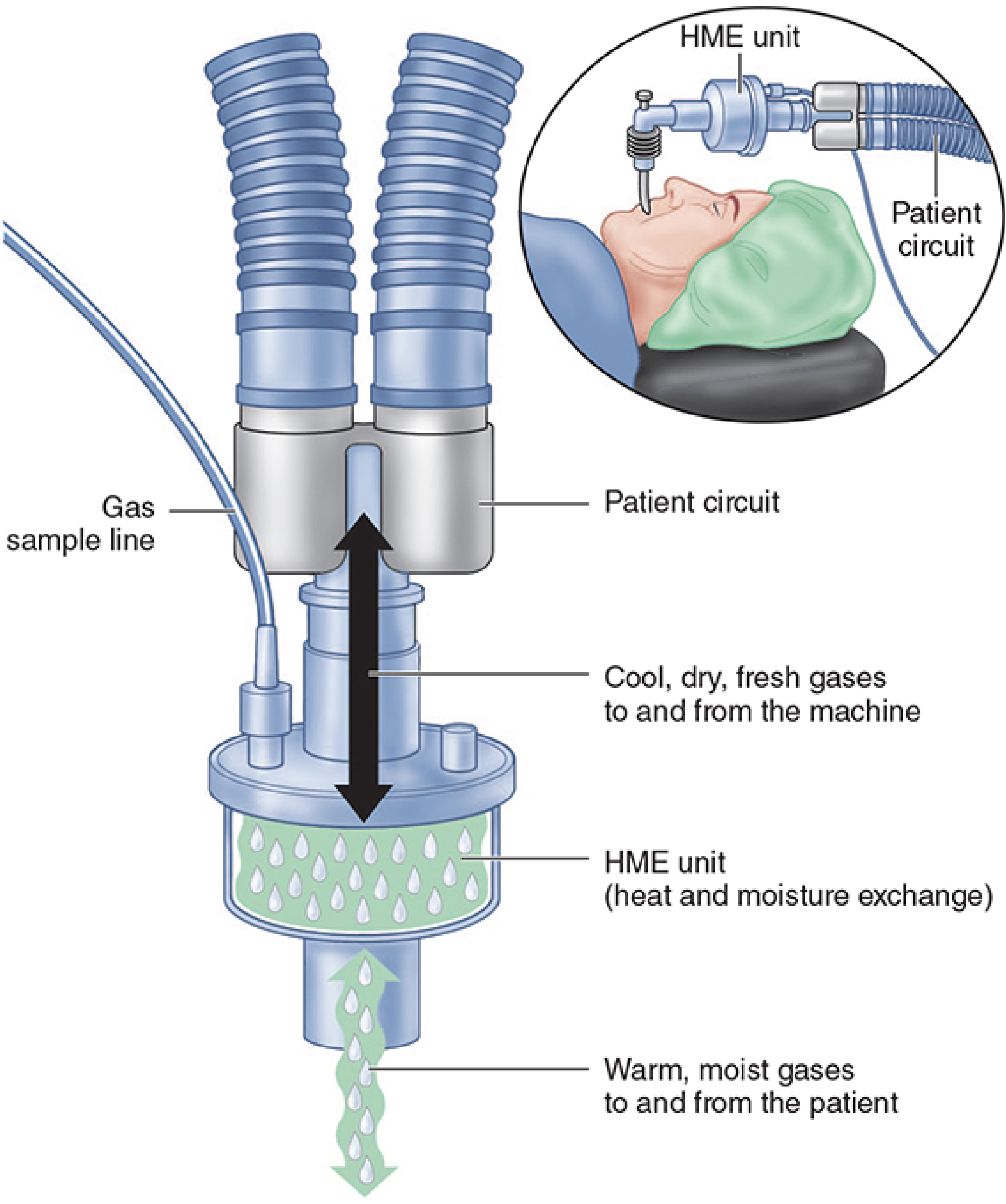
PRINCIPLE OF WORKING
During EXPIRATION:
- The patient exhales warm, moist gas (37°C, 100% RH, ~44 mg H₂O/L)
- This warm humid gas passes through the hygroscopic element of the HME
- Water vapour condenses onto the hygroscopic/hydrophobic material
- Heat and moisture are stored in the element
During INSPIRATION:
- Cool, dry fresh gas (from the anaesthesia machine/ventilator) passes through the stored element
- The stored heat re-warms the gas
- Stored moisture re-evaporates back into the inspired gas
- The patient receives warmer, more humidified gas than without the HME
Net result:
- Inspired gas temperature: ~28–32°C (not full 37°C)
- Absolute humidity of inspired gas: ~20–30 mg H₂O/L (normal = 44 mg/L)
- Efficiency: ~70% humidity retention (compared to 100% with active heated humidifier)
CONSTRUCTION / COMPONENTS
| Layer | Material | Function |
|---|---|---|
| Hygroscopic layer | Calcium chloride, lithium chloride, or paper impregnated with hygroscopic salts | Traps and releases water vapour |
| Hydrophobic layer | Polypropylene or PTFE membrane | Retains moisture; repels liquid water |
| Electrostatic filter | Charged polymer fibre | Filters bacteria and viruses |
| Housing | Plastic; standardised 15/22 mm connectors | Connects to ETT and circuit |
Types:
- Simple condenser HME - only hygroscopic material; no filtering
- HME + filter (HMEF) - combined heat-moisture exchange + bacterial/viral filtration
- Electrostatic HMEF - best filtration; preferred when infection risk is high
PERFORMANCE CHARACTERISTICS
| Parameter | Value |
|---|---|
| Temperature of inspired gas | 28–32°C |
| Absolute humidity of inspired gas | 20–30 mg H₂O/L |
| Humidity efficiency | ~70% (compared to active humidifier) |
| Added dead space | > 60 mL (significant in paediatrics) |
| Added resistance to breathing | Low at normal flows; increases at high minute volumes |
| Bacterial filtration efficiency | 99.9% (HMEF type) |
| Viral filtration efficiency | Up to 99.999% (electrostatic type) |
| Recommended change interval | 24 hours (some units rated for 48-72 h) |
INDICATIONS FOR HME USE
- Routine short-term ventilation - intraoperative use (procedures > 1 hour)
- ICU short-term ventilation - prevents VAP; recommended in VAP prevention bundle (Level 1 evidence)
- Tracheostomy patients - replaces upper airway humidification function ("Swedish nose")
- Post-laryngectomy patients - compensates for loss of nasal humidification
- Infection control - protects breathing circuit and anaesthesia machine from bacterial/viral cross-contamination; critical in patients with:
- Active respiratory infections (TB, influenza, COVID-19)
- Immunocompromised status
- Known MDR respiratory pathogens
- Transport ventilation - lightweight, no electricity needed
ADVANTAGES OF HME
- Simple and passive - no electricity, no water reservoir, no heating element needed
- Cost-effective - cheaper than active heated humidifiers
- Compact and lightweight - suitable for transport ventilation
- Bidirectional filtration - protects both patient and anaesthesia machine circuit
- Reduces VAP risk - Level 1 RCT evidence in VAP prevention bundle (Fishman's Pulmonary Diseases)
- Prevents inspissation of secretions - maintains mucosal moisture
- Reduces heat loss - especially relevant in longer procedures
- No risk of thermal injury - unlike active humidifiers (which can cause thermal lung injury if gas exceeds 41°C)
- No circuit flooding - active humidifiers risk circuit waterlogging; HME does not
- Easy to change - disposable, low contamination risk
DISADVANTAGES / LIMITATIONS
- Increased apparatus dead space - > 60 mL; causes significant CO₂ rebreathing in:
- Paediatric patients (where dead space is relatively large compared to tidal volume)
- Patients with small tidal volumes
- Increased airway resistance - increases work of breathing during spontaneous ventilation; may tire weaning patients
- Obstruction risk - excessive saturation with water or secretions can obstruct the breathing circuit (medical emergency)
- Less effective than active humidifiers - only ~70% humidity efficiency; insufficient for:
- Paediatric patients (especially neonates)
- Patients with thick secretions (cystic fibrosis, bronchiectasis)
- Prolonged ventilation (> 96 hours)
- Does not add heat or moisture - only conserves; in hypothermic patients, initial inspired gas is already cold
- Must be bypassed during nebulisation - aerosol medication cannot pass through the filter membrane efficiently
- Must be bypassed during heated humidification - two cannot be used simultaneously
CONTRAINDICATIONS
| Contraindication | Reason |
|---|---|
| Neonates and small infants | Dead space > 60 mL is unacceptably large relative to tidal volume |
| Spontaneously breathing patients who are tiring / weaning | Increased resistance increases work of breathing |
| Thick, copious secretions | Risk of HME obstruction by secretions |
| Patients needing nebulised medications | Filter impedes aerosol delivery |
| Exhaled tidal volume < 70% of set tidal volume (large leak) | HME efficiency is reduced with large ETT cuff leaks |
| Active haemoptysis | Blood clots obstruct the filter |
HME vs ACTIVE HEATED HUMIDIFIER
| Feature | HME (Passive) | Active Heated Humidifier |
|---|---|---|
| Power supply | Not needed | Required |
| Humidity efficiency | ~70% | ~100% |
| Inspired gas temperature | 28–32°C | 37°C (adjustable) |
| Dead space added | > 60 mL | Minimal (circuit level) |
| VAP prevention | Level 1 evidence | Level 1 evidence (both equivalent) |
| Risk of thermal injury | None | Yes (if > 41°C) |
| Risk of circuit flooding | None | Yes |
| Filtration | Yes (HMEF type) | No |
| Cost | Low | High |
| Use in neonates | Contraindicated | Preferred |
| Transport ventilation | Ideal | Impractical |
HME IN VAP PREVENTION
- Reduces colonisation of the breathing circuit by acting as a bacterial/viral filter
- Maintains mucosal moisture and mucociliary function, reducing bacterial adhesion
- Prevents microaspiration of contaminated condensate (active humidifiers accumulate warm-water condensate in the circuit which is a bacterial growth medium)
- Equivalent to heated humidification for VAP rates in most adult ICU patients
PRACTICAL POINTS FOR EXAM
- Changed every 24 hours in ICU (or when visibly soiled, wet, or obstructed)
- Must be removed and bypassed during nebulisation therapy
- Not used with active humidifier simultaneously (placing both in circuit is an error)
- In paediatric patients: use active heated humidifier instead due to dead space concerns
- During anaesthesia for infectious patients (TB, COVID): HME + viral filter (HMEF) placed at the ETT to protect the breathing circuit
- The "Swedish nose" is a long-term variant used in tracheostomy/laryngectomy patients for home use
CONCLUSION
Fire in the ett during surgery how to diagnose criteria and mange from standard textbook of anaesthetia
FIRE IN THE ENDOTRACHEAL TUBE (ETT) / AIRWAY FIRE DURING SURGERY
Exam Answer | Barash Clinical Anesthesia 9e / Morgan & Mikhail 7e
INTRODUCTION
THE FIRE TETRAHEDRON — THE ESSENTIAL CONCEPT
        IGNITION SOURCE
           (Laser, ESU)
               ▲
               │
  FUEL ────────┼──────── OXIDISER
 (ETT, tissue,           (O2, N2O)
  drapes, gauze)
               │
         CHAIN REACTION
      (sustains the combustion)
| Element | Source in Airway Surgery |
|---|---|
| Ignition source | CO₂ laser, KTP laser, Nd:YAG laser, electrosurgery unit (ESU/cautery), static electricity |
| Fuel | PVC endotracheal tube (ignites in 25% O₂), rubber/silicone tubes, dry gauze, drapes, charred tissue, tracheal hair |
| Oxidiser | Oxygen (especially FiO₂ > 30%), Nitrous oxide (also supports combustion) |
| Chain reaction | Sustained by abundant fuel + high O₂ concentration |
COMMON CLINICAL SCENARIOS FOR AIRWAY FIRE
- Laser airway surgery (laryngoscopy, vocal cord surgery) — most common cause
- Tracheostomy (electrosurgery + oxygen-enriched tracheal environment at tracheal entry)
- Tonsillectomy (electrocautery + oxygen leak around uncuffed ETT)
DIAGNOSIS — HOW TO RECOGNISE AN AIRWAY FIRE
Signs and Symptoms of ETT Fire:
| Sign | Description |
|---|---|
| Visible flame or smoke | Most obvious — flame visible at the ETT (blowtorch appearance), smoke rising from the airway |
| Unusual sounds | Popping, crackling, or "whooshing" sound from the airway |
| Abnormal capnography | Sudden loss of ETCO₂ waveform (ETT melted/obstructed) or unexpected waveform change |
| Airway obstruction | Sudden increase in peak airway pressure, then complete loss of ventilation |
| Burning smell | Acrid smell of burning plastic, rubber, or tissue |
| Discoloration of ETT | Surgeon notes charring, melting, or deformity of ETT in the operative field |
| SpO₂ drop | Sudden unexplained desaturation |
| Methylene blue leak | If cuffs filled with tinted saline — coloured fluid visible signals cuff rupture (fire warning sign) |
The "BLOWTORCH" appearance:
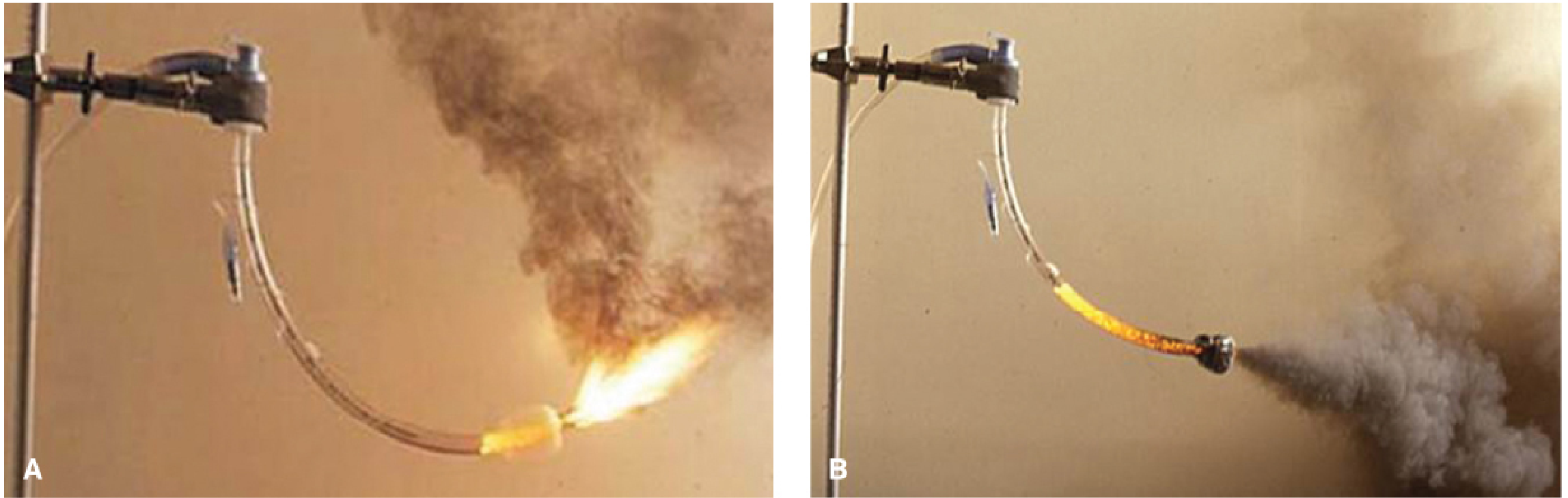
MANAGEMENT OF ETT FIRE — STEP-BY-STEP PROTOCOL
IMMEDIATE STEPS (Do simultaneously — within seconds):
"STOP — DISCONNECT — REMOVE — EXTINGUISH — REVENTILATE — ASSESS"
STEP 1 — STOP VENTILATION IMMEDIATELY
- Stop all gas flow from the anaesthesia machine
- Remove the breathing circuit from the ETT
- Disconnect the circuit from the machine (prevents further oxygen delivery fuelling the fire)
STEP 2 — REMOVE THE BURNING ETT
- Immediately remove the ETT from the patient's airway
- Do NOT attempt to extubate gently — pull it out rapidly
- The entire burning tube is removed as one unit
STEP 3 — EXTINGUISH THE FIRE
- Pour saline (or water) into the airway directly via a syringe
- Submerge the burning ETT in a water basin to extinguish it
- Remove all burning or smouldering material (gauze, pledgets, drapes) from the airway
STEP 4 — RE-ESTABLISH VENTILATION
- Ventilate with face mask using room air (21% O₂) initially — NOT 100% O₂, as this risks re-ignition if any smouldering material remains
- Once fire confirmed extinguished, reintubate
- Use a new ETT; if airway oedema is present, consider a smaller size or awake technique
STEP 5 — INSPECT AND ASSESS AIRWAY DAMAGE
- Rigid or flexible bronchoscopy — mandatory to:
- Assess extent of thermal mucosal injury (erythema, charring, sloughing)
- Confirm no ETT fragments, ash, or foreign body debris remain in trachea/bronchi
- Determine depth of subglottic and tracheobronchial injury
- Chest X-ray — baseline; serial X-rays to monitor for pulmonary oedema, pneumothorax
- Arterial blood gas (ABG) — assess oxygenation, ventilation, and carboxyhaemoglobin (CO poisoning)
- Check carboxyhaemoglobin (COHb) level — toxic products of PVC combustion include HCl gas and carbonyl compounds
STEP 6 — POST-FIRE MANAGEMENT IN ICU
- ICU admission is mandatory for all confirmed airway fires
- Consider systemic steroids (dexamethasone) to reduce airway oedema
- Consider bronchial lavage to remove carbonaceous debris and toxic combustion products
- Humidified oxygen (100% initially for CO poisoning if present)
- Monitor for:
- Progressive airway oedema causing obstruction (may need surgical tracheostomy)
- Acute lung injury / ARDS from inhalation injury
- Pneumonia (secondary infection of burned mucosa)
- Keep patient intubated and sedated until airway oedema resolves (may take 24–72 hours)
- Pulmonology and ENT/thoracic surgery consultation
AIRWAY FIRE PROTOCOL — SUMMARY TABLE (Morgan & Mikhail 7e, Table 37-3)
| Step | Action |
|---|---|
| 1 | Stop ventilation; remove tracheal tube |
| 2 | Turn off O₂; disconnect circuit from machine |
| 3 | Submerge tube in water |
| 4 | Ventilate with face mask; reintubate |
| 5 | Assess airway damage — bronchoscopy, serial CXR, ABG |
| 6 | Consider bronchial lavage and steroids |
PREVENTION — HOW TO PREVENT ETT FIRES
A. Minimise the Oxidiser
| Measure | Detail |
|---|---|
| Limit FiO₂ to ≤ 30% | Most important single measure; use air/O₂ mix; many patients tolerate FiO₂ of 21% |
| Never use N₂O during laser/ESU surgery | Nitrous oxide supports combustion; absolutely contraindicated |
| Wait for expired O₂ to equilibrate | Before activating laser or ESU, wait for expired O₂ to reach ≤ 30% |
| Cuffed ETT to seal subglottic space | Prevents oxygen from leaking up into the hypopharynx around the tube |
B. Use Laser-Resistant ETT (Modify the Fuel)
| ETT Type | Properties | Limitations |
|---|---|---|
| PVC (standard) | Cheap, non-reflective | Highly combustible in ≥25% O₂ — DO NOT USE |
| Red rubber | Puncture-resistant | Combustible; produces toxic fumes |
| Silicone | Non-reflective | Combustible; turns to toxic ash |
| Metal (Laser-Flex®, Mallinckrodt) | Most fire-resistant; corrugated stainless steel | Thick-walled; reflects laser onto adjacent tissue; flammable PVC cuff and tip remain |
| Metallic tape-wrapped PVC | Available if commercial tube not present | FDA unapproved; rough edges; no cuff protection; adhesive may itself ignite |
C. Fill ETT Cuffs with Saline (Not Air)
- Saline-filled cuffs absorb laser thermal energy
- Add methylene blue to saline — coloured leak = visible warning of cuff puncture/fire risk
- If proximal cuff is struck and deflates, the distal cuff continues to seal the airway
- Use double-cuffed laser ETT for maximum protection
D. Modify the Ignition Source
- Use lowest effective laser power and shortest pulse duration
- Use pulsed mode (not continuous) — continuous lasing > 5 seconds increases fire risk by a factor of 72 (Barash 9e)
- Avoid lasing charred tissue — lasing charred tissue increases flame risk by a factor of 98
- Bipolar cautery preferred over monopolar (less sparking)
- Surgeon must use only a knife (not cautery) to enter the trachea during tracheostomy
E. Reduce Available Fuel
- Only use wet, saline-saturated gauze pledgets (never dry gauze) in the airway
- Keep exterior of ETT moistened (use hydratable coating e.g., Merocel)
- Minimise flammable draping material near the operative field
F. Avoid ETT Altogether (If Possible)
- Intermittent apnoea technique (mask ventilate, pause for lasering)
- Jet ventilation via rigid laryngoscope/Sanders injector
- High-frequency jet ventilation (HFJV)
- Spontaneous ventilation with topical anaesthesia
- These techniques eliminate the fuel from the airway entirely
G. Emergency Preparedness
- A 60 mL water-filled syringe and basin must be immediately available at the head end at ALL times during laser/ENT surgery
- Assign a team member specifically for fire vigilance
- All OR staff must know and practise the airway fire protocol
- Institutional fire safety drills should include airway fire scenarios
SPECIAL SCENARIOS
Tracheostomy Fire
- Enter the trachea with a knife, not cautery
- Do not reapply cautery until tracheostomy cuff is inflated and airway is resecured
- The window between tracheal entry and cuff inflation is the period of highest risk
- Increase FiO₂ just before entering trachea for apnoea tolerance — then immediately reduce after airway is secured
Tonsillectomy Fire
- Use a cuffed ETT (an uncuffed ETT allows O₂ to leak around it into the pharynx)
- FiO₂ ≤ 30%; in healthy children, room air (21%) is acceptable while cautery is in use
- Wait for expired O₂ concentration to fall to target before activating ESU
COMPLICATIONS OF ETT FIRE
| Complication | Timing | Features |
|---|---|---|
| Thermal burns of lips, oral mucosa, pharynx | Immediate | Erythema, blistering, sloughing |
| Subglottic/tracheal burns | Immediate-early | Oedema, potential for complete obstruction |
| ETT fragment foreign bodies | Immediate | Retained ash/debris in airways — risk of obstruction, abscess |
| Inhalation of toxic combustion products | Immediate | HCl from PVC, carbon monoxide, carbonyl compounds |
| Carboxyhaemoglobin toxicity | Early | CO poisoning; treat with 100% O₂ |
| Acute lung injury / ARDS | Early (12–48 h) | From toxic inhalation |
| Secondary pneumonia | Late | Infected burns, impaired mucociliary clearance |
| Airway stenosis | Late (weeks) | Subglottic/tracheal stenosis from scar formation |
CONCLUSION
- Prevention — break the fire tetrahedron proactively by limiting FiO₂ ≤ 30%, avoiding N₂O, using laser-resistant ETT with saline-filled cuffs, and limiting laser power/duration
- Recognition — visible flame, smoke, burning smell, capnograph change, airway pressure change
- Response — immediate execution of the 6-step protocol: Stop ventilation → Disconnect circuit → Remove ETT → Extinguish with saline → Reventilate → Bronchoscopy + ICU