
Stroke and it's management in obstetrics for 25 marks
"stroke"[MeSH Terms] AND "pregnancy"[MeSH Terms]
acute ischemic stroke in pregnancy thrombolysis alteplase tenecteplase thrombectomy management guideline
Stroke in Pregnancy and the Puerperium (25-mark answer)
1. Definition and epidemiology
- Pregnancy-associated stroke is any cerebrovascular event (ischaemic, haemorrhagic or venous) during pregnancy or up to 6 weeks postpartum.
- Reported incidence varies widely, from 4.3 to 210 per 100,000 deliveries. Pregnancy clearly increases the risk of cerebral infarction. The risk is highest in the third trimester and postpartum (Creasy & Resnik's Maternal-Fetal Medicine, p. 1633).
- Adams and Victor's Principles of Neurology (12th ed) reports that most of the risk for both infarction and intracerebral haemorrhage lies in the 6 weeks after delivery. Arterial occlusion clusters in the 2nd and 3rd trimesters and the first week postpartum. Venous occlusion tends to occur 1 to 4 weeks postpartum.
- Risk rises with age. In Rochester, NY, the incidence was 6.2 per 100,000 and doubled with each age band from 25 to 49 years.
- Intracranial haemorrhage causes a disproportionate 4 to 7% of maternal deaths.
- A 2025 systematic review and meta-analysis of over 270 million women covers global incidence and risk factors of pregnancy-related stroke.
2. Why pregnancy predisposes to stroke (pathophysiology)
- Hypercoagulable state: raised factors V, VII, VIII, IX, X, XII and fibrinogen. Protein S falls, fibrinolysis is reduced, and platelet aggregation increases.
- Haemodynamic and vascular changes: hypertension, and endothelial injury in pre-eclampsia.
- Venous stasis from the gravid uterus. Pelvic and leg vein clots can cause paradoxical embolism through a patent foramen ovale because right heart pressures rise.
- Peripartum events: hypotension from haemorrhage or anaesthesia causes watershed infarcts. Valsalva during labour raises pressure.
3. Classification and causes
| Type | Causes in obstetrics |
|---|---|
| Arterial ischaemic stroke | Thrombus on atheromatous plaque; cardioembolism (rheumatic or ischaemic heart disease, endocarditis, myxoma, peripartum cardiomyopathy, prosthetic valve); carotid or vertebral dissection (late pregnancy or early postpartum); paradoxical embolus; amniotic fluid, air or fat embolism; reversible cerebral vasoconstriction syndrome (RCVS) or postpartum angiopathy; thrombophilias (protein C, protein S, antithrombin III deficiency; factor V Leiden; antiphospholipid syndrome; hyperhomocysteinaemia); sickle cell disease; TTP; choriocarcinoma; vasculitis; moyamoya |
| Cerebral venous thrombosis (CVT) | Third trimester or puerperium; after abortion; pre-eclampsia; thrombophilia; anaemia; dehydration; infection; caesarean section |
| Haemorrhagic stroke | Pre-eclampsia or eclampsia and gestational hypertension (the main risk factor); ruptured aneurysm (more likely in the second half of pregnancy); AVM; RCVS; coagulopathy, DIC, HELLP, thrombocytopenia; mycotic aneurysm; metastatic choriocarcinoma |
| Other | Sheehan syndrome (pituitary infarction); PRES and eclampsia (mimics and overlaps) |
4. Clinical features
- Ischaemic stroke: sudden focal deficit such as hemiparesis, aphasia, visual loss or ataxia. A preceding TIA is possible.
- CVT: headache, seizures (focal or generalised), drowsiness, confusion, papilloedema, mild pyrexia, and fluctuating hypertension. Signs of meningeal irritation may be present.
- SAH: sudden severe "thunderclap" headache, vomiting, neck stiffness, and a depressed level of consciousness.
- ICH: focal deficit depending on the site.
- RCVS: thunderclap headache. It can be complicated by infarction or haemorrhage.
- Differential diagnosis: eclampsia and PRES, migraine with aura, hypoglycaemia, postdural puncture headache, seizure with Todd paralysis, demyelination, and conversion disorder. CVT is easily mistaken for eclampsia, but eclampsia is preceded by pre-eclampsia signs.
5. Investigations
- Immediate bedside: ABC, blood glucose, BP, ECG, fetal heart rate (CTG or ultrasound).
- Bloods:
- FBC, platelets, coagulation profile (PT, aPTT), glucose, electrolytes, and renal and liver function (HELLP).
- Lipid profile and HbA1c.
- Urine protein.
- Neuroimaging:
- Non-contrast CT head is the first test to separate infarct from haemorrhage. Fetal dose is negligible with abdominal shielding.
- MRI with DWI is preferred if available in a timely manner, because there is no radiation. Gadolinium is avoided.
- CT angiography is the test of choice for large-vessel occlusion (LVO) and thrombectomy eligibility. CT perfusion is used for the 6 to 24 hour window.
- MR venography if CVT is suspected. Catheter angiography is used for SAH, AVM or aneurysm.
- Cardiac work-up: ECG, echocardiography (transthoracic, with bubble study for PFO), and Holter monitoring.
- Vascular: carotid and vertebral duplex for dissection or stenosis.
- Aetiology (young woman):
- Thrombophilia screen: protein C, protein S, antithrombin, factor V Leiden, antiphospholipid antibodies, homocysteine. Interpret with caution during pregnancy.
- Sickle screen, ESR, and autoimmune markers.
- Lumbar puncture: only after imaging. It is used if SAH is suspected with a negative CT.
6. Management
A. General principles
- A multidisciplinary team (stroke neurologist, obstetrician, maternal-fetal medicine, anaesthetist, neuroradiologist and neonatologist) manages the patient. This is the Canadian Stroke Best Practice recommendation.
- Treat the mother first. Maternal stabilisation is the best fetal treatment.
- Admit to a stroke unit or HDU. Position the patient with a left lateral tilt in later pregnancy.
- Maintain oxygenation (SpO2 above 94%) and treat hypoglycaemia and hyperglycaemia. Control fever.
- Fetal monitoring with continuous CTG if viable.
- Blood pressure:
- In ischaemic stroke, avoid aggressive lowering. Permissive hypertension applies unless thrombolysis is planned (below 185/110 mmHg) or there is pre-eclampsia.
- In pre-eclampsia, use labetalol, nifedipine or hydralazine. Avoid ACE inhibitors, ARBs and sodium nitroprusside.
- Seizures (eclampsia or CVT): magnesium sulphate for eclampsia. Use levetiracetam or lamotrigine for other seizures. Avoid valproate.
- Raised ICP: head elevation, osmotherapy, and decompressive surgery if needed.
B. Acute ischaemic stroke
- Pregnancy is not an absolute contraindication. Alteplase has been used safely and effectively, and its large molecular weight (about 59 kDa) means it does not cross the placenta (Creasy & Resnik, p. 1634).
- Time window: within 4.5 hours of onset (3 hours is the US label). Use it for disabling deficits in otherwise eligible patients.
- Risks include miscarriage, placental abruption and haemorrhage. These are greater near term, so weigh the risks case by case. A normal pregnancy at 10 weeks carries a different risk from a pregnancy at 36 weeks with abruption.
- Postpartum: caution for uterine and surgical-site bleeding.
- Fewer than 10 days after delivery: consider individually, with extremely limited evidence.
- 10 days or more after delivery: alteplase can be considered for disabling stroke. This is ESO expert consensus, quoted via the Stroke Manual.
- If bleeding risk is high and the patient has an LVO, direct thrombectomy may be preferred.
- Tenecteplase has only isolated case reports in pregnancy, so safety and comparative efficacy are not established.
- Contraindications are the usual ones: ICH, recent major surgery, active bleeding, BP above 185/110 despite treatment, and platelets below 100,000.
- Indicated for proximal LVO. Pregnancy is not a contraindication, and it is a viable option within 6 hours.
- Selected patients with a large penumbra on CT perfusion benefit up to 24 hours.
- Minimise fetal radiation by shielding the abdomen and limiting fluoroscopy.
- Aspirin within 48 hours of ischaemic stroke or TIA. Low-dose aspirin is safe in pregnancy. Clopidogrel has limited data, so use it only with a clear indication.
- Reserved for AF, mechanical heart valves, certain hypercoagulable states, and venous occlusive disease.
- LMWH (subcutaneous, with anti-Xa monitoring) is the agent of choice. It is stopped at the onset of labour and restarted about 12 hours after vaginal delivery or 24 hours after caesarean section.
- Warfarin crosses the placenta. It carries teratogenicity risk (especially in the first trimester), fetal wastage and bleeding. In select patients with mechanical valves it may be used in the second and early third trimesters.
- DOACs cross the placenta and are avoided.
- Lactation: warfarin and heparin are compatible. DOACs are avoided.
- Carotid endarterectomy for severe symptomatic stenosis (70 to 99%). Moderate stenosis (50 to 69%) is possible but the benefit in women is unclear.
- Decompressive hemicraniectomy for malignant MCA infarction.
- Dissection: antithrombotic therapy. RCVS: remove vasoactive drugs and give a calcium-channel blocker. Do not use immunosuppression.
C. Cerebral venous thrombosis
- Anticoagulation with LMWH or UFH is the treatment, even in the presence of haemorrhagic infarction (Creasy & Resnik, p. 1635).
- Continue anticoagulation after delivery for at least 3 to 6 months. LMWH may continue through the puerperium.
- Treat seizures, raised ICP and any underlying infection.
- Consider endovascular therapy if the patient deteriorates despite anticoagulation.
- Prognosis: about 15% death or dependency, although up to 80% make an excellent recovery. Recurrence risk is about 1%. CVT is not a contraindication to future pregnancy, but the risk of early miscarriage is higher.
D. Haemorrhagic stroke
- ICH:
- Control BP and correct coagulopathy.
- Reverse anticoagulants: protamine for heparin, and vitamin K with PCC for warfarin.
- Give platelet transfusion if thrombocytopenic.
- Refer to neurosurgery for haematoma evacuation or EVD.
- Treat the cause: pre-eclampsia, HELLP, DIC.
- SAH:
- Bed rest, BP control, nimodipine, and analgesia.
- Early securing of a ruptured aneurysm by coiling or clipping. Management is the same as in non-pregnant women.
- A known unruptured AVM should not preclude pregnancy.
- Eclampsia and PRES: magnesium sulphate, BP control and delivery.
E. Obstetric management (timing and mode of delivery)
- Depends on gestation, maternal neurological status and fetal condition.
- Vaginal delivery is preferred unless obstetric indications exist (Creasy & Resnik, p. 1634). Use epidural analgesia and assisted second stage to limit Valsalva.
- Caesarean section is indicated for obstetric reasons, deteriorating maternal condition or raised ICP, fetal compromise, or CVT or thrombosis just before or during labour.
- Pre-eclampsia with stroke: stabilise, then deliver after maternal stabilisation. Delivery is the definitive treatment.
- Coordinate anticoagulation, regional anaesthesia timing and neonatal team presence.
- After a brain-dead mother, somatic support may continue to allow fetal maturation. This is individualised and ethically complex.
- Postpartum: continue anticoagulation or antiplatelets, rehabilitation (PT, OT, speech therapy), VTE prophylaxis, and mood and depression screening.
F. Prevention and pre-pregnancy counselling
- Primary prevention: control hypertension, diabetes, obesity and smoking. Treat migraine. Screen for thrombophilia in high-risk women.
- Low-dose aspirin (75 to 150 mg from 12 weeks) for women at high risk of pre-eclampsia. Add LMWH prophylaxis for APS or thrombophilia as indicated.
- Prior stroke:
- Pre-conception counselling. The aim is a stable, optimised condition before pregnancy.
- Pregnancy is not absolutely contraindicated after a prior stroke.
- Continue aspirin, and consider LMWH for specific conditions.
- Avoid oestrogen-containing contraception, which carries risk with migraine with aura, hypertension and thrombophilia. Use progestin-only or non-hormonal methods.
- Monitor closely in antenatal visits (BP, growth scans, uterine artery Doppler).
- Follow-up after delivery with neurology.
7. Prognosis and complications
- Maternal mortality is higher with haemorrhagic stroke and with CVT complicated by herniation.
- Fetal outcomes: preterm birth, miscarriage, IUGR, and neonatal complications from maternal drugs or hypoxia.
- Long-term: residual disability, epilepsy, depression, and increased future cardiovascular risk. A 2026 meta-analysis found preterm delivery associated with long-term maternal stroke risk.
8. Recent evidence update (PubMed, last 2 years)
Quick exam summary (flowchart)
- Ischaemic with disabling deficit within 4.5 h → IV alteplase ± thrombectomy
- LVO → thrombectomy
- CVT → LMWH
- ICH or SAH → BP control, reverse coagulopathy, neurosurgery
- Pre-eclampsia or eclampsia → MgSO4, antihypertensives, delivery
- Always multidisciplinary; vaginal delivery preferred unless obstetric indication.
Psoriasis easy way
plaque psoriasis silvery scales erythematous plaques
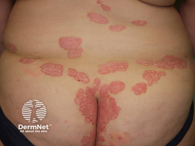
This is a high-resolution clinical dermatology photograph illustrating chronic plaque psoriasis affecting the gluteal region and lower back in an adult patient. Imaging modality: standard digital skin photography; view: posterior, dorsal aspect of buttocks and lower lumbar area. The image shows multiple well-demarcated, erythematous plaques with characteristic silvery-white scales, coalescing into larger plaques in the intergluteal cleft and along the lower back. Surfaces are slightly raised with visible scale and oozing not evident. The distribution is bilateral and symmetric over extensor-like surfaces of the trunk and buttocks, consistent with plaque-type psoriasis (psoriasis vulgaris). The plaques display typical morphology: erythematous plaques, sharp margins, thick hyperkeratosis, and adherent silvery scales. Differential includes eczema, seborrheic dermatitis, dermatophytosis, pityriasis rosea; clinical correlation required. The diagnostic significance: image supports a cutaneous psoriasis diagnosis; helps guide topical corticosteroid therapy, vitamin D analogs (calcipotriene), and phototherapy planning; indicates chronicity with possible Koebner response in friction-bearing regions. Potential clinical use cases: dermatology education, tele-dermatology triage, treatment monitoring, drug safety assessment, and research on plaque psoriasis distribution and phenotype. For histology confirmation, biopsy would show acanthosis, parakeratosis, thinning of the granular layer, Munro microabscesses. These features collectively reinforce psoriasis vulgaris as the leading clinical diagnosis in typical anatomical sites.
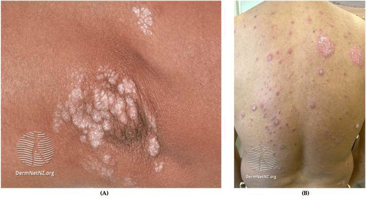
This composite clinical photograph illustrates plaque psoriasis (psoriasis vulgaris) across different skin phototypes. Image A shows a high-magnification view of a chronic plaque on an elbow or knee. It demonstrates characteristic well-demarcated, erythematous, and elevated plaques covered with thick, micaceous (silvery-white) scales. The morphology is confluent with visible fissuring. Image B displays a wider view of a patient’s back, highlighting the presentation in skin of color. In this context, the plaques may appear more violaceous or hyperpigmented rather than bright red. The distribution is widespread and symmetric, consisting of numerous discrete erythematous papules and larger circular plaques. Some lesions exhibit characteristic scaling, while others appear as smoother, indurated areas. The contrast between the silvery scale and the underlying skin is less pronounced on darker skin tones compared to lighter ones. These images collectively teach the diagnostic features of plaque psoriasis: symmetry, sharp demarcation, and scale morphology.
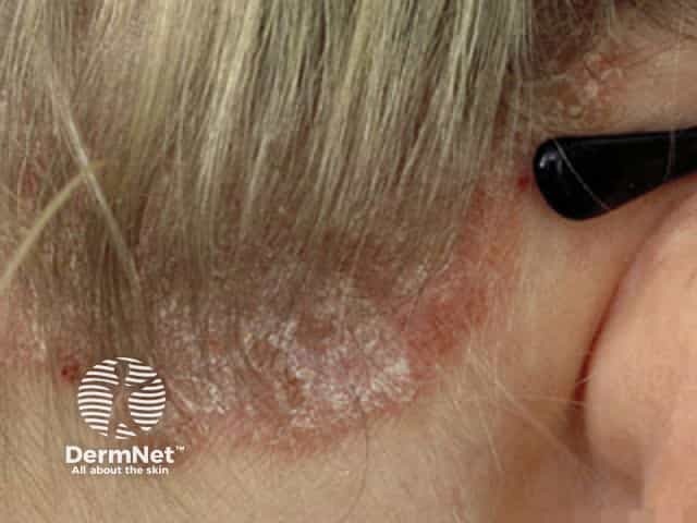
Clinical photography of dermatologic disease. This is a close-up, lateral view of the posterior neck and occipital scalp near the hairline, showing erythematous, well-demarcated plaques with thick, silvery scales consistent with active plaque psoriasis. Lesions extend along the nuchal region and behind the ear, with mild induration and irregular borders in places. The skin surface exhibits adherent scale and subtle fissuring; surrounding skin is relatively normal. The color is red-erythematous with a metallic sheen from the scales, and there is minimal lichenification. This plaque morphology—sharply demarcated erythematous plaques with overlying silvery scale—reflects classic psoriasis pattern, commonly seen on scalp, neck, and extensors. No pustules or erosions are evident. The image demonstrates typical disease distribution in the head and neck area and highlights the keratotic scale with underlying epidermal hyperplasia. Clinically, this appearance supports the diagnosis of plaque psoriasis; differential considerations include seborrheic dermatitis, atopic dermatitis, tinea capitis/corporis, and pityriasis rosea in variant presentations. Diagnostic significance lies in recognizing plaque psoriasis morphology for treatment planning, including topical corticosteroids or vitamin D analogues, calcineurin inhibitors in selected cases, and consideration of phototherapy for extensive disease. Educationally, this image aids lesion recognition, distribution mapping, and patient counseling. It is suitable for teaching reference.
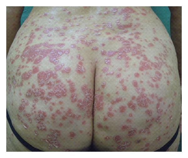
Clinical photograph of the lumbosacral and gluteal regions demonstrating characteristic manifestations of plaque psoriasis. The image shows multiple, well-demarcated erythematous plaques and papules of varying sizes. The lesions are covered with thick, silvery-white scales, a hallmark of the disease. The distribution is symmetric across both buttocks, with smaller guttate-like papules peripheral to larger, coalescing nummular plaques. The underlying skin exhibits significant erythema, and the scales appear dry and adherent. This visual illustrates the typical morphology of chronic plaque-type psoriasis in a common predilection site, highlighting the hallmark features of inflammatory erythema and hyperkeratotic scaling essential for dermatologic diagnosis and medical student education.
"psoriasis"[MeSH Terms] AND "therapeutics"[MeSH Terms]
Psoriasis (easy way)
What is it?
Why does it happen?
- Genes: HLA-Cw6 is the classic association.
- Immune cells: Th1 and Th17 cells release IL-17, IL-23 and TNF-α. These make keratinocytes grow too quickly. This is why biologics target these cytokines.
- Triggers:
- Trauma, which causes the Koebner phenomenon (new lesions at injury sites).
- Streptococcal infection (guttate type).
- Drugs (β-blockers, lithium, antimalarials, NSAIDs, steroid withdrawal).
- Stress, smoking, alcohol, obesity.
What does the skin show?
- Acanthosis, meaning a thick epidermis. The rete ridges look like "test tubes in a rack".
- Parakeratosis, meaning nuclei retained in the stratum corneum.
- Thin or absent granular layer.
- Munro microabscesses, which are neutrophils in the stratum corneum.
- Dilated dermal capillaries, which explain the Auspitz sign.
Types
| Type | Key feature |
|---|---|
| Plaque (vulgaris) | Most common. Well-demarcated red plaques with silvery scale on elbows, knees, scalp and lower back |
| Guttate | "Raindrop" small papules after a strep throat, mainly in children and young adults |
| Inverse (flexural) | Skin folds. Smooth, shiny, red, and no scale because of friction |
| Pustular | Sterile pustules. Can be localised (palms and soles) or generalised (von Zumbusch, an emergency) |
| Erythrodermic | More than 90% of body surface red and scaly. An emergency |
| Nail | Pitting, "oil drop" sign, onycholysis, thickening |
| Palmoplantar | Thick, scaly, fissured palms and soles |
| Scalp | Thick plaques that extend past the hairline |
Signs to remember
- Auspitz sign: scraping the scale gives pinpoint bleeding.
- Candle grease sign: scraping gives scale like wax.
- Koebner phenomenon: lesions at sites of trauma.
- Nail pitting is the most common nail sign.
Associated problems (comorbidities)
- Psoriatic arthritis in up to about 10 to 30% of patients. Features are asymmetric oligoarthritis, DIP involvement, dactylitis ("sausage digit"), enthesitis and sacroiliitis.
- Metabolic syndrome, obesity, diabetes, hypertension and cardiovascular disease. Psoriasis creates a chronic inflammatory state that raises death risk from heart disease (Robbins).
- Depression, uveitis, Crohn disease, and NAFLD.
Diagnosis
Treatment (stepladder)
- Mild disease (BSA below 5 to 10%): topicals
- Emollients.
- Topical corticosteroids.
- Vitamin D analogues (calcipotriol).
- Coal tar, salicylic acid, dithranol.
- Topical calcineurin inhibitors (face and folds).
- Moderate to severe disease:
- Phototherapy: narrowband UVB or PUVA.
- Conventional systemic drugs: methotrexate, ciclosporin, acitretin (avoid in women who may become pregnant).
- Biologics and targeted therapy:
- TNF inhibitors: etanercept, adalimumab, infliximab.
- IL-12/23: ustekinumab.
- IL-17: secukinumab, ixekizumab, brodalumab.
- IL-23: guselkumab, risankizumab, tildrakizumab.
- Oral small molecules: apremilast (PDE4), deucravacitinib (TYK2).
- Never give systemic steroids in plaque psoriasis. Withdrawal can trigger pustular or erythrodermic flares.
- Screen for TB and hepatitis before biologics.
- Choose drugs by comorbidity. For example, TNF or IL-17 inhibitors suit psoriatic arthritis. Avoid TNF inhibitors in heart failure.
- Avoid IL-17 inhibitors in active inflammatory bowel disease.
- Methotrexate needs liver and blood count monitoring, and folic acid.
Recent evidence
One-line exam recall
Create a one-page PDF cheat sheet on psoriasis
~/
mkdir -p /tmp/workspace/psoriasis-cheatsheet && cat > /tmp/workspace/psoriasis-cheatsheet/make.py <<'EOF' from reportlab.lib.pagesizes import A4 from reportlab.lib import colors from reportlab.lib.styles import ParagraphStyle from reportlab.lib.units import mm from reportlab.platypus import BaseDocTemplate, Frame, PageTemplate, Paragraph, Spacer, Table, TableStyle, FrameBreak OUT = "/tmp/workspace/psoriasis-cheatsheet/Psoriasis_Cheat_Sheet.pdf" W, H = A4 M = 10*mm NAVY = colors.HexColor("#1f3a5f"); RED = colors.HexColor("#b3261e"); LIGHT = colors.HexColor("#eef3f9") body = ParagraphStyle("b", fontName="Helvetica", fontSize=7.6, leading=9.4) bul = ParagraphStyle("bu", parent=body, leftIndent=8, bulletIndent=0) cell = ParagraphStyle("c", parent=body, fontSize=7.2, leading=8.6) cellb = ParagraphStyle("cb", parent=cell, fontName="Helvetica-Bold") hdr = ParagraphStyle("h", fontName="Helvetica-Bold", fontSize=9, leading=11, textColor=colors.white) warn = ParagraphStyle("w", parent=body, textColor=RED, fontName="Helvetica-Bold") def H_(t): tb = Table([[Paragraph(t, hdr)]], colWidths=[colw]) tb.setStyle(TableStyle([("BACKGROUND",(0,0),(-1,-1),NAVY),("TOPPADDING",(0,0),(-1,-1),2),("BOTTOMPADDING",(0,0),(-1,-1),2),("LEFTPADDING",(0,0),(-1,-1),4)])) return [Spacer(1,3), tb, Spacer(1,2)] def B(items): return [Paragraph(i, bul, bulletText="\u2022") for i in items] gap = 5*mm colw = (W - 2*M - gap)/2 story = [] # LEFT COLUMN story += H_("WHAT IS IT") story += B([ "Chronic <b>T-cell mediated</b> inflammatory skin disease with keratinocyte hyperproliferation. 1-3% of people; M = F; onset peaks ~20s and ~55 yrs; ~1/3 family history.", "<b>Genes:</b> HLA-Cw6 (strongest). <b>Cells:</b> Th1 and Th17. <b>Cytokines:</b> IL-17, IL-23, TNF-alpha.", "<b>Triggers:</b> trauma (<b>Koebner</b>), strep throat (guttate), drugs (beta-blockers, lithium, antimalarials, NSAIDs), steroid withdrawal, stress, smoking, alcohol, obesity, HIV."]) story += H_("HISTOLOGY") story += B([ "<b>Acanthosis</b> with elongated rete ridges (\"test tubes in a rack\")", "<b>Parakeratosis</b>; thin/absent granular layer", "<b>Munro microabscesses</b> (neutrophils in stratum corneum)", "Dilated dermal capillaries (explains Auspitz sign)"]) story += H_("TYPES") data = [[Paragraph("Type",cellb),Paragraph("Key point",cellb)], [Paragraph("Plaque (vulgaris)",cell),Paragraph("Commonest. Red plaques, silvery scale; elbows, knees, scalp, sacrum",cell)], [Paragraph("Guttate",cell),Paragraph("Raindrop papules after strep infection; children/young adults",cell)], [Paragraph("Inverse",cell),Paragraph("Folds (groin, axilla). Shiny, red, NO scale",cell)], [Paragraph("Pustular",cell),Paragraph("Sterile pustules. Palmoplantar or generalised (von Zumbusch = emergency)",cell)], [Paragraph("Erythrodermic",cell),Paragraph(">90% BSA red and scaly. EMERGENCY",cell)], [Paragraph("Nail",cell),Paragraph("Pitting, oil-drop sign, onycholysis, thickening",cell)], [Paragraph("Palmoplantar",cell),Paragraph("Thick, fissured palms and soles",cell)]] t = Table(data, colWidths=[colw*0.28, colw*0.72]) t.setStyle(TableStyle([("GRID",(0,0),(-1,-1),0.3,colors.grey),("BACKGROUND",(0,0),(-1,0),LIGHT),("VALIGN",(0,0),(-1,-1),"TOP"), ("TOPPADDING",(0,0),(-1,-1),1.5),("BOTTOMPADDING",(0,0),(-1,-1),1.5),("LEFTPADDING",(0,0),(-1,-1),3),("RIGHTPADDING",(0,0),(-1,-1),3)])) story += [t] story += H_("CLASSIC SIGNS") story += B([ "<b>Auspitz sign:</b> scale removal gives pinpoint bleeding", "<b>Candle grease sign:</b> scraping gives waxy scale", "<b>Koebner phenomenon:</b> new lesions at sites of injury", "Nail pitting = commonest nail sign. Darker skin: plaques look violaceous or grey, scale less obvious"]) story += H_("DIAGNOSIS AND SEVERITY") story += B([ "Mostly <b>clinical</b>; biopsy only if atypical.", "Severity: <b>PASI</b>, <b>BSA</b>, DLQI. Moderate-severe: BSA >10%, PASI >10, or special sites (face, genitals, nails, palms/soles)."]) story.append(FrameBreak()) # RIGHT COLUMN story += H_("COMORBIDITIES (SCREEN FOR THESE)") story += B([ "<b>Psoriatic arthritis:</b> asymmetric oligoarthritis, DIP, dactylitis (sausage digit), enthesitis, sacroiliitis", "Metabolic syndrome, obesity, diabetes, hypertension, <b>cardiovascular disease</b> (chronic inflammation raises CV death risk)", "Depression, uveitis, Crohn disease, NAFLD"]) story += H_("TREATMENT LADDER") story += B([ "<b>1. Mild (BSA <5-10%): topicals.</b> Emollients, topical steroids, vitamin D analogues (calcipotriol), tar, salicylic acid, dithranol. Calcineurin inhibitors for face and folds.", "<b>2. Moderate-severe: phototherapy.</b> NB-UVB or PUVA.", "<b>3. Conventional systemics:</b> methotrexate, ciclosporin, acitretin.", "<b>4. Biologics / targeted:</b>"]) bio = [[Paragraph("Target",cellb),Paragraph("Drugs",cellb)], [Paragraph("TNF",cell),Paragraph("Etanercept, adalimumab, infliximab",cell)], [Paragraph("IL-12/23",cell),Paragraph("Ustekinumab",cell)], [Paragraph("IL-17",cell),Paragraph("Secukinumab, ixekizumab, brodalumab",cell)], [Paragraph("IL-23",cell),Paragraph("Guselkumab, risankizumab, tildrakizumab",cell)], [Paragraph("Oral",cell),Paragraph("Apremilast (PDE4), deucravacitinib (TYK2)",cell)]] t2 = Table(bio, colWidths=[colw*0.22, colw*0.78]) t2.setStyle(TableStyle([("GRID",(0,0),(-1,-1),0.3,colors.grey),("BACKGROUND",(0,0),(-1,0),LIGHT),("VALIGN",(0,0),(-1,-1),"TOP"), ("TOPPADDING",(0,0),(-1,-1),1.5),("BOTTOMPADDING",(0,0),(-1,-1),1.5),("LEFTPADDING",(0,0),(-1,-1),3),("RIGHTPADDING",(0,0),(-1,-1),3)])) story += [Spacer(1,2), t2] story += H_("DRUG PEARLS AND CAUTIONS") story += B([ "<b>Methotrexate:</b> LFT and CBC monitoring, give folic acid; teratogenic.", "<b>Ciclosporin:</b> nephrotoxicity, hypertension; short courses.", "<b>Acitretin:</b> teratogenic; avoid in women who may conceive (contraception for 3 yrs after).", "<b>PUVA:</b> skin cancer risk. <b>TNF inhibitors:</b> avoid in heart failure, demyelination. <b>IL-17 inhibitors:</b> avoid in active IBD; candidiasis risk.", "Screen for <b>TB and hepatitis B/C</b> before biologics.", "Choose by comorbidity: PsA -> TNF, IL-17, IL-23 (or methotrexate)."]) story.append(Spacer(1,3)) bx = Table([[Paragraph("NEVER give systemic corticosteroids in plaque psoriasis: withdrawal can trigger pustular or erythrodermic flare.", warn)]], colWidths=[colw]) bx.setStyle(TableStyle([("BOX",(0,0),(-1,-1),1,RED),("BACKGROUND",(0,0),(-1,-1),colors.HexColor("#fdecea")),("TOPPADDING",(0,0),(-1,-1),3),("BOTTOMPADDING",(0,0),(-1,-1),3)])) story += [bx] story += H_("EMERGENCIES") story += B([ "<b>Erythroderma</b> and <b>generalised pustular psoriasis:</b> fluid and temperature loss, infection, high-output failure. Admit; ciclosporin, infliximab or acitretin; supportive care."]) story += H_("30-SECOND RECALL") rec = Table([[Paragraph("Th17 / IL-17 / IL-23 driven hyperproliferation. Red plaques with silvery scale on extensors. Auspitz, Koebner, nail pitting. Linked to arthritis and heart disease. Treat stepwise: topicals, then light, then methotrexate or biologics. No systemic steroids.", body)]], colWidths=[colw]) rec.setStyle(TableStyle([("BACKGROUND",(0,0),(-1,-1),LIGHT),("BOX",(0,0),(-1,-1),0.6,NAVY),("TOPPADDING",(0,0),(-1,-1),3),("BOTTOMPADDING",(0,0),(-1,-1),3)])) story += [rec] def deco(c, d): c.saveState() c.setFillColor(NAVY); c.rect(0, H-22*mm, W, 22*mm, fill=1, stroke=0) c.setFillColor(colors.white); c.setFont("Helvetica-Bold", 20); c.drawString(M, H-12.5*mm, "PSORIASIS CHEAT SHEET") c.setFont("Helvetica", 8.5); c.drawString(M, H-18*mm, "Chronic immune-mediated papulosquamous disease: pathogenesis, types, signs, treatment") c.setFillColor(colors.grey); c.setFont("Helvetica-Oblique", 6.5) c.drawString(M, 6*mm, "Sources: Goldman-Cecil Medicine; Robbins & Kumar Basic Pathology; standard dermatology teaching. For revision only, not a substitute for clinical guidelines or local protocols.") c.restoreState() top = H - 25*mm; bottom = 10*mm doc = BaseDocTemplate(OUT, pagesize=A4, leftMargin=M, rightMargin=M, topMargin=M, bottomMargin=M, title="Psoriasis Cheat Sheet", author="Orris") f1 = Frame(M, bottom, colw, top-bottom, leftPadding=0, rightPadding=0, topPadding=0, bottomPadding=0, id="l") f2 = Frame(M+colw+gap, bottom, colw, top-bottom, leftPadding=0, rightPadding=0, topPadding=0, bottomPadding=0, id="r") doc.addPageTemplates([PageTemplate(id="p", frames=[f1,f2], onPage=deco)]) doc.build(story) from pypdf import PdfReader print("pages:", len(PdfReader(OUT).pages)) EOF python3 /tmp/workspace/psoriasis-cheatsheet/make.py
cd /tmp/workspace/psoriasis-cheatsheet && python3 -c " import pdfplumber with pdfplumber.open('Psoriasis_Cheat_Sheet.pdf') as p: pg=p.pages[0] pg.to_image(resolution=80).save('preview.png') ws=pg.extract_words() print(max(w['bottom'] for w in ws if w['x0']<300 and w['bottom']<800), max(w['bottom'] for w in ws if w['x0']>300 and w['bottom']<800)) "; ls -la
cd /tmp/workspace/psoriasis-cheatsheet && sed -i 's/fontSize=7.6, leading=9.4/fontSize=9.3, leading=11.6/; s/fontSize=7.2, leading=8.6/fontSize=8.7, leading=10.6/; s/fontSize=9, leading=11, textColor/fontSize=10.5, leading=12.5, textColor/; s/leftIndent=8, bulletIndent=0/leftIndent=9, bulletIndent=0/' make.py && python3 make.py && python3 -c " import pdfplumber with pdfplumber.open('Psoriasis_Cheat_Sheet.pdf') as p: pg=p.pages[0]; ws=pg.extract_words() print(pg.height, max(w['bottom'] for w in ws if w['x0']<300 and w['bottom']<820), max(w['bottom'] for w in ws if w['x0']>300 and w['bottom']<820)) "
cd /tmp/workspace/psoriasis-cheatsheet && sed -i 's/fontSize=9.3, leading=11.6/fontSize=10.4, leading=13/; s/fontSize=8.7, leading=10.6/fontSize=9.7, leading=11.9/; s/fontSize=10.5, leading=12.5, textColor/fontSize=11.5, leading=13.5, textColor/' make.py && python3 make.py && python3 -c " import pdfplumber with pdfplumber.open('Psoriasis_Cheat_Sheet.pdf') as p: pg=p.pages[0]; ws=pg.extract_words() print(max(w['bottom'] for w in ws if w['x0']<300 and w['bottom']<820), max(w['bottom'] for w in ws if w['x0']>300 and w['bottom']<820)) "
Psoriasis Cheat Sheet
PDF Document · PDF
- What it is: Th1/Th17 cells, IL-17, IL-23 and TNF-alpha, HLA-Cw6, and the common triggers.
- Histology: acanthosis ("test tubes in a rack"), parakeratosis, Munro microabscesses.
- A table of the eight types.
- Classic signs: Auspitz, candle grease, Koebner.
- Diagnosis and severity scoring with PASI, BSA and DLQI.
- Comorbidities to screen for, including psoriatic arthritis and cardiovascular disease.
- The treatment ladder, from topicals to phototherapy, conventional systemics, and a biologics table by target.
- Drug cautions: methotrexate, ciclosporin, acitretin, PUVA, TNF inhibitors, IL-17 inhibitors, and screening for TB and hepatitis before biologics.
- A red warning box: never give systemic steroids, because withdrawal can trigger a pustular or erythrodermic flare.
- The emergencies (erythrodermic and generalised pustular psoriasis) and a 30-second recall box.