
i have to present a ppt in my college for PG2. and i got the topic: physiotherapy management of disorders of peripheral nerves. Kindly, give me details on this topic for my presentation
peripheral nerve injury physiotherapy rehabilitation
peripheral nerve injury anatomy classification Seddon Sunderland diagram
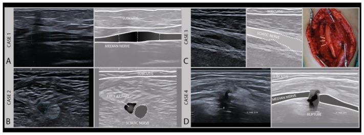
This composite educational graphic illustrates various degrees of axonal nerve damage using high-frequency musculoskeletal ultrasound (MSKUS) and intraoperative photography. The content is divided into four clinical cases, categorized by Sunderland's classification of nerve injury. Case 1 (A) shows a longitudinal US image of a median nerve demonstrating focal swelling and hypoechogenicity with intact fascicles (Sunderland Grade II). Case 2 (B) presents a transverse US image of a sciatic nerve (peroneal/fibular part) showing a heterogeneous, enlarged lesion. Case 3 (C) includes a longitudinal US and an intraoperative photograph showing severe sciatic nerve damage (Sunderland Grade III), where the intraoperative view confirms structural alterations corresponding to sonographic irregularities. Case 4 (D) displays a longitudinal US image of a median nerve featuring a complete loss of continuity and a visible rupture (Sunderland Grade V), typical of neurotmesis. Each case is accompanied by an anatomical schematic overlay identifying the subcutis and the specific nerve pathology. This resource is designed for intermediate to advanced medical learners focusing on neurology, radiology, and trauma surgery.
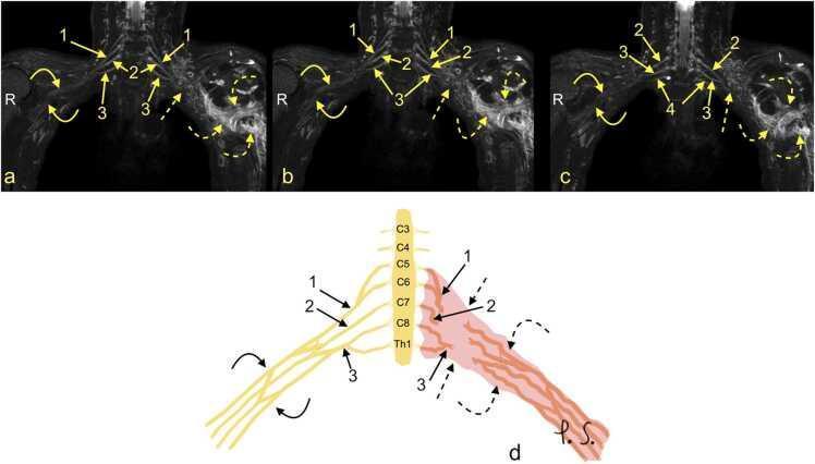
This composite educational resource comprises three Magnetic Resonance Neurography (MRN) coronal views (a, b, c) and a correlating schematic anatomical diagram (d) illustrating a severe traumatic brachial plexus injury. The right side of the images displays normal brachial plexus anatomy, with continuous nerve structures including the upper trunk (1), middle trunk (2), C8 root (3), and Th1 root (4). In contrast, the left side demonstrates a total rupture of the brachial plexus (dashed arrows). This injury is characterized by the complete discontinuity of the neural trunks and cords, accompanied by a large axillary cavity hematoma (curved dashed arrows). The schematic (d) highlights the side-to-side difference, showing the organized right-sided roots (C5-Th1) and trunks versus the disorganized, hemorrhagic area on the left. This material is designed for advanced medical education in radiology and neurosurgery, focusing on diagnostic imaging features of high-grade nerve trauma, specifically Seddon grade 3-5 injuries (neurotmesis) and the identification of postganglionic plexopathy.
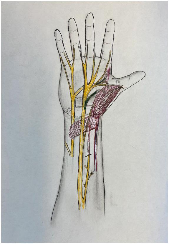
Anatomical diagram of the human hand and distal forearm in a palmar view, illustrating peripheral nerve anatomy and muscle transposition for pronator quadratus opponensplasty. The diagram features color-coded structures overlaid on a skeletal and soft tissue outline. Major peripheral nerves, including the median and ulnar nerves, are highlighted in yellow, showing their proximal courses and distal branching into digital nerves. The recipient anterior interosseous nerve (AIN) motor branch is highlighted in red, indicating its path toward the transposed muscle. The donor recurrent median nerve and donor deep ulnar nerve branches are highlighted in green, demonstrating potential donor sites for nerve coaptation to restore thenar function. The pronator quadratus muscle is shaded in red, shown in a transposed position toward the thumb (thenar eminence) to simulate its role as an opponens substitute. This illustration is designed for surgical planning in cases of chronic carpal tunnel syndrome or atrophic thenar muscle dysfunction, focusing on the spatial relationships required for effective nerve transfer and muscle mobilization.
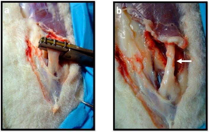
This clinical photograph set illustrates the induction of an experimental axonotmesis (crush injury) on a peripheral nerve, likely the common peroneal nerve. Panel (a) shows the intraoperative procedure where a specialized metallic, non-serrated clamp with a flat crushing surface is applied perpendicular to the long axis of a taut, whitish nerve bundle. The clamp is used to apply a controlled compressive force to disrupt the axons while maintaining the continuity of the connective tissue sheaths (epineurium and perineurium). Panel (b) depicts the nerve immediately after the removal of the instrument. A white arrow highlights the resulting pathology: a distinct, flattened, and narrowed region corresponding to the crush site. This morphology demonstrates the visual characteristics of a Seddon's second-degree nerve injury. The surgical site shows surrounding muscular tissue and minor hemorrhage, providing a clear demonstration of nerve trauma models used to study peripheral nerve regeneration and functional recovery.
Wallerian degeneration nerve regeneration stages diagram
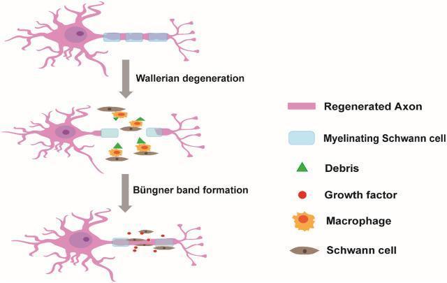
A pathophysiology diagram illustrating the stages of peripheral nerve injury (PNI) repair and the role of Schwann cells. The process is depicted in three vertical steps. Step 1 shows an intact regenerated axon (pink) with segmented myelinating Schwann cells (light blue). Step 2, labeled 'Wallerian degeneration,' shows axonal fragmentation in the distal stump, myelin breakdown into debris (green triangles), and the recruitment of macrophages (orange). Step 3, labeled 'Büngner band formation,' demonstrates the regenerative phase where activated Schwann cells (brown ovals) align to form a supportive path, secreting growth factors (red dots) to guide the re-growth of the axon from the proximal stump toward the distal target. The diagram highlights the transition of Schwann cells from a myelinating state to a repair-promoting state, facilitating clearance of debris and subsequent axonal guidance through specialized cellular columns known as bands of Büngner.
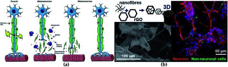
This composite educational graphic illustrates the pathophysiology of nerve injury and modern tissue engineering repair strategies. Panel (a) is a Pathophysiology Diagram showing a timeline of nerve regeneration: initial 'Injury' to an axon and its Schwann cell sheath; 'Degeneration' characterized by axonal breakdown, debris, and macrophage infiltration; and 'Regeneration' featuring proliferating Schwann cells and axonal regrowth toward the muscle target. Panel (b) focuses on Nanomaterial-based therapeutics, presenting a schematic of nanofibers combined with reduced graphene oxide (rGO) to create a 3D scaffold. This is supported by a scanning electron microscope (SEM) image showing the microscale architecture of the GO-based scaffold and a fluorescence microscopy image (50 μm scale) demonstrating cellular integration. In the fluorescence image, neurons are labeled in red as elongated fibers, while non-neuronal cells and nuclei are visible in green and blue. The visual highlights the intersection of neurology and regenerative medicine, specifically the use of conductive carbon-based nanomaterials to enhance neural progenitor cell viability and differentiation.
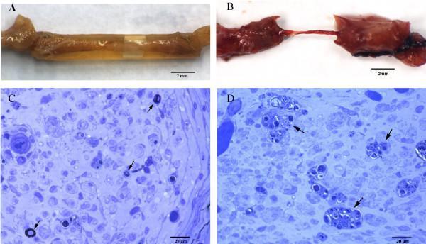
A multi-panel figure illustrating macroscopic and microscopic findings of peripheral nerve regeneration in a rat model following tubulization. Panels A and B are macroscopic clinical photographs of sciatic nerve segments 70 days post-injury. Panel A shows a yellowish-tan regeneration cable traversing a 0.4% agarose gel tube, maintaining continuity between proximal and distal stumps. Panel B shows a significantly thinner, reddish filiform cable in a DMEM-treated control. Panels C and D are light microscopy images of semithin distal nerve sections stained with Toluidine blue. Panel C (20 µm scale) demonstrates sparse, regenerating myelinated fibers (indicated by arrows) characterized by dark, circular axonal rings. Panel D (20 µm scale) highlights the endoneurial area showing clear evidence of Wallerian degeneration, including digestion chambers (arrows) containing accumulated myelin debris and disorganized cellular architecture. This sequence is used in neurology and pathology to compare the efficacy of biomaterials in supporting nerve repair and axonal regrowth.
carpal tunnel syndrome median nerve compression clinical signs
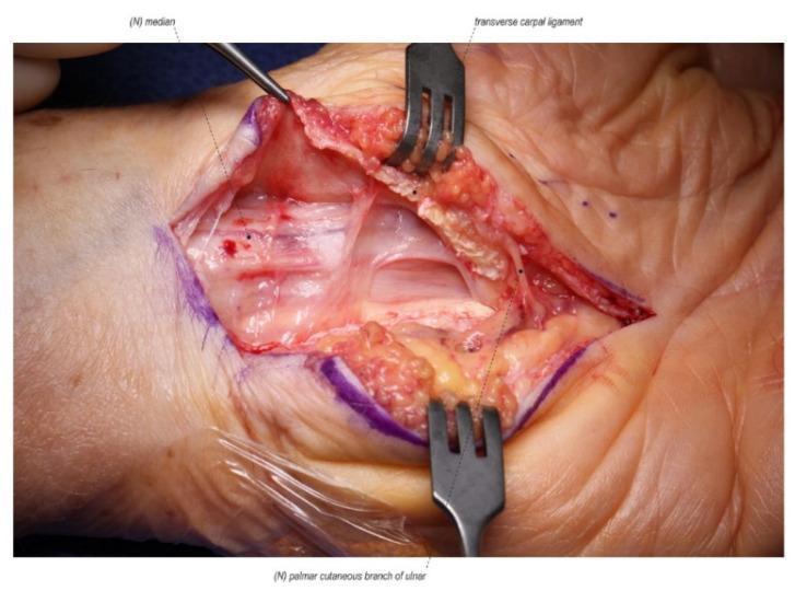
This clinical photograph captures a revision open carpal tunnel release surgical procedure on a left hand. The intraoperative field shows the median nerve, labeled as '(N) median', which exhibits signs of chronic compression, including proximal bruising, edema, and a scarred, thickened texture. The nerve is visibly adherent to the undersurface of the radial leaf of the transverse carpal ligament. The transverse carpal ligament itself is shown partially divided, with its radial and ulnar leaves retracted. Surrounding the surgical site, there is evidence of extensive postoperative fibrosis and scarring from the prior intervention, as well as localized tissue inflammation and disrupted anatomical planes. A surgical probe is used to identify the pathological tethering between the median nerve and the ligamentous tissue. The palmar cutaneous branch is also annotated in the distal surgical field. This image serves as a clinical example of recurrent or persistent carpal tunnel syndrome caused by incomplete primary release and subsequent perineural scar formation.
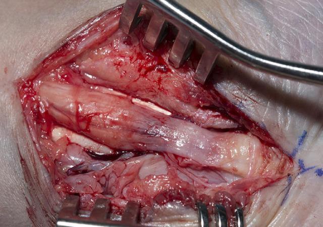
This clinical photograph captures an intraoperative view of a carpal tunnel decompression surgery, focusing on the median nerve. The image illustrates classic pathological changes associated with chronic nerve compression: a proximal bulbous swelling (pseudoneuroma) of the nerve followed by significant distal thinning (hourglass deformity) beneath the site of the flexor retinaculum. The median nerve appears pale and glistening, while the surrounding surgical field shows signs of inflammation, hemorrhage, and extensive fibrosis. Retractors are positioned to expose the nerve and underlying flexor tendons, which appear infiltrated by scar tissue and thickened subsynovial connective tissue. This visual serves as an educational example of severe carpal tunnel syndrome and the anatomical landmarks encountered during surgical release, including the relationship between the nerve, tendons, and surrounding fibrotic changes.
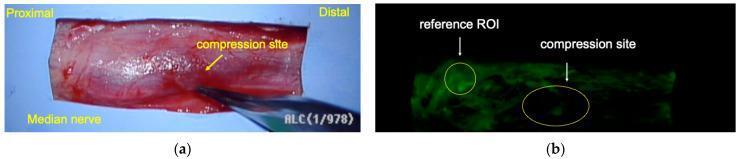
This figure demonstrates clinical and diagnostic imaging of the median nerve in a patient with Carpal Tunnel Syndrome (CTS). Image (a) is a macro clinical photograph showing a surgically exposed segment of the median nerve, with the proximal and distal ends labeled; surgical forceps point to the specific compression site, which exhibits focal indentation and surface vascular congestion. Image (b) shows the corresponding fluorescein angiography (FAG) of the same nerve segment. This diagnostic image highlights differences in microvascular perfusion through fluorescein uptake. Two Regions of Interest (ROI) are marked: a 'reference ROI' at a non-compressed segment and a 'compression site' ROI. Visually, the reference area shows higher green luminance, suggesting normal blood flow, while the compression site displays significantly reduced luminance, indicating focal ischemia or decreased perfusion due to chronic nerve compression. This comparison is used to quantify the degree of vascular impairment in neuropathic conditions.
Physiotherapy Management of Disorders of Peripheral Nerves
SLIDE 1 - Title Slide
- Presented by: [Your Name]
- Institution | Department of Physiotherapy
- Date
SLIDE 2 - Overview / Contents
- Anatomy of Peripheral Nerves
- Pathophysiology - Degeneration & Regeneration
- Classification of Nerve Injuries
- Types of Peripheral Nerve Disorders
- Clinical Assessment
- Physiotherapy Management - Principles
- Condition-Specific Management
- Outcome Measures
- Recent Evidence
SLIDE 3 - Anatomy of Peripheral Nerves
- Endoneurium - innermost connective tissue sleeve around individual axons
- Perineurium - surrounds a fascicle (bundle of axons)
- Epineurium - outermost sheath enclosing the entire nerve trunk
| Fiber | Myelination | Function |
|---|---|---|
| A-alpha | Thick myelinated | Motor, proprioception |
| A-beta | Myelinated | Touch, pressure |
| A-delta | Thin myelinated | Pain, temperature |
| C fibers | Unmyelinated | Slow pain, autonomic |
SLIDE 4 - Pathophysiology: Degeneration & Regeneration
- Axon and myelin sheath fragment and degenerate
- Macrophages phagocytose the debris
- Schwann cells dedifferentiate and proliferate to form Bands of Büngner - guiding columns for regenerating axons
- Focal damage to myelin without axon disruption
- Results in conduction block (neurapraxia)
- Remyelination occurs within 1-3 months
- Axons regenerate at ~1 mm/day (or ~1 inch/month clinically)
- Rate influenced by patient age, distance from target, fibrosis, and blood supply
- Tinel's sign advances distally as regeneration progresses
SLIDE 5 - Classification of Nerve Injuries
Seddon's Classification (1942) - 3 types:
| Seddon | Pathology | Recovery |
|---|---|---|
| Neurapraxia | Segmental demyelination; axon intact | Complete; 1-3 months |
| Axonotmesis | Axon disrupted; connective tissue intact | Slow but often complete |
| Neurotmesis | Complete nerve disruption | Surgical repair needed |
Sunderland's Classification (1991) - 5 Grades:
| Grade | Seddon Equivalent | Structures Injured | Wallerian Degeneration | Treatment |
|---|---|---|---|---|
| I | Neurapraxia | Myelin only | No | Spontaneous recovery |
| II | Axonotmesis | Axon + myelin (endoneurium intact) | Yes | Often recovers spontaneously |
| III | Axonotmesis | Axon + endoneurium damaged (perineurium intact) | Yes | Fair prognosis; may need surgery |
| IV | Axonotmesis | Axon + endo + perineurium (epineurium intact) | Yes | Poor prognosis; usually needs surgery |
| V | Neurotmesis | Complete nerve transection | Yes | Surgical repair mandatory |
| VI (MacKinnon) | Mixed | Multiple grades in same nerve | Variable | Complex reconstruction |
SLIDE 6 - Types / Causes of Peripheral Nerve Disorders
A. Traumatic Nerve Injuries
- Compression (Saturday night palsy - radial nerve, prolonged tourniquet)
- Traction / Stretch (brachial plexus injuries in motorcyclists - Erb's palsy, Klumpke's palsy)
- Laceration (knife wounds, glass, surgical injury)
- Crush (blunt trauma)
- Injection injuries (intramuscular injections near sciatic nerve)
- Avulsion (rootlet pulled from spinal cord - worst prognosis)
B. Entrapment / Compression Neuropathies
- Carpal Tunnel Syndrome (CTS) - median nerve at wrist
- Cubital Tunnel Syndrome - ulnar nerve at elbow
- Tarsal Tunnel Syndrome - tibial nerve at ankle
- Meralgia Paresthetica - lateral femoral cutaneous nerve
- Thoracic Outlet Syndrome - brachial plexus/subclavian vessels
C. Polyneuropathies
- Diabetic peripheral neuropathy (most common metabolic cause)
- Guillain-Barre Syndrome (GBS) - acute immune-mediated demyelinating polyneuropathy
- Chronic Inflammatory Demyelinating Polyneuropathy (CIDP)
- Hereditary neuropathies (Charcot-Marie-Tooth disease)
- Alcohol-related neuropathy
D. Peripheral Nerve Palsies
- Radial nerve palsy - wrist drop
- Ulnar nerve palsy - claw hand (ring & little finger), Froment's sign
- Median nerve palsy - ape hand (thenar wasting), loss of precision grip
- Common peroneal nerve palsy - foot drop
- Long thoracic nerve palsy - winging of scapula
SLIDE 7 - Clinical Assessment
History:
- Mechanism and duration of injury
- Distribution of weakness/sensory loss
- Autonomic features (dryness, color change)
- Functional limitations (ADL, work, sport)
Motor Assessment:
- MRC grading (0-5) of affected muscle groups
- Pattern of weakness maps to specific nerve territory
- Look for wasting, fasciculations
Sensory Assessment:
- Light touch, pin prick, temperature
- Two-point discrimination (2PD): Static and moving. Normal 2PD = 6 mm or less. If nerve is transected, patient cannot discriminate between 1 and 2 points. Markedly widened 2PD seen in partial injuries. (Campbell's Operative Orthopaedics)
- Proprioception, vibration (tuning fork 128 Hz)
- Semmes-Weinstein Monofilaments
Special Tests:
- Tinel's Sign - tapping over nerve; advancing Tinel = regeneration
- Phalen's Test - for CTS
- Froment's Sign - for ulnar nerve (weak adductor pollicis)
- Wartenberg's Sign - little finger abduction in ulnar neuropathy
- Finkelstein's Test - for De Quervain's (differentiate)
Electrodiagnostic Studies:
- Nerve Conduction Studies (NCS): Measure conduction velocity and amplitude. Useful from 3 weeks post-injury. Shows demyelination vs. axon loss patterns
- Electromyography (EMG): Detects fibrillation potentials and denervation at 3 weeks; monitors reinnervation
- Both NCS and EMG are valuable for monitoring recovery progress (Sabiston Textbook of Surgery)
Functional Assessment:
- Grip and pinch strength (dynamometry)
- Dexterity tests (9-hole peg test, Purdue Pegboard)
- DASH or QuickDASH (upper extremity disability)
SLIDE 8 - Physiotherapy Management: General Principles
- Prevent secondary complications (contractures, muscle wasting, stiff joints)
- Maintain joint range of motion
- Protect denervated skin and insensate areas
- Facilitate nerve regeneration
- Re-educate sensorimotor function
- Restore functional independence
- Phase 1 - Acute / Denervation phase: Protection, oedema control, splinting, passive exercises
- Phase 2 - Regeneration phase: Re-education begins, progressive active exercises
- Phase 3 - Reinnervation / Recovery phase: Sensory re-education, strengthening, functional training
SLIDE 9 - Physiotherapy Management: Specific Interventions
1. Splinting / Orthoses
- Used to prevent deformity during denervation
- Wrist drop (radial nerve): Cock-up wrist splint (wrist in 30-45° extension)
- Claw hand (ulnar nerve): Lumbrical bar splint (blocks MP hyperextension)
- Foot drop (peroneal nerve): Ankle-foot orthosis (AFO) in neutral position
- Median nerve palsy: Opponens splint to maintain thumb web space
2. Exercise Therapy
- Passive range of motion (PROM): Prevents contracture of denervated muscles; maintain full joint ROM daily
- Active-assisted exercises: As reinnervation begins
- Progressive resistive exercises: Once MRC grade 3+ is achieved
- Neuromuscular electrical stimulation (NMES): Can be used on denervated muscle to reduce atrophy while awaiting reinnervation
3. Sensory Re-education
- Begins when protective sensation returns (moving touch perceived)
- Early phase: Localization exercises with eyes closed/open; graded textures
- Late phase: Object identification (stereognosis), textured discrimination
- Mirror therapy for sensory reorganization
- Goal: Cortical remapping to restore functional sensory perception
4. Electrotherapy / Physical Agents
- Transcutaneous Electrical Nerve Stimulation (TENS): Pain management
- Neuromuscular Electrical Stimulation (NMES/FES): Retard denervation atrophy
- Ultrasound therapy: Promotes tissue healing, reduce fibrosis around nerve
- LASER / Low Level Light Therapy (LLLT): Some evidence for accelerating nerve regeneration
- Extracorporeal Shock Wave Therapy (ESWT): Recent systematic review (2024) shows improvement in nerve conduction velocity (PMID: 39650239)
- Hydrotherapy: Useful in Guillain-Barre for buoyancy-assisted exercise
5. Scar Management (post-surgical cases)
- Scar massage, silicone gel, desensitization techniques
- Prevent perineural adhesions that restrict nerve gliding
6. Nerve Gliding Exercises
- Gentle tensioning and sliding of the nerve within its bed
- Reduces intraneural oedema, prevents adhesion
- Used in entrapment neuropathies (CTS, cubital tunnel)
- Median nerve glides (wrist, elbow, shoulder positions)
- Ulnar nerve glides
- Neural tension exercises based on Butler's neurodynamics
7. Education & Activity Modification
- Posture correction (e.g. avoiding sustained elbow flexion in cubital tunnel)
- Ergonomic advice (keyboard/workstation for CTS)
- Skin care for insensate areas - daily inspection, padded footwear
- Avoidance of pressure over vulnerable sites
SLIDE 10 - Condition-Specific Physiotherapy
A. Carpal Tunnel Syndrome (CTS - Median Nerve at Wrist)
- Neutral wrist splint at night (first-line for mild-moderate CTS)
- Nerve and tendon gliding exercises
- Ergonomic modification
- Ultrasound/TENS for pain
- Post-surgical: scar management, nerve gliding, grip strengthening
B. Ulnar Nerve Palsy (Cubital Tunnel / Guyon's Canal)
- Elbow extension splint at night (cubital tunnel)
- Lumbrical bar splint to correct claw deformity
- Intrinsic muscle strengthening
- Sensory re-education (little/ring finger and ulnar palm)
- Tendon transfer rehab if surgery performed
C. Radial Nerve Palsy (Wrist Drop)
- Cock-up wrist splint (dorsal; wrist in 30-45° extension)
- PROM of wrist and finger joints
- NMES to wrist/finger extensors
- Progressive tenodesis exercises
- Functional retraining; dynamic splint as recovery progresses
D. Foot Drop (Common Peroneal Nerve Palsy)
- AFO in neutral/slight dorsiflexion
- PROM ankle, toe joints
- NMES to tibialis anterior and peronei
- Gait re-education (hip hiking elimination)
- Balance and proprioceptive training
- FES (functional electrical stimulation) during walking
E. Guillain-Barre Syndrome (GBS)
- Acute ICU phase: Positioning, chest physiotherapy, passive ROM, DVT prevention
- Subacute phase: Tilt table for orthostatic hypotension, progressive mobilization, hydrotherapy
- Rehabilitation phase: Strengthening, gait training, ADL retraining, fatigue management
- Monitor for autonomic instability during exercise
- Respiratory physiotherapy - breathing exercises, secretion clearance
- Energy conservation strategies (fatigue is a major long-term issue)
F. Brachial Plexus Injury
- PROM all joints of the affected limb daily
- Splinting to prevent contractures (shoulder external rotation, elbow flexion, etc.)
- NMES, biofeedback to facilitated returning muscle activity
- Proprioceptive training
- Post-surgical rehab (nerve repair/grafting/transfer)
- Functional re-education; compensatory strategies for permanent deficits
SLIDE 11 - Outcome Measures
| Domain | Tool |
|---|---|
| Motor strength | MRC Scale (0-5) |
| Sensation | Semmes-Weinstein Monofilaments, Static/Moving 2PD |
| Function (UL) | DASH / QuickDASH, ABILHAND |
| Function (LL) | WHOQOL, 10MWT, BERG Balance Scale |
| Pain | VAS / NRS / NPRS |
| Quality of Life | SF-36 |
| Nerve regeneration | EMG, NCS |
| Dexterity | 9-Hole Peg Test, Purdue Pegboard |
SLIDE 12 - Prognosis & Factors Affecting Recovery
- Young age
- Neurapraxia / Grade I-II injuries
- Short distance from injury to target organ
- Clean laceration (vs. crush/avulsion)
- Early treatment
- Good vascular supply
- Avulsion injuries (rootlet from cord)
- Neurotmesis (Grade V)
- Elderly patients
- Long distance to reinnervation target
- Delay in management
- Systemic diseases (diabetes, vasculopathy)
SLIDE 13 - Recent Evidence (2024-2025)
-
ESWT for peripheral nerve disorders - A 2024 systematic review (PMID 39650239) found extracorporeal shock wave therapy significantly improves nerve conduction velocity in peripheral nerve disorders.
-
Intraoperative electrical stimulation - A 2025 meta-analysis (PMID 40199698) investigated whether short-term intraoperative electrical stimulation enhances nerve regeneration following peripheral nerve repair.
-
Nerve stimulation for neuropathic pain - A 2025 systematic review (PMID 40108850) assessed brachial plexus nerve stimulation for refractory neuropathic pain in upper extremities.
SLIDE 14 - Take-Home Points
- Peripheral nerve disorders result from trauma, entrapment, metabolic, or immune-mediated causes
- Seddon (3 types) and Sunderland (5 grades) classifications guide prognosis and management
- Wallerian degeneration occurs distal to injury; axons regenerate at ~1 mm/day
- Physiotherapy is the cornerstone of non-surgical management: splinting, exercises, electrotherapy, sensory re-education, and nerve gliding
- Outcome depends on severity of injury, distance, age, and timeliness of intervention
- EMG/NCS are essential for monitoring regeneration
- Multidisciplinary approach (PT + OT + surgeon + neurologist) is ideal
SLIDE 15 - References
- Bradley and Daroff's Neurology in Clinical Practice (8th ed.)
- Campbell's Operative Orthopaedics (15th ed., 2026)
- Sabiston Textbook of Surgery (21st ed.)
- Miller's Review of Orthopaedics (9th ed.)
- Current Surgical Therapy (14th ed.)
- Yang L et al. Effect of ESWT on nerve conduction: A systematic review. Front Neurol. 2024. PMID: 39650239
- Koopman JE et al. Intraoperative electrical stimulation and nerve regeneration. J Plast Reconstr Aesthet Surg. 2025. PMID: 40199698
Additional Images for Your Slides
- Structure it exactly as above: anatomy → pathology → classification → conditions → assessment → management → outcomes
- Use the classification table (Seddon vs Sunderland) as a key diagram - examiners love that
- For each condition, use the format: nerve affected → muscles/sensory lost → deformity → splint → exercises
- Emphasize the 1 mm/day regeneration rate and Tinel's sign progression - these are high-yield clinical pearls
- Include sensory re-education as a distinct section - it is often under-covered and will set your presentation apart
Physiotherapy management in more detail week wise
peripheral nerve injury rehabilitation protocol week
physiotherapy week wise protocol peripheral nerve injury rehabilitation phases
https://www.bssh.ac.uk/_userfiles/pages/files/professionals/…
Week-Wise Physiotherapy Management of Peripheral Nerve Disorders
BIOLOGICAL TIMELINE - Why the phases exist
| Time | Biology | Clinical correlate |
|---|---|---|
| 0-72 hrs | Inflammatory response | Pain, swelling, risk of tension at repair |
| Day 3-14 | Wallerian degeneration (distal) | EMG shows fibrillations from day 21 |
| Day 1-21 | Axon sprouting from proximal stump begins | No clinical recovery yet |
| Week 3 onwards | Axon regenerating at ~1 mm/day | Advancing Tinel's sign |
| Week 3-12 | Muscle atrophy accelerates without stimulation | Muscle wasting, fibrosis risk |
| 3-6 months | First clinical signs of reinnervation (MRC 1-2) | Exercise becomes effective |
| 6-18 months | Functional recovery if nerve supply reaches muscle | Sensory re-education critical here |
| >18-24 months | Irreversible fibrosis of denervated muscle | Time window for recovery closes |
PHASE 1: ACUTE / PROTECTION PHASE
Week 0-1 (Days 1-7) - Immediate Post-Injury or Post-Surgery
- Protect nerve repair from tension and stress
- Control pain and oedema
- Prevent stiffness of joints proximal and distal to injury
- Patient education
- Apply protective splint to offload tension from repair site (positioned to shorten nerve - e.g., elbow flexed for ulnar nerve repair at elbow, wrist neutral for median/ulnar at wrist)
- Full-time splint wear at this stage
- Monitor for pressure areas given loss of protective sensation
- No active exercise of denervated muscles
- Passive ROM of all joints in the affected limb: gentle, full arc, twice daily (prevents stiffness)
- Active ROM of joints not in the zone of injury (proximal and distal uninvolved segments)
- Shoulder and elbow pendulum exercises (upper limb injuries) - prevent frozen shoulder
- Elevate limb to control oedema
- Cryotherapy (ice packs 10-15 min) for acute pain and oedema control
- TENS for pain management (conventional mode, 80-100 Hz)
- Wound dressings as appropriate
- Protect insensate skin - inspect daily, avoid heat/cold/pressure
- Positioning during rest and sleep (e.g., elevate hand above heart level)
- Do NOT try to use the paralyzed muscles forcefully
- Realistic expectation-setting on recovery timeline
Week 1-3 (Days 7-21) - Early Protection Phase
- Maintain protection while beginning cautious mobilization
- Begin edema reduction
- Joint mobility maintenance
- Continue protective splint (3-4 weeks of immobilization typical after nerve repair) (BSSH Guidelines, Hunter's Rehabilitation of the Hand)
- Begin checking for pressure areas, adjust splint as swelling reduces
- Continue passive ROM of all affected joints twice daily - full range, gentle
- Begin place-and-hold exercises for unaffected muscles (patient uses uninvolved hand to position, then attempts minimal isometric hold)
- Shoulder/elbow circumduction if upper limb (avoid frozen shoulder/stiff elbow)
- Ankle pumps and toe exercises (lower limb - foot drop)
- NMES / Faradic stimulation of denervated muscles can begin from Day 7-10:
- Indication: Prevents muscle fibre atrophy and fibrosis during the denervation period
- Parameters: Exponential current, 1:5 duty cycle (1 sec on: 5 sec off), 10-15 min/day
- Continue until voluntary contraction is possible (MRC 1-2)
- TENS continues for pain
- Retrograde massage from distal to proximal
- Compression bandaging/gloves (upper limb)
- Elevation
PHASE 2: REGENERATION / FACILITATION PHASE
Week 3-6 - Early Active Phase (Post-immobilization)
- Recover ROM lost during immobilization
- Begin nerve gliding
- Initiate sensory re-education (early phase)
- Commence scar management
- Transition from immobilization splint to functional/resting splint
- Dynamic splints may be introduced to assist weak muscles while protecting repair:
- Radial nerve: dynamic wrist extension splint (cock-up)
- Ulnar nerve: lumbrical bar splint
- Foot drop: AFO in neutral
- Gradually wean wear time during day - full-time use at night
- Begin active-assisted ROM for affected joints
- Avoid composite positions that tension the nerve repair excessively (e.g., avoid composite wrist + finger flexion in radial nerve repairs until week 8)
- Nerve gliding exercises commence at Week 3 (BSSH Guidelines):
- Median nerve glides: wrist extension with finger extension (tensioner) vs. wrist flexion with finger flexion (slider)
- Ulnar nerve glides: elbow flex/extend with shoulder ABD
- Sciatic/peroneal: ankle dorsiflexion with knee extension (neural tensioner)
- Do 10 repetitions, 3 sets, twice daily - pain-free range only
- Tendon gliding exercises (hand injuries): hook fist, straight fist, full fist - prevents intrinsic tightness
- Begin scar massage over healed incision - circular friction technique
- Silicone gel sheets over hypertrophic scar (wear 23 hrs/day)
- Goal: prevent perineural adhesions limiting nerve mobility
- Localization of moving touch: apply light moving touch to skin; patient (eyes open) identifies area; then eyes closed and repeats
- Grade up: cotton wool → fingertip → eraser end
- Vibration (30 Hz tuning fork): first modality to return after reinnervation
- Spend 5-10 minutes per session, 2-3 sessions/day
- Begin with least aversive textures: cotton, soft towel
- Progress through textures: velvet → flannel → terrycloth → denim → Velcro
- Progress to immersion: rice, beans, sand
- Each texture 10 minutes, 3x/day; replace when no longer uncomfortable
- Continue NMES on denervated muscles
- Therapeutic ultrasound over repair site (pulsed mode, 1 MHz, 0.5-1.0 W/cm², 5 min) - reduces fibrosis
- Low-level laser therapy (LLLT): over nerve repair site - may accelerate regeneration
Week 6-12 - Active Rehabilitation Phase
- Restore full ROM
- Progressive muscle strengthening as reinnervation begins
- Advance sensory re-education
- Begin functional tasks
- Dynamic splint continues to assist weak muscles functionally
- Begin reducing day wear time as strength improves (MRC 3 = against gravity)
- Wean splint progressively - use only for demanding activities/work
- Active ROM exercises - full range without assistance
- Progressive resisted exercises commence once MRC grade reaches 3 (against gravity):
- Week 6-8: Putty exercises (light resistance, grading by putty hardness)
- Begin with light open-chain exercises, progress to closed-chain
- Isometric → isotonic → isokinetic
- Intrinsic muscle exercises (hand): finger spreading, pinch strengthening with pegs
- Proprioceptive exercises: Weight-bearing through hand (upper limb), single-leg stance progression (lower limb)
- Balance training for foot drop cases: single leg stance on foam → unstable surfaces
- Functional task practice: writing, dressing, pouring (upper limb); walking on uneven surfaces (lower limb)
- Gait training in AFO: normal heel-toe pattern
- Proprioceptive taping (kinesiology tape) as adjunct
- Progress to walking without AFO indoors as dorsiflexors recover (MRC 3+)
- Object identification (stereognosis): identify coins, keys, shapes with eyes closed
- Texture discrimination: fine sandpapers (graded from coarse to fine)
- Functional sensory tasks: manipulating coins, buttoning, identifying objects in a bag
- Mirror therapy as adjunct for cortical remapping
- Progress 5-10 minutes per session, 3x/day
- Biofeedback (surface EMG): once MRC grade 1 appears, use EMG biofeedback to teach patient to selectively activate returning motor units - very effective for cortical reorganization
- NMES transitions to functional electrical stimulation (FES): triggered by voluntary intent
- TENS for any residual neuropathic pain
- Continue LLLT if available
PHASE 3: FUNCTIONAL RESTORATION / RETRAINING PHASE
Week 12-24 (3-6 Months)
- Maximize muscle strength and endurance
- Restore normal movement patterns and coordination
- Develop functional independence in ADL and occupational tasks
- Address compensatory movement patterns
- Dynamic splint use only during demanding occupational/sport activities
- Night resting splint discontinued if ROM is maintained
- Reassess need for orthoses based on recovery
- Progressive resisted exercises: theraband, free weights, resistance machines
- 3 sets × 10-15 reps, 3x/week; progress by 10% per week (ACSM principle)
- Endurance training: cycling, walking, swimming (low-impact, high proprioceptive benefit)
- Task-specific motor training:
- UL: pegboards, blocks, handwriting retraining, keyboard use, simulated ADL
- LL: step-ups, stairs, running progression, sports-specific drills
- Coordination and dexterity drills:
- 9-hole peg test practice
- Coin manipulation
- Instrumental activities (tying laces, using cutlery)
- Proprioceptive training (advanced):
- Bosu ball, wobble board (lower limb)
- Unstable surface hand weight-bearing (upper limb)
- TENS (burst mode or AL-TENS for neuropathic pain)
- Desensitization (if allodynia persists)
- Graded motor imagery / mirror therapy
- Pain neuroscience education
- Grip and pinch dynamometry (compare to normative values / contralateral side)
- Functional capacity evaluation
- Work simulation exercises
- Ergonomic modification advice
- Progressive return to work or sport activities
PHASE 4: LONG-TERM MAINTENANCE (6+ Months)
Week 24 onwards
- Maximize functional outcome within limits of nerve recovery
- Manage permanent deficits
- Prevent secondary complications
- Continue progressive strengthening (no ceiling - push to maximum recovery)
- Sensory re-education continues for up to 2 years (cortical plasticity persists)
- Monitor EMG/NCS every 3-6 months
- Assess Tinel's sign advancement at each review
- Compensatory strategies: adapted grips, one-handed techniques, assistive devices
- Long-term splinting: static splints for positioning (e.g., AFO for permanent foot drop)
- Strengthening of intact muscles to compensate (e.g., wrist extensors for ulnar intrinsic palsy)
- Home exercise program maintenance
CONDITION-SPECIFIC WEEK-WISE VARIATIONS
Carpal Tunnel Syndrome (Non-surgical / Post-surgical)
| Week | Management |
|---|---|
| Week 0-3 (non-surgical) | Night wrist splint (neutral); nerve + tendon glides 3x/day; ergonomic modification |
| Week 0-3 (post-surgical) | Bulky dressing → light splint; elevation; gentle finger ROM; scar protection |
| Week 3-6 | Scar massage begins; wrist ROM exercises; TENS for pain |
| Week 6-12 | Progressive grip strengthening (putty, squeeze ball); full activity return |
| Week 12+ | Return to normal activity; workstation re-assessment |
Radial Nerve Palsy (Wrist Drop) - Non-surgical
| Week | Management |
|---|---|
| Week 0-3 | Cock-up wrist splint full-time; PROM wrist/finger joints; NMES to extensors; elevation |
| Week 3-6 | Active-assisted wrist/finger extension in splint; nerve glides; NMES continues |
| Week 6-12 | Active resisted exercises when MRC 3+ appears; dynamic splint; functional tasks |
| Week 12+ | Progressive strengthening; wean splint; grip/pinch dynamometry; return to work |
Ulnar Nerve Palsy (Cubital Tunnel)
| Week | Management |
|---|---|
| Week 0-3 | Elbow extension splint at night; lumbrical bar splint; PROM; oedema control |
| Week 3-6 | Nerve glides (ulnar); intrinsic muscle NMES; scar management (post-surgical) |
| Week 6-12 | Intrinsic strengthening (finger abduction/adduction); sensory re-education ring/little finger |
| Week 12+ | Pinch strength, key pinch, coordination; functional ADL training |
Foot Drop (Common Peroneal Nerve)
| Week | Management |
|---|---|
| Week 0-3 | AFO full-time; PROM ankle/toes; NMES tibialis anterior 2x/day; crutch-assisted gait |
| Week 3-6 | Gait re-education in AFO; balance training (bilateral, stable surface) |
| Week 6-12 | Progressive dorsiflexion strengthening; FES during walking; proprioceptive exercises |
| Week 12+ | Stair/ramp training; running if recovery allows; sport-specific rehab |
Guillain-Barré Syndrome (GBS)
| Phase | Timing | Management |
|---|---|---|
| ICU / Acute | Week 1-4 | Chest PT, secretion clearance; PROM all 4 limbs 2x/day; positioning; DVT prophylaxis; tilt table for autonomic instability; communication aids |
| Subacute | Week 4-8 | Hydrotherapy (buoyancy assists exercise); passive to active-assisted transitions; sitting balance; low-intensity strengthening (avoid overwork fatigue) |
| Early rehab | Week 8-16 | Progressive ambulation; transfer training; ADL retraining; fatigue management (pacing strategy) |
| Late rehab | Month 4-12 | Community mobility; return to work prep; aerobic conditioning; sensory re-education; psychosocial support |
PROGRESSION CRITERIA (Advance to Next Phase When:)
| Milestone | Criteria |
|---|---|
| Start active exercise | Wound healed; immobilization period complete; MRC ≥ 1 |
| Start resisted exercise | MRC ≥ 3 (movement against gravity) |
| Wean splint during day | MRC ≥ 3+ in target muscles |
| Discontinue splint | Full ROM maintained; MRC ≥ 4 |
| Start late sensory re-education | Static 2PD returns; protective sensation present |
| Return to work | MRC ≥ 4; adequate sensory return; grip ≥ 75% of contralateral |
| Discharge | Plateau in recovery (3 consecutive sessions no progress); home program established |
KEY MONITORING PARAMETERS (Record at Each Visit)
- MRC grade of target muscles
- 2PD (static and moving) - record in mm
- Tinel's sign location (mark on skin diagram - advancing = regenerating)
- ROM (goniometry)
- Grip/pinch strength (dynamometry)
- VAS/NRS for pain
- Oedema (volumetry or circumferential measurement)
- Functional outcome score (DASH, QuickDASH, WHOQOL)
- EMG/NCS every 3 months until recovery plateau