
Ankylosing spondyitis
ankylosing spondylitis bamboo spine X-ray
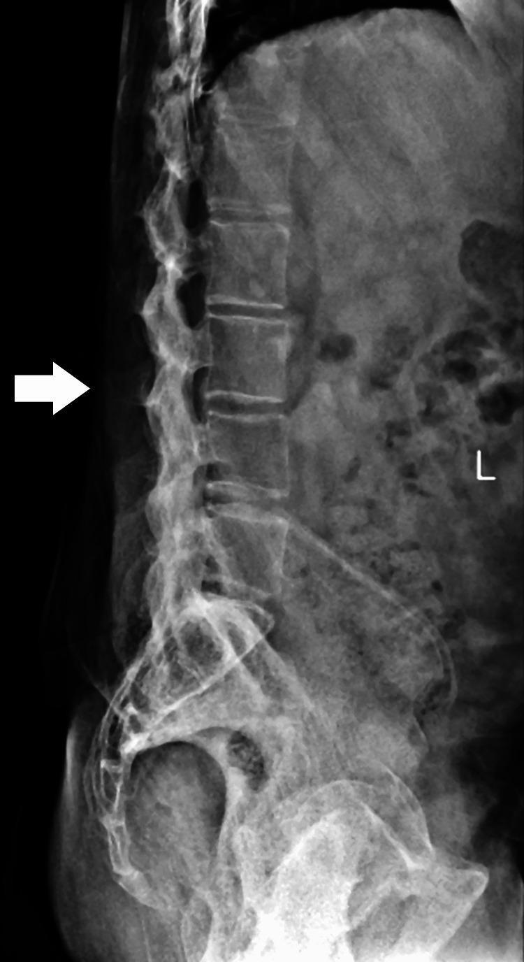
A lateral view X-ray of the lumbosacral spine demonstrating advanced radiographic features of Ankylosing Spondylitis. The image shows the classic 'bamboo spine' appearance, characterized by the formation of thin, vertical marginal syndesmophytes that bridge the intervertebral spaces. There is significant bony ankylosis (fusion) of the vertebral bodies, with blurring and near-complete loss of distinct intervertebral disc spaces. The vertebral endplates appear indistinct and merged. Additional findings include 'squaring' of the lumbar vertebrae and ossification of the spinal ligaments, specifically the interspinous and supraspinous ligaments (indicated by a white arrow pointing to the fused posterior elements). There is a notable loss of the normal lumbar lordosis, suggesting severe spinal stiffness and reduced mobility. The image serves as a definitive educational example of end-stage axial spondyloarthritis involving the lumbar region and sacrum.
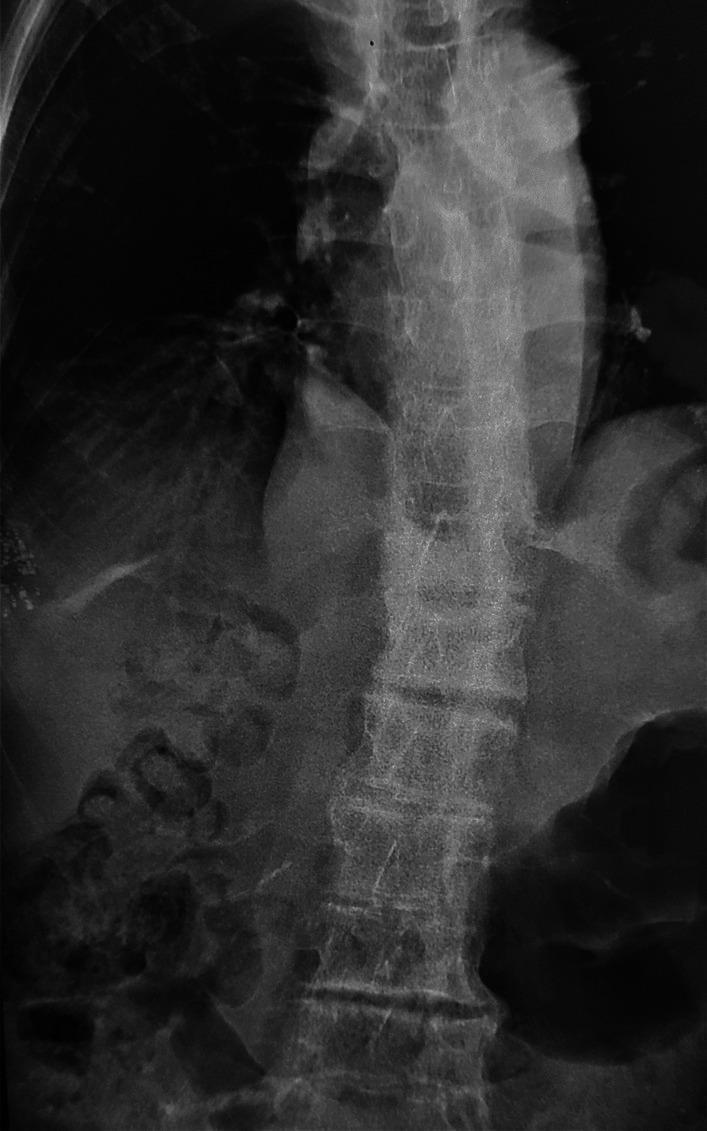
Frontal (anteroposterior) X-ray of the thoracolumbar spine demonstrating classic features of ankylosing spondylitis. The most prominent finding is the 'bamboo spine' appearance, characterized by diffuse, thin, vertical syndesmophytes that bridge the intervertebral disc spaces along the lateral aspects of the vertebral bodies. These osseous bridges are the result of ossification of the outer fibers of the annulus fibrosus. The vertebral bodies exhibit a 'squaring' morphology with irregular endplates and narrowed intervertebral spaces. There is evidence of diffuse bone demineralization (osteopenia), resulting in a generalized decrease in radiodensity and a more translucent appearance of the trabecular bone. The image also captures a mild scoliotic curvature and loss of the normal spinal contours. This diagnostic image illustrates the chronic progression of an inflammatory spondyloarthropathy, emphasizing the fusion of spinal segments and associated metabolic bone changes often seen in long-term clinical cases.
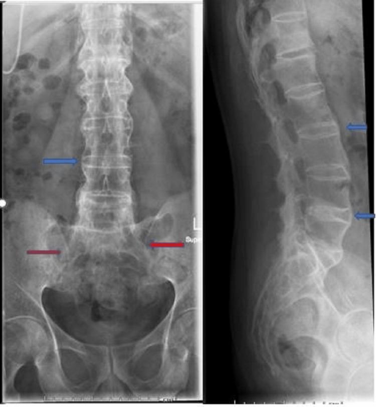
This diagnostic imaging shows anteroposterior (AP) and lateral X-ray views of the lumbar spine and pelvis, demonstrating classic radiographic hallmarks of advanced ankylosing spondylitis. The lumbar spine exhibits 'bamboo spine' morphology, characterized by the ossification of the annulus fibrosus and the formation of syndesmophytes. These vertical bony bridges connect adjacent vertebral bodies (highlighted by blue arrows), resulting in a continuous, straightened, and fused vertebral column with significant loss of normal intervertebral disc space. In the pelvic region, the sacroiliac (SI) joints show evidence of bilateral ankylosis (indicated by red arrows), with complete obliteration of the joint spaces, subchondral sclerosis, and bony fusion. These findings are clinically significant as they represent the end-stage structural damage typical of axial spondyloarthritis, leading to spinal rigidity and decreased mobility. The image serves as a definitive educational example of progressive inflammatory arthritis affecting the axial skeleton.
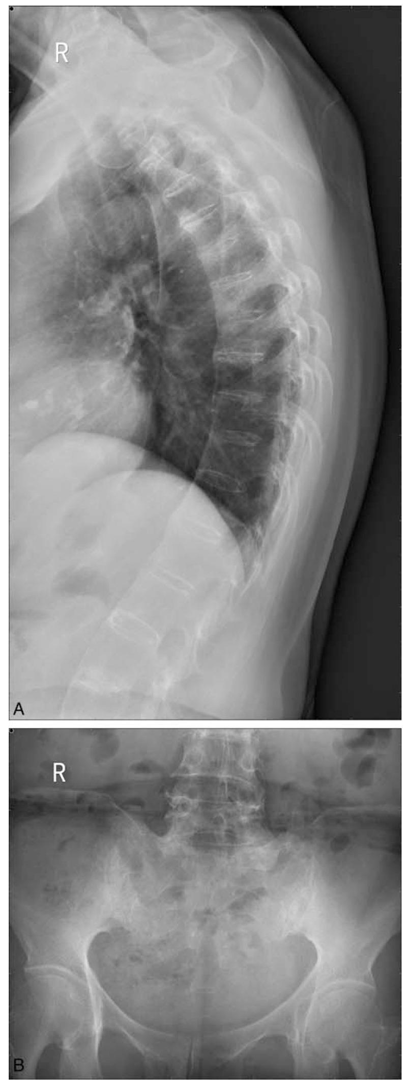
This composite of two X-ray images demonstrates classic radiographic signs of Ankylosing Spondylitis (AS). Panel A is a lateral chest/thoracic spine radiograph showing characteristic 'bamboo spine' appearance. This is characterized by extensive syndesmophyte formation, where bony bridges fuse the thoracic vertebral bodies together, creating a continuous, rigid vertical column. Notably, the intervertebral disc spaces remain relatively preserved in height despite the fusion. Panel B is an anteroposterior (AP) pelvic radiograph focusing on the sacroiliac joints. It shows Grade III bilateral sacroiliitis, evidenced by significant subchondral sclerosis (increased bone density) and narrowing with irregular joint margins. The sacrum and iliac wings exhibit focal areas of erosion and fusion. These findings are hallmarks of seronegative spondyloarthropathies and are used clinically to assess disease progression and staging in patients presenting with chronic inflammatory back pain and HLA-B27 positivity.
"ankylosing spondylitis"[MeSH Terms] AND treatment
"axial spondyloarthritis" AND biologics
Ankylosing Spondylitis (AS)
Definition and Classification
Epidemiology
- Occurs in approximately 0.2% of the general population
- Found in ~2% of HLA-B27-positive individuals, rising to ~20% if they also have an affected family member
- Most common onset age: 15-25 years (can also occur in adolescence)
- Male-to-female ratio historically 2.5:1 to 5:1, though more recent data suggest a lower ratio
- HLA-B27 is present in 90-95% of White patients with AS; prevalence of B27 varies widely by ethnicity (absent in Australian aboriginals, 1% in Japan, 7% in northern Europeans)
Pathobiology
- Molecular mimicry - sequence homology between B27 and gram-negative enteric bacteria (e.g., Klebsiella) may trigger cross-reactive immune responses
- Unfolded protein response (UPR) - B27 is distinctive in its tendency to misfold in the endoplasmic reticulum, inducing a pro-inflammatory cascade
- Heavy-chain homodimers - B27 can form homodimers at the cell surface, potentially activating NK and CD4+ T cells
Clinical Manifestations
Axial Disease (core)
- Inflammatory back pain - the hallmark: insidious onset, worse with rest/at night, improves with activity (distinguishes from mechanical back pain)
- Morning stiffness lasting >1 hour
- Sacroiliitis - bilateral, symmetrical; tenderness over SI joints
- Progressive spinal stiffness, loss of lumbar lordosis, increased thoracic kyphosis
- Reduced chest expansion (<2.5 cm at 4th intercostal space)
- Enthesitis - inflammation at tendon and ligament insertion sites (Achilles, plantar fascia, spinous processes)
Peripheral Joint Involvement
- Up to 30% of patients
- Hip joint is the most commonly affected peripheral joint after the SI joints
- Entheseal new bone forms around the femoral head
Extra-Articular Manifestations
| System | Feature |
|---|---|
| Eye | Anterior uveitis (iritis) - most common extra-articular feature; acute, recurrent, unilateral |
| Cardiac | Aortitis, aortic regurgitation, conduction defects |
| Pulmonary | Restrictive lung disease (rib cage fixation); fibrobullous upper lobe disease (1-4%) |
| Skin | Psoriasis |
| GI | Inflammatory bowel disease (Crohn's, UC) |
| Neurological | Cauda equina syndrome (late complication); atlantoaxial subluxation |
| Renal | IgA nephropathy; amyloidosis |
| Bone | Osteoporosis - significant complication of chronic inflammation |
Imaging Findings
Plain Radiography
- Sacroiliitis: erosions, subchondral sclerosis, joint space loss progressing to ankylosis
- Romanus lesions: sclerotic "shiny corners" at vertebral body corners (enthesitis at Sharpey fibers)
- Vertebral squaring: loss of normal concave anterior vertebral border
- Syndesmophytes: thin vertical bony outgrowths at Sharpey fiber insertions (distinguish from the coarser, more horizontal osteophytes of psoriatic or reactive arthritis)
- Bamboo spine: complete fusion of vertebral bodies with bridging syndesmophytes - the end-stage appearance
MRI
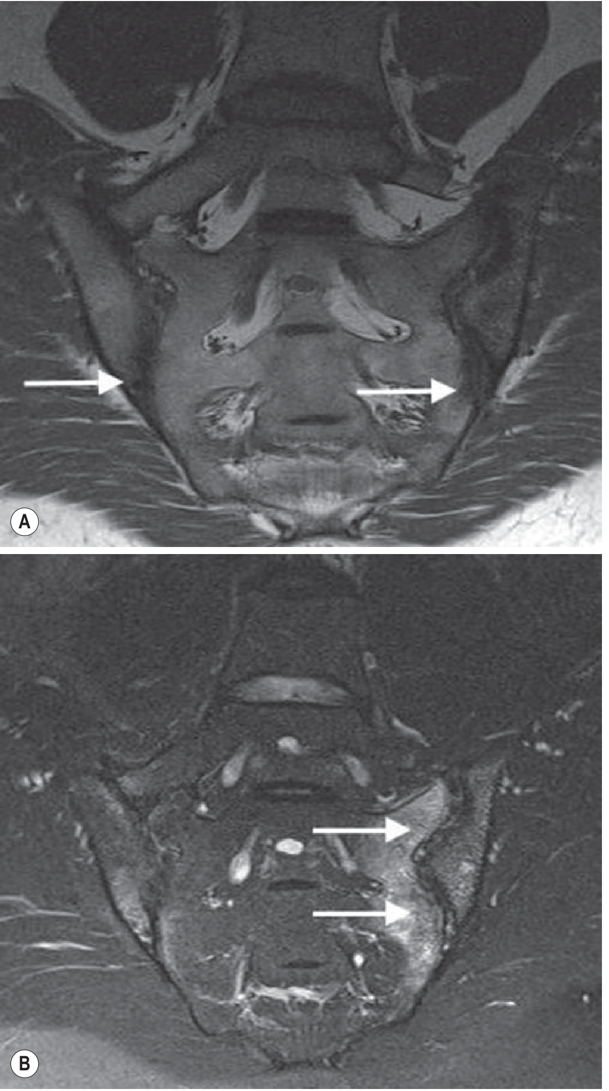
- Most sensitive for early sacroiliitis - detects subchondral bone marrow oedema before radiographic changes
- MRI equivalent of Romanus lesions: bone oedema at vertebral corners
- STIR sequences show active inflammation at costovertebral joints and entheseal sites
Important Complication
Diagnostic Criteria
- Low back pain >3 months, improves with exercise, not relieved by rest
- Limited lumbar spine motion in sagittal AND frontal planes
- Reduced chest expansion relative to normal
- Radiographic sacroiliitis (Grade ≥2 bilateral or Grade 3-4 unilateral)
Laboratory Findings
- HLA-B27: positive in ~90% of cases (not diagnostic alone; present in ~7% of general population)
- ESR and CRP: elevated in ~50-70% of active cases; can be normal
- RF and ANA: negative (seronegative arthritis)
- Mild normochromic anaemia in active disease
- Elevated alkaline phosphatase occasionally
Treatment
Non-pharmacological
- Exercise and physiotherapy programs are fundamental - improve cardiorespiratory fitness, spinal mobility
- Patient education, posture training
- Aquatic therapy, hydrotherapy
Step 1: NSAIDs
- First-line therapy for pain and stiffness
- Continuous NSAID use may slow radiographic progression (debated)
- Full anti-inflammatory doses required (e.g., naproxen, indomethacin, diclofenac)
- Cochrane evidence (Grade A) supports NSAIDs for axial SpA
Step 2: Local injections
- Corticosteroid injections into SI joints or peripheral entheses
- Systemic corticosteroids do NOT prevent progression and are not recommended for spinal disease
Step 3: Biologics (when NSAIDs fail after 6 weeks)
| Drug Class | Examples | Target |
|---|---|---|
| TNF-α inhibitors | Etanercept, Adalimumab, Infliximab, Certolizumab, Golimumab | TNF-α |
| IL-17A inhibitors | Secukinumab, Ixekizumab | IL-17A |
| IL-12/23 inhibitor | Ustekinumab | IL-12/23 |
| JAK inhibitors | Tofacitinib, Upadacitinib | JAK pathway |
- TNF-α inhibitors are the most established; Cochrane evidence confirms efficacy (Grade A)
- Secukinumab (IL-17A) is highly effective, especially for radiographic AS
- NB: Screen for latent TB before starting anti-TNF therapy
- DMARDs (methotrexate, sulfasalazine) have limited efficacy for axial disease but may help peripheral arthritis
ACR/SAA/SPARTAN 2019 Guidelines framework:
Prognosis and Complications
- Clinical course is highly variable - some patients have mild, stable disease; others develop severe deformity
- Strongest predictor of new syndesmophyte formation: presence of syndesmophytes at baseline
- Late complications:
- Cauda equina syndrome
- Osteoporotic vertebral fractures
- Spondylodiscitis (Andersson lesion)
- Restrictive lung disease
- Aortic regurgitation
- Renal amyloidosis
- Ankylosing spondylitis rarely leads to pulmonary disability unless fibrobullous disease develops
- Work disability and quality-of-life impact are significant, especially with late diagnosis
Recent Evidence (2025-2026)
Ankylosing spondyitis 15mrk
ankylosing spondylitis bamboo spine sacroiliitis X-ray
This composite of two X-ray images demonstrates classic radiographic signs of Ankylosing Spondylitis (AS). Panel A is a lateral chest/thoracic spine radiograph showing characteristic 'bamboo spine' appearance. This is characterized by extensive syndesmophyte formation, where bony bridges fuse the thoracic vertebral bodies together, creating a continuous, rigid vertical column. Notably, the intervertebral disc spaces remain relatively preserved in height despite the fusion. Panel B is an anteroposterior (AP) pelvic radiograph focusing on the sacroiliac joints. It shows Grade III bilateral sacroiliitis, evidenced by significant subchondral sclerosis (increased bone density) and narrowing with irregular joint margins. The sacrum and iliac wings exhibit focal areas of erosion and fusion. These findings are hallmarks of seronegative spondyloarthropathies and are used clinically to assess disease progression and staging in patients presenting with chronic inflammatory back pain and HLA-B27 positivity.
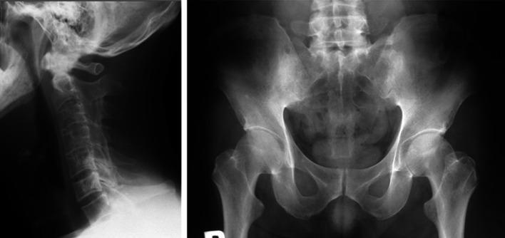
This composite diagnostic image features two X-ray radiographs illustrating the hallmark skeletal manifestations of advanced ankylosing spondylitis (SpA). The left panel is a lateral radiograph of the cervical spine showing 'bamboo spine' morphology. It demonstrates extensive marginal syndesmophytes and ankylosis leading to the fusion of vertebral bodies and complete ossification of the intervertebral disc spaces. The result is a continuous, rigid vertebral column with loss of the normal cervical lordosis. The right panel is an anteroposterior (AP) radiograph of the pelvis focusing on the sacroiliac (SI) joints. Both SI joints show Grade 4 sacroiliitis according to the Modified New York criteria, characterized by total ankylosis (fusion), significant subchondral sclerosis, and loss of visible joint space. These findings represent end-stage chronic inflammation in axial spondyloarthritis. The image is intended for medical education regarding the progression of rheumatological conditions and the interpretation of radiological signs of spinal fusion and sacroiliitis.
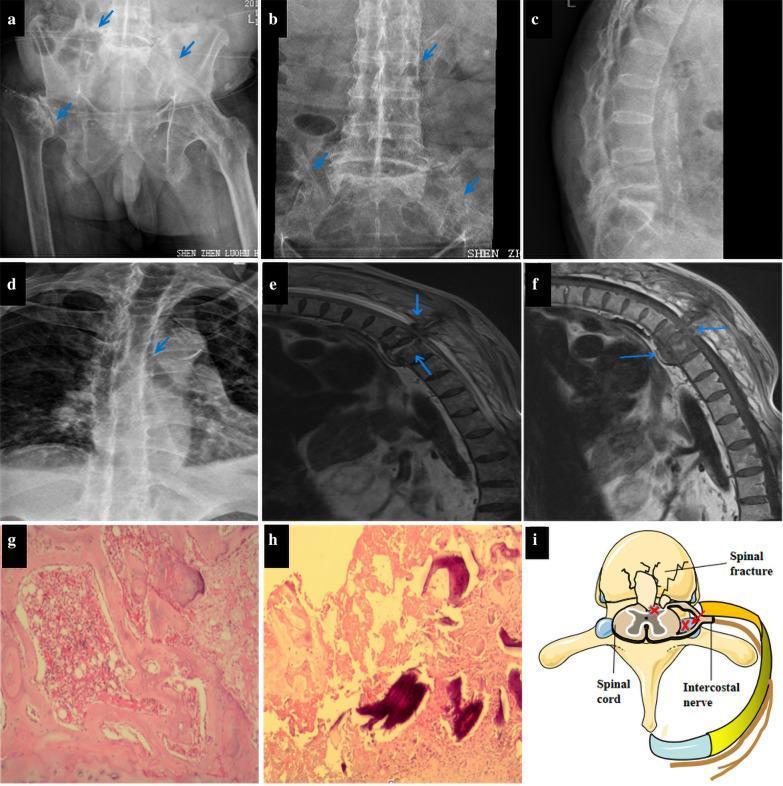
This composite figure presents clinical imaging and pathology related to advanced ankylosing spondylitis (AS). (a-d) X-ray radiographs demonstrate classic musculoskeletal manifestations: bilateral sacroiliitis with joint space narrowing and articular sclerosis, a 'bamboo spine' appearance due to extensive syndesmophyte formation, and thoracic kyphosis. Osteonecrosis of the right femoral head is also visible. (e-f) T2-weighted sagittal MRIs of the thoracolumbar spine reveal destruction at the T9-10 level with disc denaturation, abnormal high signal intensity indicating hemorrhage or edema, and suspected interspinous ligament rupture. (g-h) H&E-stained histopathology (40x) shows necrotic bone fragments, granulation tissue, and fibrous connective tissue with inflammatory infiltration, ruling out malignancy or tuberculosis. (i) A schematic diagram illustrates the pathophysiology of intercostal neuralgia, depicting how a thoracic vertebral fracture can cause mechanical compression of the spinal cord and spinal nerve roots. This collection serves as an educational resource for recognizing the late-stage radiological features of AS and its potential neurological complications.
ANKYLOSING SPONDYLITIS - 15 Mark Answer
1. DEFINITION (1 mark)
2. EPIDEMIOLOGY (1 mark)
- Prevalence: ~0.2% of general population; ~2% of HLA-B27-positive individuals
- Age of onset: most commonly 15-25 years (young adults/adolescents)
- Sex: male predominance (M:F = 2.5-5:1, though gap is narrowing)
- Race: highest in White/Northern European populations; rare in Japanese and Australian aboriginal populations
- HLA-B27 positive in 90-95% of White patients with AS
3. ETIOPATHOGENESIS (2 marks)
- HLA-B27 is the strongest genetic association; present in ~90% of patients
- B27 subtypes B2705 (primordial) confer susceptibility; B2706 and B*2709 do not
- Non-MHC genes (ERAP1, IL-23R) also contribute
- Molecular mimicry - sequence homology between B27 and gram-negative enteric bacteria (e.g., Klebsiella pneumoniae) triggers cross-reactive immune responses
- Unfolded protein response (UPR) - B27 misfolds in the endoplasmic reticulum, inducing a pro-inflammatory cascade (IL-23/IL-17 axis activation)
- Heavy-chain homodimers - B27 forms homodimers at the cell surface, aberrantly activating NK and CD4+ T cells
- Primary lesion is enthesitis - inflammation at the insertion of ligaments, tendons, and joint capsules into bone (especially at Sharpey fibers of the annulus fibrosus)
- Chronic inflammation → erosion → reactive new bone formation
- Syndesmophytes develop → bridging → ankylosis (fusion)
4. CLINICAL FEATURES (3 marks)
A. Spinal and Axial (Core Features)
| Feature | Details |
|---|---|
| Inflammatory back pain | Insidious onset, age <40, >3 months duration, worse at rest and at night, improves with activity and NSAIDs |
| Morning stiffness | Lasting >1 hour; hallmark feature |
| Sacroiliitis | Bilateral deep buttock pain; tenderness on sacroiliac compression |
| Reduced spinal mobility | Loss of lumbar lordosis; measured by Schober's test (<5 cm rise on forward flexion) |
| Reduced chest expansion | <2.5 cm at 4th intercostal space; due to costovertebral/sternoclavicular fusion |
| Progressive kyphosis | "Question mark" posture in advanced disease |
| Bamboo spine | End-stage complete vertebral fusion |
B. Peripheral Involvement
- ~30% of patients develop peripheral arthritis - asymmetrical, large joints (hips, knees, ankles)
- Hip joint is the most common peripheral joint involved
- Enthesitis at Achilles tendon insertion, plantar fascia, tibial tuberosity
C. Extra-Articular Manifestations (remember: A COPE)
| System | Manifestation |
|---|---|
| Acute anterior uveitis | Most common extra-articular feature; recurrent, unilateral, painful red eye with photophobia |
| Cardiac | Aortitis, aortic regurgitation, conduction defects (AV block) |
| Ocular | Also conjunctivitis |
| Pulmonary | Restrictive lung disease (rib cage fixation); fibrobullous upper lobe disease (1-4%) |
| Enteropathy | IBD (Crohn's, UC); subclinical gut inflammation in ~60% |
| Also: | Psoriasis, IgA nephropathy, amyloidosis (renal), cauda equina syndrome (late) |
5. INVESTIGATIONS (2 marks)
Laboratory
| Test | Finding |
|---|---|
| HLA-B27 | Positive in ~90% (not diagnostic alone) |
| ESR, CRP | Raised in ~50-70%; can be normal |
| Rheumatoid Factor | Negative (seronegative) |
| ANA | Negative |
| FBC | Mild normochromic anaemia in active disease |
| Alkaline phosphatase | Occasionally elevated |
Imaging
- Sacroiliac joints: erosions, subchondral sclerosis, joint space narrowing → eventual fusion (Grade 0-4 by New York criteria)
- Romanus lesion: sclerotic "shiny corners" of vertebral bodies (earliest spinal radiograph sign)
- Vertebral squaring: loss of normal concave anterior border
- Syndesmophytes: thin, vertical bony outgrowths at Sharpey fiber insertions
- Bamboo spine: complete spinal fusion with bridging syndesmophytes
- Most sensitive for early sacroiliitis before X-ray changes
- Subchondral bone marrow oedema on STIR/T2 fat-suppressed sequences = active inflammation
- Detects enthesitis at costovertebral joints, spinous processes, facet joints
- Preferred in young patients to avoid radiation
6. DIAGNOSIS (2 marks)
Modified New York Criteria (1984) - Still widely used clinically
- Low back pain and stiffness >3 months, improves with exercise, not relieved by rest
- Limited lumbar spine motion in sagittal AND frontal planes
- Limited chest expansion (relative to normal for age and sex)
Definite AS = radiological criterion + at least 1 clinical criterion
ASAS Criteria for Axial SpA (2009) - Captures earlier/non-radiographic disease
- Imaging arm: Sacroiliitis on X-ray or MRI + ≥1 SpA feature
- Clinical arm: HLA-B27 positive + ≥2 SpA features
7. DIFFERENTIAL DIAGNOSIS (1 mark)
| Condition | Distinguishing Feature |
|---|---|
| Mechanical back pain | No morning stiffness, no response to NSAIDs, worsens with activity |
| Psoriatic arthritis (axial) | Asymmetric sacroiliitis, coarser skip syndesmophytes, skin/nail changes |
| Reactive arthritis | Preceded by infection (urogenital/enteric), usually self-limiting |
| Enteropathic arthritis | Associated with IBD |
| Diffuse idiopathic skeletal hyperostosis (DISH) | Flowing ossification, no sacroiliitis, older patients |
| Septic sacroiliitis | Unilateral, fever, raised WBC |
8. TREATMENT (3 marks)
A. Non-pharmacological (Always first-line)
- Physiotherapy is the cornerstone - spinal mobility exercises, posture training
- Swimming and hydrotherapy - ideal low-impact exercise
- Deep breathing exercises (to maintain chest expansion)
- Avoid smoking (worsens pulmonary restriction)
- Correct sleep posture (flat, straight back; no pillow under knees)
- Patient education and psychosocial support
B. NSAIDs (First-line pharmacotherapy)
- Indomethacin, diclofenac (up to 150 mg/day), or naproxen (up to 1000 mg/day)
- Continuous use may slow radiographic progression
- Goal: sufficient pain relief to enable exercise
- Use with caution in patients with IBD
C. Local Corticosteroid Injections
- For peripheral joints, SI joints, entheseal sites
- Systemic corticosteroids have no role in axial AS; do NOT prevent progression
D. DMARDs
- Sulfasalazine - for peripheral arthritis only (no benefit for axial disease)
- Methotrexate - limited evidence for AS
E. Biologic Therapies (when NSAIDs fail after 4-6 weeks)
| Drug Class | Examples | Route | Dose |
|---|---|---|---|
| TNF-α inhibitors (first-line biologic) | Infliximab | IV | 5 mg/kg at 0, 2, 6 wks, then every 8 wks |
| Adalimumab | SC | 40 mg every 2 weeks | |
| Etanercept | SC | 50 mg/week | |
| Certolizumab, Golimumab | SC | Various | |
| IL-17A inhibitors | Secukinumab | SC | 150 mg/month |
| Ixekizumab | SC | 80 mg every 4 wks | |
| JAK inhibitors (tsDMARDs) | Tofacitinib, Upadacitinib | Oral | Various |
Important: Screen for latent TB before initiating anti-TNF therapy
F. Surgery
- Total hip arthroplasty for severe hip disease
- Corrective osteotomy for severe fixed kyphosis
- Spinal surgery for fracture/instability
9. COMPLICATIONS AND PROGNOSIS (1 mark)
| Complication | Note |
|---|---|
| Spinal fracture | Rigid osteopenic spine fractures with minimal trauma; highly unstable |
| Osteoporosis | From chronic inflammation + abnormal biomechanics |
| Cauda equina syndrome | Late complication; chronic arachnoiditis |
| Spondylodiscitis (Andersson lesion) | Pseudarthrosis at unfused levels; mimics infection |
| Aortic regurgitation/heart block | Cardiac conduction disease |
| Fibrobullous lung disease | Upper lobe cysts; ~1-4%; may cause massive haemoptysis |
| Amyloidosis (AA) | Renal involvement |