
Make notes on cardiovascular system examinations in detailed from Hutchison clinical book (like all the of pulse and heave with demonstration images , how to perform) correlate with the disease
Reading File
Finding Sources
Finding Sources
Finding Sources
Finding Sources
Finding Sources
Finding Sources
Finding Sources
Reading File
Reading File
Reading File
Reading File
Reading File
Reading File
Reading File
Reading File
Reading File
Loading Image
Loading Image
Loading Image
Loading Image
Reading File
Loading Image
Reading File
Note on sources: Hutchison's Clinical Methods is not currently in the available library. The notes below are compiled from the three most authoritative alternatives available: Harrison's Principles of Internal Medicine 22E, Fuster and Hurst's The Heart 15th Edition, and Goldman-Cecil Medicine - all of which cover the same Hutchisonian physical examination framework in even greater depth.
Cardiovascular System Examination - Comprehensive Notes
OVERVIEW: SEQUENCE OF EXAMINATION
| Step | Component |
|---|---|
| 1 | General inspection - appearance, cyanosis, clubbing, edema |
| 2 | Blood pressure measurement (both arms) |
| 3 | Arterial pulse - rate, rhythm, volume, character |
| 4 | Jugular venous pressure (JVP) and waveform analysis |
| 5 | Precordial inspection and palpation (apex beat, heaves, thrills) |
| 6 | Cardiac auscultation (heart sounds, added sounds, murmurs) |
| 7 | Peripheral vascular assessment |
PART 1: GENERAL INSPECTION
Before touching the patient, observe:
- Cyanosis - central (lips, tongue) vs peripheral (fingers)
- Central: Eisenmenger's, Fallot's, pulmonary hypertension
- Peripheral: low cardiac output, heart failure
- Clubbing - infective endocarditis, cyanotic congenital heart disease
- Malar flush - mitral stenosis (MS) with low cardiac output
- Pallor - anaemia (high-output states), cardiogenic shock
- Xanthelasma / xanthomata - hypercholesterolaemia, premature IHD
- Corneal arcus - if <50 years, suggests dyslipidaemia
- Marfanoid habitus - tall, arachnodactyly, high arched palate - aortic root dilatation, AR, MVP
- Edema - bilateral pitting pedal edema + elevated JVP = right heart failure / cor pulmonale
PART 2: BLOOD PRESSURE
Technique
- Patient seated, arm at heart level, supported, back supported, feet on floor
- Wait 5-10 minutes before measuring
- Cuff bladder: length 80%, width 40% of arm circumference (use correct cuff size - too small overestimates BP)
- Inflate 30 mmHg above expected systolic, deflate at 2-3 mmHg/sec
- Systolic = first Korotkoff sound (Phase I); Diastolic = disappearance (Phase V)
- Measure both arms - normal difference <10 mmHg
Abnormal Findings
| Finding | Disease Correlation |
|---|---|
| Both arms differ >10 mmHg | Subclavian artery stenosis, aortic dissection, coarctation of aorta |
| Wide pulse pressure | Severe AR, hypertension (elderly), PDA, AV fistula |
| Narrow pulse pressure | Shock, severe AS, cardiac tamponade |
| Leg BP lower than arm | Coarctation of aorta (normally legs are 20 mmHg higher) |
Pulsus Paradoxus
A fall in systolic BP >10 mmHg on inspiration.
How to measure:
- Inflate cuff 15-20 mmHg above systolic
- Deflate slowly while patient breathes normally
- Note the pressure at which sounds first appear (expiratory) and the pressure at which sounds are heard continuously through both phases
- Difference >10 mmHg = pulsus paradoxus
Diseases: Cardiac tamponade (classic), constrictive pericarditis, massive PE, severe asthma, tension pneumothorax, hemorrhagic shock
Pulsus Alternans
Beat-to-beat variation in pulse amplitude - every other Korotkoff sound is audible as the cuff is slowly deflated.
Disease: Severe LV systolic dysfunction (heart failure)
PART 3: ARTERIAL PULSE
Technique
Palpate the carotid artery for character and contour (most reliable). Alternatively palpate the brachial artery - rest the patient's elbow on your palm, palpate the vessel with your thumb; brachial pulse is deeper.
Assess simultaneously:
- Rate and rhythm
- Volume (amplitude)
- Character (upstroke velocity, waveform contour)
- Peripheral pulse equality
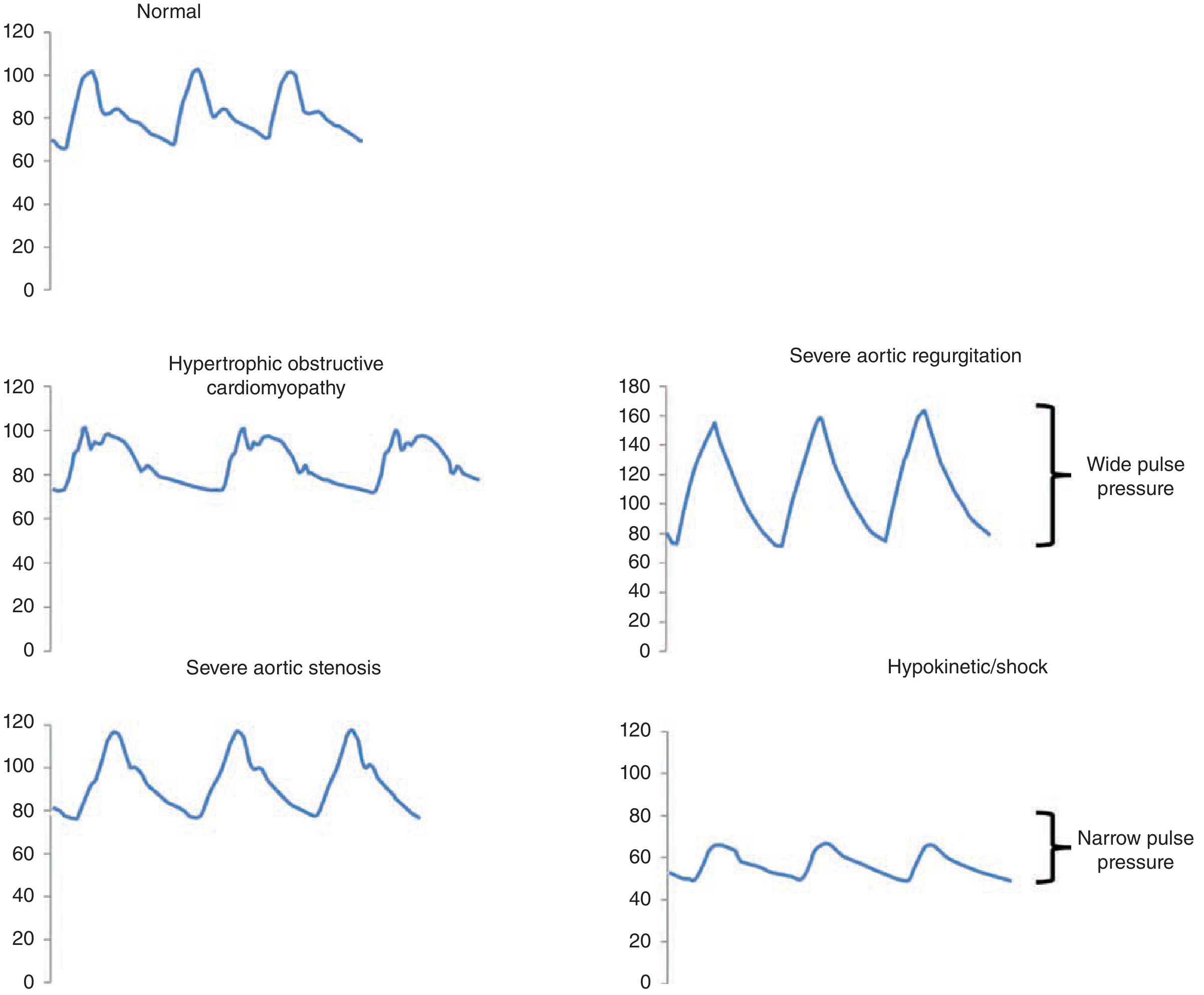
Named Pulse Abnormalities and Disease Correlations
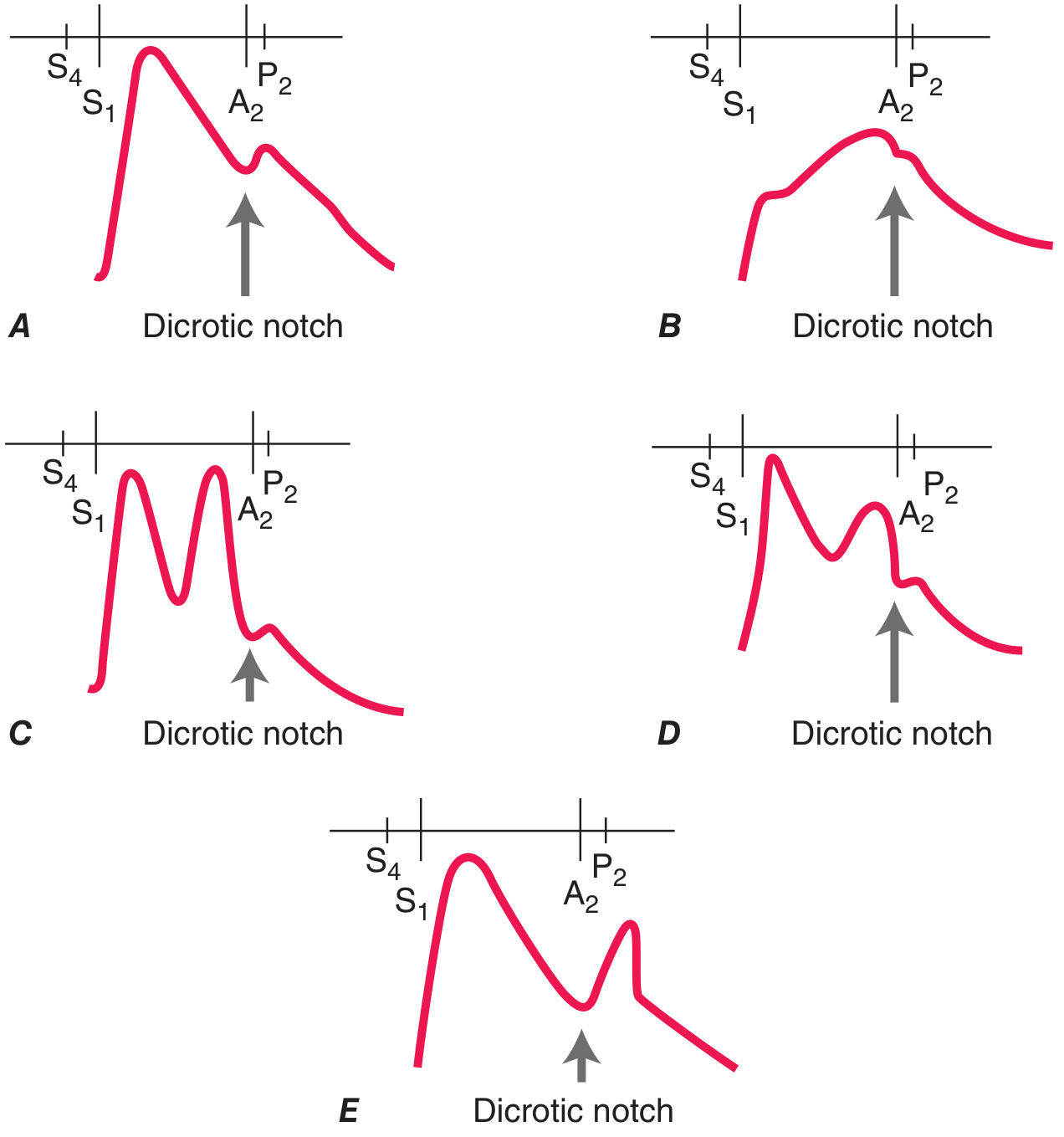
| Pulse Type | Character | Disease |
|---|---|---|
| Pulsus parvus et tardus | Weak volume + slow delayed upstroke | Severe aortic stenosis (AS) |
| Anacrotic pulse | Notch on ascending limb; slow interrupted upstroke | Severe aortic stenosis |
| Corrigan's / Water-hammer pulse | Sharp rapid rise with sudden collapse | Severe aortic regurgitation (AR) |
| Bisferiens pulse (AR) | Two systolic peaks; bounding | Severe AR (or mixed aortic disease) |
| Bisferiens pulse (HOCM) | Spike-and-dome pattern: initial rapid peak then secondary wave | Hypertrophic obstructive cardiomyopathy |
| Dicrotic pulse | Secondary peak in diastole (exaggerated dicrotic notch) | Sepsis, severe peripheral vasoconstriction, low output states |
| Pulsus paradoxus | Systolic BP falls >10 mmHg on inspiration | Cardiac tamponade, constrictive pericarditis, severe asthma |
| Pulsus alternans | Beat-to-beat amplitude variation | Severe LV systolic dysfunction |
| Collapsing pulse (water-hammer - also named) | Maximized by elevating arm above heart level - feel the pulse collapse | AR, PDA, thyrotoxicosis, severe anaemia |
| Thready/weak pulse | Low volume, barely palpable | Cardiogenic shock, severe AS, cardiac tamponade |
How to check for collapsing pulse (Corrigan's sign):
Grip the patient's wrist with your full hand, then elevate the arm above the level of the heart - a sharp percussive thump felt in the palm = positive water-hammer pulse (AR).
PART 4: JUGULAR VENOUS PRESSURE (JVP)
The JVP is the single most important bedside measurement to estimate volume status and right heart hemodynamics.
Why the Internal Jugular Vein?
- Directly in line with the superior vena cava and right atrium
- Not valved
- External jugular is valved and laterally positioned - less reliable for waveform analysis (though useful for high vs low CVP screening)
How to Measure JVP
Technique (Fuster & Hurst / Harrison's standard):
- Position patient at 45° with neck slightly extended; use 30°, 45°, or 60° depending on where pulsations are visible
- Look for the internal jugular pulsation just medial to the SCM muscle
- Identify the sternal angle (angle of Louis) - the manubriosternal junction, which lies approximately 5 cm above the center of the right atrium
- CVP (cmH₂O) = vertical height of the venous pulsation above the sternal angle + 5 cm
- Normal: venous pulsation <3 cm above the sternal angle at 45° (CVP <8 cmH₂O)
Practical grading at 90° (sitting upright):
- No pulsation visible = normal (CVP <8 cmH₂O)
- Pulsation at clavicle = ~10 cmH₂O
- Pulsation at mid-neck = ~15 cmH₂O
- Pulsation at angle of jaw = ~20 cmH₂O
Distinguishing JVP from Carotid Pulse
| Feature | JVP | Carotid |
|---|---|---|
| Obliterable with pressure | Yes (press above clavicle) | No |
| Waveform | Biphasic (a and v waves) in SR | Monophasic |
| Respiration | Normally falls with inspiration | No change |
| Visible vs palpable | Visible, NOT palpable | Palpable, NOT easily visible |
The JVP Waveform
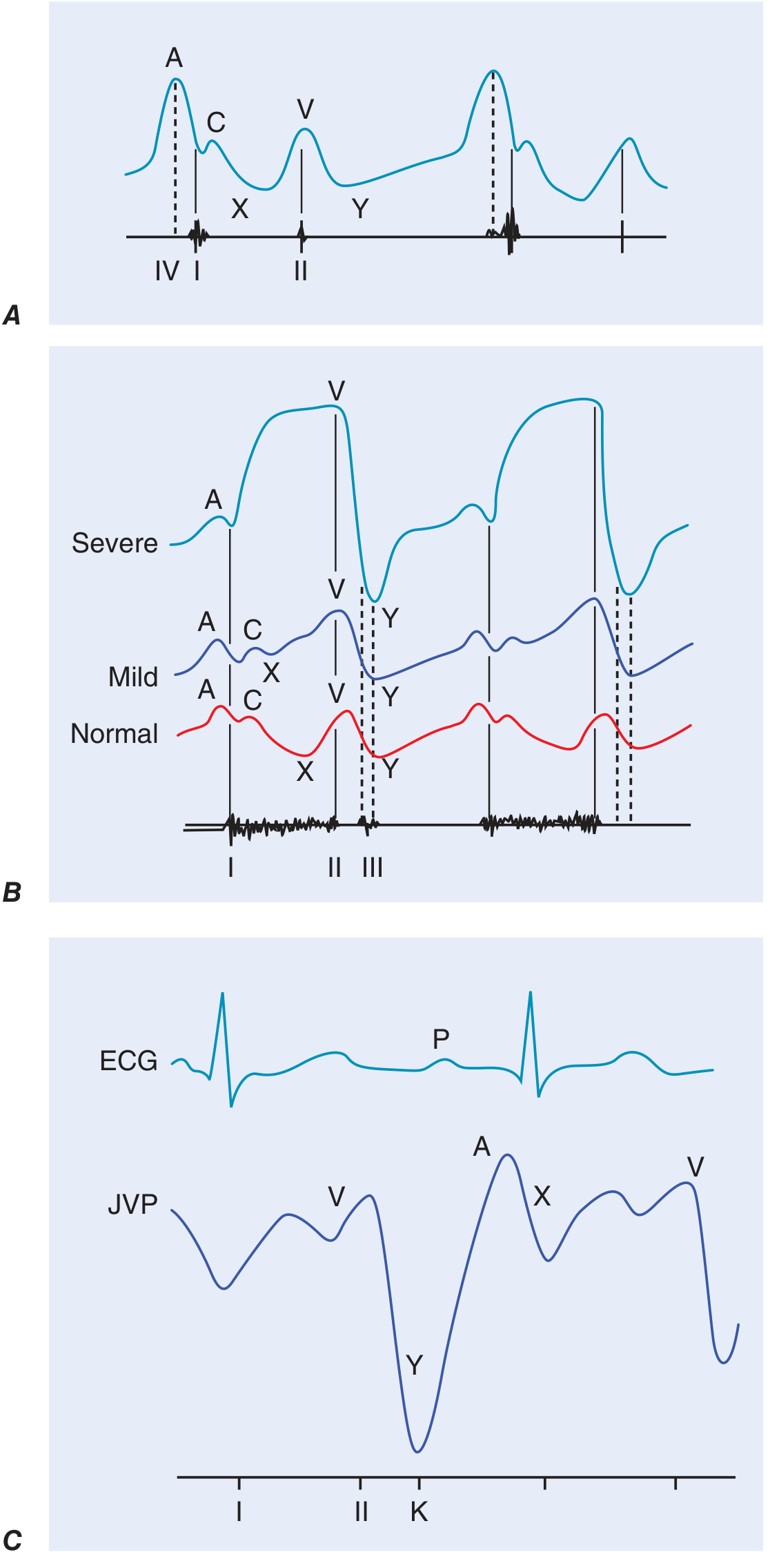
| Component | Timing | Mechanism | Abnormality |
|---|---|---|---|
| a wave | Pre-systole (after P wave) | Right atrial contraction | ↑ in reduced RV compliance (pulmonary stenosis, pulmonary HTN, RV hypertrophy); Absent in atrial fibrillation |
| Cannon a wave | Pre-systole | RA contracts against closed tricuspid valve (AV dissociation) | Complete heart block, VT |
| c wave | Early systole | Tricuspid valve bulging into RA | Usually not seen clinically |
| x descent | Systolic | RA relaxation + tricuspid valve descent | Obliterated in cardiac tamponade |
| v wave | Late systole | RA filling during ventricular systole | ↑↑ in tricuspid regurgitation (TR); "ventricularzied" waveform in severe TR |
| y descent | Early diastole | Tricuspid valve opens, RA empties | Rapid/steep in constrictive pericarditis; Slow/blunted in tricuspid stenosis, tamponade |
Special JVP Signs
| Sign | Finding | Disease |
|---|---|---|
| Kussmaul's sign | JVP rises (or fails to fall) with inspiration | Constrictive pericarditis (classic), restrictive cardiomyopathy, massive PE, RV infarction |
| Abdominojugular reflux (AJR) | Sustained JVP rise >3 cm on firm RUQ pressure for >15 sec | Volume overload, heart failure (predicts PCWP >15 mmHg) |
| Elevated JVP | >4.5 cm above sternal angle at 45° | Right heart failure, TR, tamponade, constrictive pericarditis, SVCO |
| Low JVP | Pulsation visible only when almost supine | Hypovolaemia, dehydration |
PART 5: PRECORDIAL INSPECTION AND PALPATION
Inspection
- Apex beat - normally at 5th ICS, midclavicular line in thin adults
- Visible pulsations anywhere outside this location = abnormal
- Visible chest wall heaving = enlarged or hyperdynamic LV or RV
- Right upper parasternal pulsation = consider ascending aortic aneurysm
Palpation of the Apex Beat
Technique:
- Patient supine at 30°
- Place flat of fingers over the left precordium to locate the impulse
- Enhance sensitivity by rolling the patient into the left lateral decubitus position
- Locate the outermost, lowermost point of the impulse
- Normal: <2 cm in diameter, moves quickly away from fingers, at 5th ICS MCL at end-expiration
Apex Beat Characteristics and Disease Correlations
| Character | Description | Disease |
|---|---|---|
| Normal | Small (<2 cm), brief, outward tap, well-localised | Normal |
| Displaced (down and out) | Below 5th ICS and/or lateral to MCL | LV dilatation (DCM, severe AR, severe MR, ischemic cardiomyopathy) |
| Sustained (heaving/pressure overload) | Prolonged outward thrust, does not fall away quickly | LV pressure overload: aortic stenosis, systemic hypertension, HOCM |
| Hyperdynamic (volume overload) | Vigorous and brisk, moves quickly outward and back | LV volume overload: AR, MR, VSD, PDA |
| Tapping | Palpable S1 - brief, abrupt tap | Mitral stenosis (palpable S1 from loud, snapping MV closure) |
| Double or Triple impulse | Two or three palpable components | HOCM (S4 + bisferiens systolic); LV aneurysm (dyskinetic ectopic impulse) |
| Diffuse / Not localisable | Widespread impulse | Dilated cardiomyopathy, pericardial effusion |
| Absent | Cannot feel at supine position | Obesity, emphysema, pericardial effusion, dextrocardia |
Parasternal Heave (Right Ventricular Heave)
Technique (three methods):
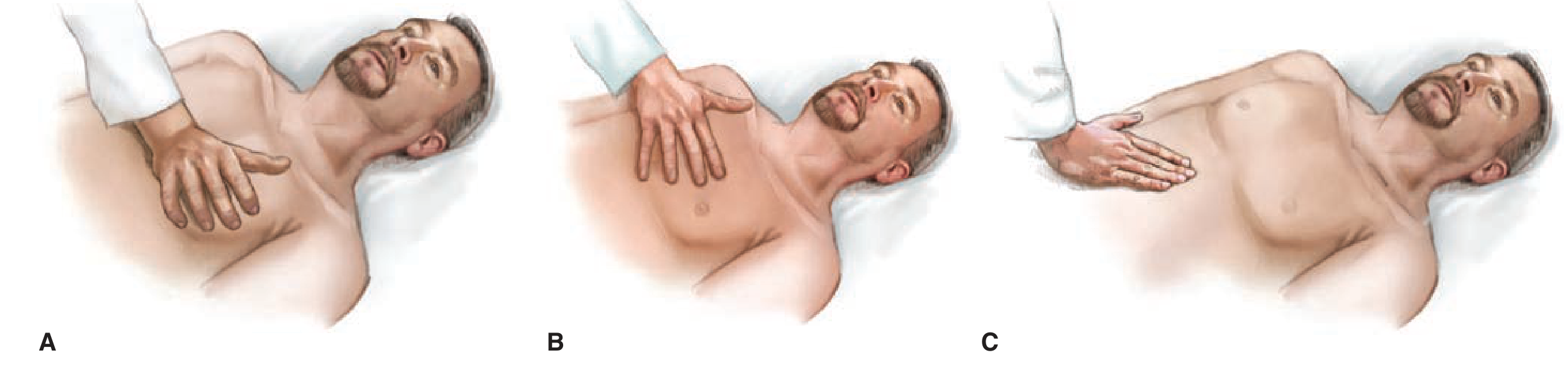
- Method A: Place thenar/hypothenar surface of right hand on the left lower sternal border
- Method B: Place right 2nd, 3rd, 4th fingers over the 3rd, 4th, 5th right intercostal spaces
- Method C: Subxiphoid approach (best in COPD with barrel chest/flat diaphragm) - feel the RV impulse rise into your palm
A parasternal (left sternal) heave = RV lift = sustained outward movement of the sternum/left parasternal region with each systole.
| Heave | Cause |
|---|---|
| Left parasternal heave (RV lift) | RV pressure overload: pulmonary hypertension, pulmonary stenosis, cor pulmonale, ASD with RV overload |
| Left parasternal heave with prominent v waves in JVP | Severe TR (RV volume overload) |
| Right parasternal pulsation | Ascending aortic aneurysm |
| Subxiphoid pulsation | RV enlargement; distinguish from pulsatile liver (hepatic in expiration, RV in inspiration) |
Thrills
A thrill = palpable murmur (grade ≥4/6). Feel with flat of hand.
| Location | Disease |
|---|---|
| Upper left sternal edge | Pulmonary stenosis, VSD |
| Upper right sternal edge | Aortic stenosis |
| Suprasternal notch | Severe AS, coarctation |
| Apex | Mitral valve disease |
PART 6: CARDIAC AUSCULTATION
Auscultatory Areas
- Aortic area: 2nd right ICS (RICS)
- Pulmonary area: 2nd left ICS (LICS)
- Tricuspid area: 4th/5th left sternal edge
- Mitral area: Cardiac apex (5th ICS, MCL)
- Erb's point: 3rd LICS - AR heard best here
Use diaphragm for high-pitched sounds (S1, S2, AS, MR, TR murmurs, pericardial rub).
Use bell (light pressure) for low-pitched sounds (S3, S4, MS rumble).
First Heart Sound (S1)
Mechanism: Mitral (M1) + Tricuspid (T1) valve closure. M1 precedes T1.
Best heard: Apex (mitral component predominates)
| S1 Intensity | Disease |
|---|---|
| Loud S1 | Mitral stenosis (early, mobile leaflets), hyperkinetic states (fever, anaemia, thyrotoxicosis), short PR interval |
| Soft S1 | Late MS (rigid/calcified leaflets), first-degree AV block (long PR), LV dysfunction, beta-blockers |
| Variable S1 | Complete heart block (AV dissociation), AF |
Second Heart Sound (S2)
Mechanism: Aortic (A2) + Pulmonic (P2) valve closure. A2 precedes P2.
Normal splitting: A2-P2 interval widens on inspiration (increased RV filling delays P2), narrows on expiration.
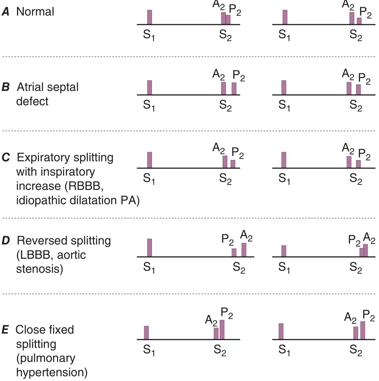
| Splitting Pattern | Disease |
|---|---|
| Wide splitting (inspiration) | RBBB (delayed P2), pulmonary stenosis, massive PE |
| Wide splitting + also present on expiration | Severe MR (early A2 due to rapid LV emptying) |
| Fixed splitting (no respiratory variation) | Atrial septal defect (ASD) - classic finding |
| Reversed (paradoxical) splitting - P2 precedes A2, splits on expiration | LBBB (delayed A2), severe AS, HOCM, RV pacing |
| Single S2 | Severe AS (A2 inaudible), Eisenmenger's (P2 = A2 in timing) |
| Loud P2 | Pulmonary hypertension (may be palpable at left 2nd ICS) |
Added Heart Sounds
Systolic Sounds
| Sound | Timing | Character | Disease |
|---|---|---|---|
| Ejection click | Early systole (with carotid upstroke) | High-pitched, sharp click at base | Bicuspid aortic valve, pulmonary valve stenosis, aortic/pulmonary root dilatation |
| Pulmonic ejection click | Early systole | Only right-sided sound that decreases with inspiration | Pulmonary stenosis (click moves closer to S1 as severity increases) |
| Mid-systolic click (non-ejection) | After start of carotid upstroke | Click at apex | Mitral valve prolapse (MVP) - moves toward S1 on standing, away from S1 on squatting |
Diastolic Sounds
| Sound | Timing | Character | Disease |
|---|---|---|---|
| Opening snap (OS) | Early diastole, shortly after S2 | High-pitched snap | Mitral stenosis (A2-OS interval inversely proportional to severity - shorter = more severe MS) |
| Pericardial knock (PK) | Slightly later than OS | High-pitched; corresponds to sudden halt of ventricular expansion | Constrictive pericarditis (coincides with deep y descent in JVP) |
| Tumor plop | Diastole | Low-pitched; position-dependent | Left atrial myxoma |
Gallop Sounds
| Sound | Mechanism | Character | Disease |
|---|---|---|---|
| S3 (Third heart sound) | Rapid early LV filling phase; abrupt check to filling | Low-pitched, best at apex with bell, louder in left lateral decubitus, louder in expiration | Pathological in adults: LV failure, dilated cardiomyopathy; Normal in young healthy adults and children |
| S4 (Fourth heart sound) | Atrial contraction against stiff LV ("atrial kick") | Low-pitched, presystolic, just before S1 | LV hypertrophy (hypertension, AS, HOCM), active myocardial ischemia; Absent in AF |
| Summation gallop | S3 + S4 overlap at high heart rate | Single loud diastolic sound | Severe heart failure with tachycardia |
Heart Murmurs
Grading (Levine scale):
| Grade | Description |
|---|---|
| 1/6 | Barely audible, requires concentration |
| 2/6 | Soft but readily heard |
| 3/6 | Moderately loud, no thrill |
| 4/6 | Loud + palpable thrill |
| 5/6 | Very loud, heard with stethoscope partially off chest |
| 6/6 | Audible without stethoscope |
Systolic Murmurs
| Murmur | Character | Location + Radiation | Disease |
|---|---|---|---|
| Aortic stenosis | Ejection systolic (ESM), crescendo-decrescendo, harsh, rough | 2nd RICS, radiates to carotids and apex (Gallavardin phenomenon at apex may sound like MR) | AS - loud murmur peaks later as severity increases |
| Pulmonary stenosis | ESM | 2nd LICS, radiates to left shoulder | PS |
| Mitral regurgitation | Pansystolic (holosystolic), blowing | Apex, radiates to axilla | MR |
| Tricuspid regurgitation | Pansystolic | Lower left sternal edge, louder with inspiration (Carvallo's sign) | TR |
| VSD | Pansystolic, harsh | Left sternal edge | VSD |
| HOCM | ESM | Left sternal edge and apex, NOT radiating to carotids | HOCM |
| MVP | Late systolic murmur after mid-systolic click | Apex | Mitral valve prolapse |
| Innocent murmur | Soft, ESM, no radiation, changes with posture | 2nd LICS or LLSB | Normal variant, especially in children |
Dynamic auscultation (bedside maneuvers):
| Maneuver | Effect on murmur | Useful for |
|---|---|---|
| Valsalva (strain phase) | Most murmurs decrease; HOCM increases; MVP click moves earlier | Differentiating HOCM |
| Squatting | Most murmurs louder; HOCM and MVP decrease/delay | Differentiating HOCM, MVP |
| Standing | HOCM and MVP murmurs louder/earlier | |
| Inspiration | Right-sided murmurs louder (TR, PS) | Carvallo's sign for TR |
| Expiration | Left-sided murmurs louder | |
| Hand grip / isometric exercise | MR, AR, VSD murmur louder; AS, HOCM quieter | Differentiating AS from MR |
Diastolic Murmurs (always pathological)
| Murmur | Character | Location | Disease |
|---|---|---|---|
| Aortic regurgitation | Early diastolic, decrescendo, high-pitched, blowing | 3rd LICS (Erb's point), radiates to apex; best heard sitting forward in expiration | AR - length proportional to severity |
| Pulmonary regurgitation | Graham Steell murmur - early diastolic | Upper left sternal edge | Pulmonary hypertension |
| Mitral stenosis | Mid-to-late diastolic, low-pitched rumble, preceded by OS; presystolic accentuation in SR | Apex, best with bell in left lateral position | MS - louder with exercise, tachycardia |
| Tricuspid stenosis | Mid-diastolic rumble | Lower sternal edge, louder with inspiration | TS (rare, seen in rheumatic/carcinoid) |
Continuous Murmurs
| Murmur | Disease |
|---|---|
| Machinery murmur (continuous systolic + diastolic), louder in systole | Patent ductus arteriosus (PDA) |
| To-and-fro murmur (systolic ejection + early diastolic) | Combined AS + AR |
| Venous hum (benign) | Normal children (disappears on gentle neck pressure) |
| Cervical venous hum | Hyperdynamic states - anaemia, thyrotoxicosis |
PART 7: PERIPHERAL VASCULAR EXAMINATION
Peripheral Pulses - Check all:
- Upper: Radial, brachial, axillary
- Lower: Femoral, popliteal, posterior tibial, dorsalis pedis
- Simultaneous radiofemoral palpation: Delay = coarctation of the aorta
Ankle-Brachial Index (ABI)
- ABI = Ankle systolic pressure / Higher brachial pressure
- Normal: 1.0-1.4
- Borderline: 0.91-0.99
- Mild PAD: 0.7-0.9
- Moderate-severe PAD: <0.5
Peripheral Signs Specific to Cardiac Diseases
| Sign | Description | Disease |
|---|---|---|
| Corrigan's sign | Visible carotid pulsation | Severe AR |
| De Musset's sign | Head nodding with each heartbeat | Severe AR |
| Quinke's sign | Nail bed capillary pulsation | Severe AR |
| Duroziez's sign | To-and-fro femoral bruit on compression | Severe AR |
| Traube's sign | Pistol-shot femoral pulse | Severe AR |
| Janeway lesions | Non-tender hemorrhagic macules (palms/soles) | Infective endocarditis |
| Osler's nodes | Tender nodules (finger pulps) | Infective endocarditis |
| Splinter hemorrhages | Linear subungual hemorrhages | IE (also trauma) |
| Roth spots | Retinal hemorrhages with pale centre | Infective endocarditis |
PART 8: SUMMARY TABLE - DISEASE CORRELATIONS
| Disease | Key Examination Findings |
|---|---|
| Aortic stenosis | Pulsus parvus et tardus; sustained apex beat (pressure overload); ESM at 2RICS radiating to carotids; soft A2; reversed splitting of S2; S4 |
| Aortic regurgitation | Water-hammer (Corrigan's) pulse; wide pulse pressure; displaced hyperdynamic apex; early diastolic murmur at Erb's point; De Musset's, Quinke's, Duroziez's signs |
| Mitral stenosis | Tapping apex beat; loud S1; OS + mid-diastolic rumble at apex; elevated JVP; AF (irregular pulse); malar flush; left parasternal heave if pulmonary HTN develops |
| Mitral regurgitation | Displaced hyperdynamic apex; pansystolic murmur at apex radiating to axilla; soft S1; S3 gallop; wide splitting of S2 |
| Pulmonary hypertension | Elevated JVP; loud/palpable P2; left parasternal heave; fixed or close fixed S2 splitting; Graham Steell murmur; TR murmur; right-sided S3/S4 |
| Heart failure (LV) | Displaced apex; S3 gallop; elevated JVP + AJR; bilateral basal crackles; pulsus alternans (severe) |
| Cardiac tamponade | Elevated JVP (with absent y descent); pulsus paradoxus >10 mmHg; tachycardia; hypotension; muffled heart sounds (Beck's triad) |
| Constrictive pericarditis | Elevated JVP; Kussmaul's sign; prominent rapid y descent; pericardial knock; normal or near-normal cardiac impulse |
| HOCM | Bisferiens pulse (spike-and-dome); jerky carotid; double/triple apex impulse; ESM at LLSB - louder with Valsalva/standing; S4; murmur decreases with squatting/handgrip |
| Atrial septal defect | Fixed splitting of S2; left parasternal heave (RV overload); ESM at upper LICS (relative PS); soft mid-diastolic murmur at tricuspid (relative TS) |
| Infective endocarditis | Fever, new murmur, Janeway, Osler's, Roth spots, splinter hemorrhages, splenomegaly, clubbing |
| Aortic dissection | Unequal arm BPs; absent peripheral pulses; AR murmur; chest/back pain |
| Coarctation of aorta | Radiofemoral delay; BP right arm > left arm > legs; ESM at left upper sternal edge and back (between scapulae); collateral vessel pulsations |
Quick Reference: The Examination in Under 5 Minutes
1. HANDS: Clubbing, splinters, cyanosis, Osler's nodes, Janeway lesions
2. RADIAL PULSE: Rate, rhythm, volume, character
3. BLOOD PRESSURE: Both arms; check for pulsus paradoxus/alternans
4. FACE: Cyanosis, malar flush, xanthelasma, corneal arcus
5. JVP: Height, waveform (a, v waves, x and y descents), Kussmaul's, AJR
6. CAROTID PULSE: Character (upstroke, volume)
7. PRECORDIUM INSPECT: Scars, deformities, visible pulsations
8. APEX BEAT: Position, size, character (tapping/sustained/hyperdynamic/displaced)
9. PARASTERNAL HEAVE: Three methods (thenar, fingers, subxiphoid)
10. THRILLS: All four areas
11. AUSCULTATION: S1, S2 (splitting), added sounds, murmurs - all areas; dynamic maneuvers
12. PERIPHERAL PULSES: Femoral, popliteal, posterior tibial, DP; radiofemoral delay
13. EDEMA: Sacral + pedal pitting
14. AUSCULTATE LUNG BASES: Crackles of pulmonary edema
Sources: Harrison's Principles of Internal Medicine 22E (2025); Fuster and Hurst's The Heart 15th Edition; Goldman-Cecil Medicine International Edition. Note: Hutchison's Clinical Methods was not available in the textbook library; the above follows the identical examination framework described therein.
This is a shared conversation. Sign in to Orris to start your own chat.