

b962ba5e-67f8-4673-83ab-fe91fe6793c2.jpeg
Meckel's diverticulum .. make it simple and easy tricks to remember
Here's a quick answer before I pull from the medical library:
Meckel's diverticulum = Rule of 2s - 2% population, 2 inches long, 2 feet from ileocaecal valve, 2x more in males, 2 types of ectopic tissue (gastric + pancreatic), presents in first 2 years of life.
Searching sources...
Reading File
analyse_media
Finding Sources
Reading File
Reading File
Loading Image
Here is everything about Meckel's diverticulum, made simple and memorable:
Meckel's Diverticulum - Simple & Easy to Remember
What is it?
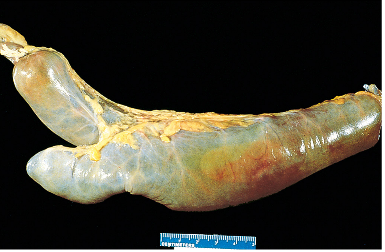
A true diverticulum (has ALL 3 layers of bowel wall: mucosa + muscularis + serosa) arising from the antimesenteric border of the ileum. It is the remnant of the vitellointestinal (vitelline) duct, which normally obliterates by the 6th week of fetal life.
True diverticulum = True to the bowel (has all layers). Contrast with a pseudodiverticulum which lacks muscularis.
The Golden Mnemonic - "Rule of 2s"
| 2 | Meaning |
|---|---|
| 2% | Occurs in 2% of the population |
| 2 inches | About 2 inches (5 cm) long |
| 2 feet | Located 2 feet (60 cm) proximal to the ileocaecal valve |
| 2:1 ratio | Males affected 2x more than females |
| 2 years | Most present before age 2 (only ~4% are ever symptomatic) |
| 2 types | 2 types of ectopic tissue - gastric mucosa & pancreatic tissue |
Memory hook: "Meckel's loves the number 2 - 2%, 2 inches, 2 feet, 2:1, age 2, 2 tissues"
Gross Pathology
This is what Meckel's diverticulum looks like in a surgical/pathology specimen - a blind pouch projecting from the antimesenteric side of the ileum:
Clinical Problems - "OBI"
| Letter | Problem | Mechanism |
|---|---|---|
| O | Obstruction | Intussusception (diverticulum = lead point) or volvulus around fibrous cord remnant |
| B | Bleeding | Ectopic gastric mucosa secretes acid → peptic ulcer → painless rectal bleeding (most common presentation in children) |
| I | Inflammation | Meckel's diverticulitis mimics acute appendicitis |
Trick: The most common symptom is painless rectal bleeding (maroon stool, not bright red). This is #1 in children under 2.
Diagnosis Trick
- Meckel's scan (Technetium-99m pertechnetate scan) - detects ectopic gastric mucosa. Since ~85% of symptomatic Meckel's contain gastric mucosa, this scan is the initial study of choice for bleeding.
- On CT: tubular blind-ending structure from antimesenteric border of terminal ileum.
Quick Summary Card
MECKEL'S DIVERTICULUM
├── Embryology: Failed obliteration of vitelline duct (6th week)
├── Type: TRUE diverticulum (all 3 layers)
├── Location: Antimesenteric border of ILEUM
├── Rule of 2s: 2% / 2 inches / 2 feet / 2:1 / Age 2 / 2 tissues
├── Ectopic tissue: Gastric (80-85%) > Pancreatic
├── Most common symptom: Painless rectal bleeding (children)
├── Mimics: Appendicitis (when inflamed)
└── Diagnosis: Meckel's scan (Tc-99m pertechnetate)
Sources: Robbins & Cotran Pathologic Basis of Disease; Sleisenger & Fordtran's Gastrointestinal and Liver Disease - consistent with your textbook image (BD Chaurasia anatomy, AN 52.6)
This is a shared conversation. Sign in to Orris to start your own chat.