
Give me a detailed history taking list in proper order for general medicine with systemic examinations
Reading File
Now let me search the medical library for history taking content.
Finding Sources
Finding Sources
Finding Sources
Finding Sources
Finding Sources
Finding Sources
Finding Sources
I found relevant internal medicine textbooks. Let me search for history taking content in Harrison's and related books.
Finding Sources
Finding Sources
Let me read the key Harrison's history-taking sections:
Reading File
Reading File
Let me look at the first section of Harrison's block 1 which typically contains the approach to the patient/clinical history:
Reading File
Reading File
Reading File
I have enough foundational knowledge from Harrison's and my comprehensive medical training to give a thorough answer. Let me now compile the complete, detailed history-taking guide with systemic examinations.
Complete History Taking in General Medicine with Systemic Examination
Based on Harrison's Principles of Internal Medicine (22nd Ed., 2025) and standard clinical medicine framework.
STRUCTURE OF CLINICAL HISTORY TAKING
1. BIOGRAPHICAL / IDENTIFICATION DATA
Recorded first to establish the patient's identity and demographic context.
| Item | Details to Record |
|---|---|
| Name | Full name |
| Age | Date of birth / actual age |
| Sex | Male / Female / Other |
| Occupation | Current and past (relevant to exposure history) |
| Address | Permanent and current residence |
| Marital status | Single / Married / Divorced / Widowed |
| Religion / Nationality | (relevant for certain genetic disorders, dietary habits) |
| Date and time of admission | - |
| Referred by | GP / specialist / self |
| Informant | Patient / relative / bystander (and reliability of history) |
2. CHIEF COMPLAINT (CC)
- State the main presenting symptom(s) in the patient's own words
- Record duration alongside each complaint
- List in chronological order if multiple complaints
- Avoid using diagnostic labels - use the symptom itself (e.g., "chest pain for 2 days", not "angina")
Example: "Fever, cough, and breathlessness for 5 days"
3. HISTORY OF PRESENT ILLNESS (HPI)
This is the most critical part. Explore the chief complaint(s) in detail using the SOCRATES mnemonic (or equivalent):
SOCRATES Framework for Each Symptom
| Letter | Aspect | What to Ask |
|---|---|---|
| S | Site | Where exactly? Does it move? |
| O | Onset | Sudden or gradual? What were you doing at onset? |
| C | Character | Nature/quality of the symptom (dull, sharp, burning, colicky, throbbing) |
| R | Radiation | Does it spread? Where to? |
| A | Associations | Other symptoms occurring with it (nausea, sweating, dyspnea) |
| T | Time course | Constant or intermittent? Getting better / worse / same? |
| E | Exacerbating / Relieving factors | What makes it better or worse? |
| S | Severity | 0-10 scale; effect on daily activities |
Additional HPI Details
- Sequence of events - how the illness evolved from first symptom to present
- Prior episodes - any similar illness in the past?
- Treatment sought - any medications or interventions already tried, and response
- Functional impact - effect on work, sleep, appetite, activities of daily living
4. PAST MEDICAL HISTORY (PMH)
Systematic review of previous illnesses, surgeries, and relevant medical events.
Medical Illnesses
- Previous diagnoses: hypertension, diabetes mellitus, asthma, TB, rheumatic fever, epilepsy, jaundice/hepatitis, peptic ulcer, renal disease, thyroid disease
- Previous hospitalisations (when, where, reason, outcome)
- Known allergies (drug, food, environmental) - always document
Surgical History
- Operations with dates, type of procedure, and any complications (including anaesthetic reactions)
Obstetric/Gynaecological History (in females)
- LMP (last menstrual period)
- Menstrual cycle: regularity, duration, flow, dysmenorrhoea
- Number of pregnancies (G), deliveries (P), abortions (A)
- Complications of pregnancy (pre-eclampsia, gestational diabetes)
- Menopausal status
Immunisation History
- Childhood vaccinations
- Adult vaccines (influenza, hepatitis B, COVID-19, pneumococcal)
5. DRUG HISTORY
- Current medications: name, dose, frequency, duration, route (include over-the-counter drugs, herbal remedies, supplements, oral contraceptives)
- Compliance: is the patient taking medications as prescribed?
- Drug allergies and adverse reactions: nature of reaction (rash, anaphylaxis, GI upset), not just "allergy" label
- Recent changes to medications
6. FAMILY HISTORY
- Health status of parents, siblings, children (alive/dead - cause of death if deceased)
- Hereditary / familial conditions: ischaemic heart disease, hypertension, diabetes mellitus, cancers, epilepsy, mental illness, haemophilia, sickle cell disease, thyroid disease, autoimmune conditions
- Consanguinity (especially relevant in populations where this is common)
- Pedigree chart if needed for complex family patterns
7. PERSONAL / SOCIAL HISTORY
Lifestyle
- Smoking: current/ex/never - type (cigarettes, bidi, pipe, chewing tobacco), pack-years (packs/day × years)
- Alcohol: units per week, pattern (binge vs. regular), duration, type
- Recreational drugs: type, route of use, duration
- Diet: vegetarian/non-vegetarian, special dietary restrictions, caloric intake if relevant
- Exercise: level of physical activity
Social Context
- Occupation: current and past, workplace exposures (dust, chemicals, radiation, asbestos, noise)
- Living conditions: housing (cramped, overcrowded), sanitation, water supply
- Marital status and domestic relationships
- Recent travel (international or to endemic areas - malaria, TB, typhoid, parasites)
- Sexual history: relevant when evaluating STIs, HIV, hepatitis B/C (asked sensitively)
- Financial / insurance status: affects treatment compliance
- Education and literacy: affects communication strategy
8. REVIEW OF SYSTEMS (ROS) / SYSTEMIC ENQUIRY
A systematic screen for symptoms in body systems not covered in the HPI. Ask specifically - do not rely on the patient volunteering these.
General / Constitutional
- Fever, chills, night sweats
- Weight loss or weight gain (quantify: how many kg over what period)
- Fatigue, malaise, lethargy
- Appetite change (anorexia)
- Sleep disturbance
Cardiovascular System
- Chest pain / discomfort (character, radiation, relationship to exertion)
- Palpitations (onset, duration, regularity, associated symptoms)
- Breathlessness / dyspnoea (at rest vs. on exertion, how many flights of stairs)
- Orthopnoea (number of pillows used)
- Paroxysmal nocturnal dyspnoea (PND)
- Ankle / leg oedema (bilateral vs. unilateral, pitting, time of day)
- Syncope / pre-syncope
- Claudication (leg pain on walking, distance)
Respiratory System
- Cough (dry vs. productive, duration)
- Sputum (colour, amount, consistency, blood-stained)
- Haemoptysis (amount, frequency)
- Dyspnoea (grading: MRC scale 1-5)
- Wheeze
- Chest pain (pleuritic - worse with breathing)
- Snoring / sleep apnoea
Gastrointestinal System
- Dysphagia (to solids, liquids, or both; progressive?)
- Heartburn / regurgitation / waterbrash
- Nausea and vomiting (content - blood / bile / faeculent)
- Haematemesis
- Abdominal pain (site, character, radiation, relation to meals, bowel habit)
- Change in bowel habit (constipation, diarrhoea, alternating)
- Stool character: colour (melaena, fresh blood, pale/steatorrhoea), consistency
- Flatulence, bloating
- Jaundice (skin/eyes/urine/stool changes)
- Pruritus (with or without jaundice)
Genitourinary System
- Dysuria, frequency, urgency
- Nocturia (how many times)
- Haematuria (frank vs. microscopic, at beginning / end / throughout stream)
- Incontinence
- Hesitancy, poor stream, terminal dribbling (males - prostatic symptoms)
- Loin pain
- Frothy urine (proteinuria), dark urine
- Urethral / vaginal discharge
Nervous System
- Headache (location, character, timing, associated nausea/photophobia/phonophobia)
- Dizziness / vertigo (true rotatory vs. light-headedness)
- Syncope / transient loss of consciousness
- Seizures / fits (type, frequency, post-ictal state, aura)
- Memory problems / confusion
- Weakness (focal vs. generalised, upper vs. lower limb)
- Sensory disturbance: numbness, tingling, paraesthesiae
- Visual disturbance (diplopia, blurring, visual field loss, amaurosis fugax)
- Speech disturbance (dysarthria, dysphasia)
- Tremor, coordination problems
- Gait disturbance
- Hearing loss, tinnitus
Musculoskeletal System
- Joint pain (which joints, bilateral/unilateral, migratory/fixed, morning stiffness duration)
- Joint swelling, warmth, redness
- Back pain (site, radiation to leg, relationship to posture)
- Muscle pain / cramps / weakness
- Bone pain
Endocrine System
- Heat/cold intolerance
- Excessive sweating
- Polydipsia, polyuria (diabetes)
- Skin/hair/nail changes
- Changes in libido or sexual function
- Growth abnormalities
Haematological System
- Easy bruising or bleeding
- Petechiae / purpura
- Prolonged bleeding from cuts
- Recurrent infections
- Lymph node enlargement (painless vs. painful, duration)
- Pallor, fatigue (anaemia symptoms)
Psychiatric / Mental Health
- Mood (depression, elation)
- Anxiety
- Hallucinations, delusions
- Sleep patterns
- Memory and cognition
Skin
- Rashes (site, character, spread, relationship to sun)
- Itching
- Changes in moles
- Hair loss, nail changes
PHYSICAL EXAMINATION
9. GENERAL PHYSICAL EXAMINATION (GPE)
Performed before systemic examination. Assess the patient from the end of the bed first ("end of the bed diagnosis").
General Appearance
- Level of consciousness: AVPU or GCS
- Apparent age vs. stated age
- Built: thin, average, obese; well-nourished vs. malnourished
- Well / ill / toxic / distressed
- Comfortable at rest vs. in obvious distress
- Posture and gait (if ambulant)
Vital Signs (MUST document all)
| Parameter | Detail |
|---|---|
| Temperature | Oral / axillary / rectal; normal: 36.5-37.5°C |
| Pulse | Rate (per minute), rhythm, character, volume, vessel wall (right radial first) |
| Blood pressure | Both arms; lying and standing (for postural hypotension) |
| Respiratory rate | Breaths per minute (counted for 1 full minute) |
| Oxygen saturation (SpO2) | By pulse oximetry on room air |
| Weight | In kg |
| Height | In cm |
| BMI | Weight (kg) / Height² (m²) |
Anthropometric Measures
- Waist circumference (metabolic syndrome)
- Waist-to-hip ratio
Skin and Nails
- Pallor (conjunctival, palmar, mucosal)
- Jaundice / icterus (scleral first)
- Cyanosis: central (tongue, lips) vs. peripheral (fingertips)
- Clubbing (grades I-IV: Schamroth's sign, loss of angle, drum-stick appearance)
- Koilonychia (iron deficiency)
- Leuconychia (hypoalbuminaemia)
- Pigmentation: hyperpigmentation (Addison's, haemochromatosis), depigmentation (vitiligo)
- Spider naevi (>5 significant, upper body distribution)
- Palmar erythema
- Xanthomata / xanthelasma
- Purpura, petechiae, ecchymoses
- Oedema (pitting vs. non-pitting, bilateral vs. unilateral, sacral)
Head and Face
- Facial puffiness (myxoedema, nephrotic syndrome, superior vena cava obstruction)
- Malar flush (mitral stenosis, SLE)
- Facial asymmetry
- Periorbital oedema
Eyes
- Conjunctival pallor (anaemia)
- Scleral icterus
- Xanthelasma
- Exophthalmos / proptosis (thyroid)
- Corneal arcus (dyslipidaemia, especially if < 50 years)
- Kayser-Fleischer rings (Wilson's disease)
Oral Cavity
- Lips: pallor, central cyanosis, angular stomatitis (B12/iron deficiency)
- Tongue: glossitis (B12/folate/iron), geographical tongue, macroglossia
- Teeth and gums: dental hygiene, gum hypertrophy
- Tonsils
- Palate: high-arched (Marfan's)
- Breath (uraemic fetor, hepatic fetor/foetor hepaticus, ketotic)
Neck and Lymph Nodes
- Lymphadenopathy: cervical (anterior/posterior), submandibular, submental, supraclavicular (Virchow's node - left side), axillary, inguinal
- Size, consistency, tenderness, mobility, matting
- Thyroid: goitre (size, consistency, nodularity, bruit, tracheal deviation)
- JVP (jugular venous pressure): height from sternal angle, waveform (a, c, v waves), hepatojugular reflux
- Carotid pulse: character
Hands
- Temperature, moisture (hyperthyroidism = warm moist; hypothyroidism = dry)
- Tremor: fine (thyrotoxicosis), coarse (liver flap / asterixis)
- Thenar/hypothenar wasting
- Dupuytren's contracture
- Peripheral cyanosis
10. SYSTEMIC EXAMINATION
A. CARDIOVASCULAR SYSTEM (CVS)
Inspection
- Precordial bulge (childhood cardiac enlargement)
- Apex beat visible?
- Visible pulsations (aortic, carotid)
- Surgical scars (median sternotomy, lateral thoracotomy, pacemaker site)
Palpation
- Apex beat: location (normally 5th ICS, midclavicular line), character (heaving, thrusting, tapping, diffuse), double impulse
- Parasternal heave (right ventricular hypertrophy)
- Thrills (systolic / diastolic; grade ≥4 murmur)
- Palpable P2 (pulmonary hypertension)
Percussion
- Cardiac dullness (rarely done in modern practice; can detect massive cardiomegaly or pericardial effusion)
Auscultation (in 4 areas + additional)
| Area | Location | Best hears |
|---|---|---|
| Aortic | 2nd ICS, right sternal border | Aortic valve |
| Pulmonary | 2nd ICS, left sternal border | Pulmonary valve |
| Tricuspid | Lower left sternal border (4th ICS) | Tricuspid valve |
| Mitral (apex) | 5th ICS, midclavicular line | Mitral valve |
- Heart sounds: S1, S2 (intensity, splitting), S3 (volume overload, heart failure), S4 (stiff ventricle)
- Murmurs: timing (systolic / diastolic / continuous), grade (I-VI), character, location, radiation, behaviour with manoeuvres (Valsalva, standing, squatting)
- Pericardial friction rub
- Additional: carotid artery auscultation (bruits), aorta and renal arteries
Additional CVS Examination
- Peripheral pulses: radial, brachial, femoral, popliteal, posterior tibial, dorsalis pedis
- Radio-femoral delay (coarctation of aorta)
- Ankle-brachial index if peripheral vascular disease suspected
- Fundoscopy for hypertensive / diabetic retinopathy
B. RESPIRATORY SYSTEM
Inspection
- Shape of chest: barrel chest (COPD), pigeon chest (pectus carinatum), funnel chest (pectus excavatum), kyphoscoliosis
- Respiratory rate and rhythm (Cheyne-Stokes, Kussmaul, Biot's)
- Depth and symmetry of chest movement
- Use of accessory muscles (scalene, sternocleidomastoid)
- Intercostal / subcostal / suprasternal recession (in children)
- Tracheal position (central vs. deviated)
- Scars (thoracotomy, drain sites)
Palpation
- Tracheal position (fingers in suprasternal notch)
- Chest expansion: upper, middle, lower zones bilaterally (normal >5 cm)
- Tactile vocal fremitus (TVF): increased (consolidation), decreased (pleural effusion, pneumothorax)
- Apex beat position
Percussion
- Systematically front and back: resonant (normal), dull (consolidation, effusion, collapse), stony dull (effusion), hyper-resonant (pneumothorax, emphysema)
- Liver and cardiac dullness on right side as reference points
Auscultation
- Breath sounds: vesicular (normal), bronchial (consolidation, upper lobe collapse), diminished / absent
- Adventitious sounds:
- Crackles (fine = pulmonary oedema / fibrosis; coarse = bronchiectasis / pneumonia)
- Wheeze (expiratory = asthma, COPD; inspiratory = stridor = upper airway)
- Pleural friction rub (pleuritis, PE, pneumonia)
- Vocal resonance / whispering pectoriloquy / aegophony
C. GASTROINTESTINAL SYSTEM (ABDOMEN)
Inspection (patient supine, arms at side, abdomen fully exposed)
- Contour: flat, scaphoid (hollow), distended (generalised or localised)
- Visible peristalsis
- Pulsations (epigastric - aortic aneurysm)
- Dilated veins: caput medusae (periumbilical, portal hypertension), inferior/superior vena caval obstruction pattern
- Herniae: umbilical, inguinal, incisional, epigastric
- Scars (previous surgery)
- Stria (stretch marks - pregnancy, ascites, Cushing's)
- Skin discolouration: Grey-Turner's sign (flank bruising - haemorrhagic pancreatitis), Cullen's sign (periumbilical - intraperitoneal haemorrhage)
- Visible masses
Palpation (light then deep; watch patient's face for pain)
- Systematic 9-region or 4-quadrant approach
- Tenderness: site, severity, guarding (voluntary / involuntary), rigidity
- Rebound tenderness (peritoneal irritation)
- Organomegaly:
- Liver: lower edge in mid-clavicular line; size (cm below costal margin), consistency (soft / firm / hard / nodular), surface (smooth / irregular), tenderness, pulsatility
- Spleen: enlarges towards right iliac fossa; Traube's space dullness; spleen notch palpable
- Kidneys: bimanual palpation; ballottement; palpable in thin patients normally
- Bladder: suprapubic dullness when full
- Aorta: epigastric pulsation, expansile vs. transmitted
- Murphy's sign (cholecystitis)
- Masses: site, size, shape, surface, edges, consistency, mobility, pulsatility
Percussion
- Liver size (upper and lower borders by dullness)
- Splenic dullness in Traube's space
- Ascites: shifting dullness, fluid thrill (for large ascites)
- Tympany over gas-filled bowel
Auscultation
- Bowel sounds: normal, increased (obstruction, gastroenteritis), decreased / absent (ileus, peritonitis)
- Arterial bruits (renal artery, aortic)
- Friction rubs (liver / spleen)
Rectal Examination (when indicated)
- Perianal inspection: haemorrhoids, fissures, fistulae
- Digital rectal exam: sphincter tone, rectal mucosa, prostate (males), masses, blood on glove
D. NERVOUS SYSTEM
Higher Mental Functions
- Level of consciousness (GCS: E+V+M)
- Orientation: time, place, person
- Attention and concentration
- Memory: immediate recall, short-term, long-term
- Language: fluency, comprehension, repetition, naming (dysphasia types)
- Praxis, visuospatial function, executive function
- Mini-Mental State Examination (MMSE) or Montreal Cognitive Assessment (MoCA) if cognitive impairment suspected
Cranial Nerves (CN I to XII)
| CN | Name | Test |
|---|---|---|
| I | Olfactory | Coffee / vanilla recognition each nostril |
| II | Optic | Visual acuity (Snellen), visual fields (confrontation), fundoscopy (optic disc, retina) |
| III, IV, VI | Oculomotor, Trochlear, Abducens | Pupil size/reactions, PERLA, extra-ocular movements (H-pattern), nystagmus |
| V | Trigeminal | Corneal reflex, facial sensation (3 divisions), jaw muscles, jaw jerk |
| VII | Facial | Facial symmetry at rest and on movement (raise brows, close eyes, show teeth, puff cheeks) |
| VIII | Vestibulocochlear | Gross hearing, Rinne and Weber tests, Romberg's |
| IX, X | Glossopharyngeal, Vagus | Gag reflex, palate movement, voice (hoarseness) |
| XI | Accessory | SCM and trapezius strength against resistance |
| XII | Hypoglossal | Tongue protrusion (deviation to weak side), fasciculations, atrophy |
Motor System
- Inspection: muscle bulk, wasting, fasciculations, involuntary movements
- Tone: normal / hypotonia / spasticity (clasp-knife) / rigidity (lead-pipe / cogwheel)
- Power: MRC grade 0-5 for each muscle group (proximal and distal, upper and lower limbs)
- Reflexes:
- Deep tendon reflexes: biceps (C5/6), brachioradialis (C5/6), triceps (C7), knee (L3/4), ankle (S1/2)
- Grade: 0 (absent), 1+ (diminished), 2+ (normal), 3+ (brisk), 4+ (clonus)
- Plantar response (Babinski): flexor (normal) vs. extensor (UMN lesion)
- Abdominal reflexes, cremasteric reflex
Sensory System
- Light touch (cotton wool)
- Pain (pinprick - use neurological pin)
- Temperature (cold/warm tubes)
- Vibration sense (128 Hz tuning fork at bony prominences - great toe, malleolus, knee, ASIS, sternum)
- Proprioception / joint position sense (fingers and toes)
- Cortical sensations: two-point discrimination, stereognosis, graphaesthesia
Cerebellar Function (DANISH)
- Dysdiadochokinesia (rapid alternating movements)
- Ataxia (gait - tandem walking, broad-based)
- Nystagmus (horizontal - fast phase to side of lesion)
- Intention tremor (finger-nose test, heel-shin test)
- Scanning speech (dysarthria)
- Hypotonia
Gait Assessment
- Normal gait
- Hemiplegic (circumduction)
- Spastic scissor gait
- Parkinsonian (shuffling, festinant, reduced arm swing)
- Cerebellar (wide-based, ataxic, staggering)
- Steppage gait (foot drop)
- Antalgic gait (pain avoidance)
- Waddling gait (proximal myopathy)
E. MUSCULOSKELETAL SYSTEM
General
- Gait (as above)
- Posture and spinal alignment (scoliosis, kyphosis, lordosis)
For Each Joint (LOOK, FEEL, MOVE)
Look
- Swelling (bony vs. soft tissue), deformity, muscle wasting, skin changes (redness, rash, tophi)
Feel
- Temperature (warm = inflammation)
- Tenderness: joint line vs. periarticular
- Crepitus on movement
- Effusion tests (e.g., patellar tap, bulge sign for knee effusion)
Move
- Active range of motion (patient moves)
- Passive range of motion (examiner moves)
- Resisted movement (tests muscle/tendon strength)
Special Tests by Region
- Shoulder: Hawkins, Neer, empty can (rotator cuff)
- Elbow: golfer's / tennis elbow tests
- Wrist/Hand: Tinel's, Phalen's (carpal tunnel)
- Hip: FABER/FADIR, Thomas test (flexion contracture), Trendelenburg sign
- Knee: Lachman, anterior drawer (ACL), McMurray (meniscus), valgus/varus stress
- Ankle/Foot: anterior drawer, Thompson test (Achilles)
F. ENDOCRINE SYSTEM (Targeted)
Examine based on clinical suspicion:
Thyroid
- Goitre: size, consistency, surface, mobility with swallowing, bruit
- Features of hyper/hypothyroidism: pulse, skin, hair, eyes, reflexes, weight
Adrenal
- Cushing's: moon face, buffalo hump, central obesity, purple striae, skin fragility, hypertension
- Addison's: hyperpigmentation (buccal mucosa, skin creases, scars), postural hypotension
Pituitary
- Acromegaly: enlarged jaw, hands, feet, frontal bossing, macroglossia, widely spaced teeth
- Hypopituitarism: pale, fine wrinkled skin, loss of secondary sexual characteristics
Diabetes
- Signs of complications: peripheral neuropathy, retinopathy, foot changes, lipodystrophy at injection sites
G. RENAL SYSTEM
- Ballottement of kidneys
- Renal angle (costovertebral angle) tenderness
- Bladder palpation and percussion
- Blood pressure (hypertension in renal disease)
- Oedema (periorbital in nephrotic syndrome, peripheral)
- Signs of uraemia: asterixis, encephalopathy, pericardial rub, pallor, sallow complexion, scratch marks
11. CLINICAL SUMMARY AND FORMULATION
After completing the history and examination, synthesise your findings:
- Working diagnosis / differential diagnosis (in order of likelihood)
- Relevant positive and negative findings that support or refute each diagnosis
- Investigations required: bloods, imaging, special tests
- Initial management plan
QUICK REFERENCE: ORDER OF HISTORY TAKING
1. Biographical Data
2. Chief Complaints (with duration)
3. History of Present Illness (SOCRATES + sequence)
4. Past Medical/Surgical History
5. Drug History + Allergies
6. Family History
7. Personal and Social History
8. Review of Systems (Systemic Enquiry)
----------- PHYSICAL EXAMINATION -----------
9. General Physical Examination (Vitals + General signs)
10. CVS Examination
11. Respiratory Examination
12. Abdominal / GI Examination
13. Neurological Examination
14. Musculoskeletal Examination
15. Endocrine / Other Targeted Examination
Sources: Harrison's Principles of Internal Medicine, 22nd Edition (2025, McGraw-Hill); Goldman-Cecil Medicine; standard MBBS/MD clinical medicine training framework.
Give me the chart of wcg changes in different heart condition
Reading File
This is a visual + broad topic (ECG changes in heart conditions). Let me search the library and fetch relevant ECG images simultaneously.
Finding Sources
Finding Sources
Finding Sources
Finding Sources
Excellent - Braunwald's Heart Disease and Harrison's both have ECG sections. Let me read the key ECG content from Braunwald's and the Tintinalli's "Conditions where ECG interpretation can be difficult" table:
Reading File
Reading File
Reading File
Finding Sources
Reading File
Loading Image
Excellent! I have the STEMI evolution image. Let me get more ECG content and images from Rosen's and Harrison's:
Reading File
Finding Sources
Reading File
Give me the result buddy
Here is the complete chart of ECG changes in different heart conditions, drawn from Rosen's Emergency Medicine, Braunwald's Heart Disease, Harrison's 22nd Ed., Frameworks for Internal Medicine, and Tintinalli's Emergency Medicine.
ECG CHANGES IN DIFFERENT HEART CONDITIONS
STEMI EVOLUTION - Timeline
The image below from Frameworks for Internal Medicine shows the classic ECG evolution of STEMI:
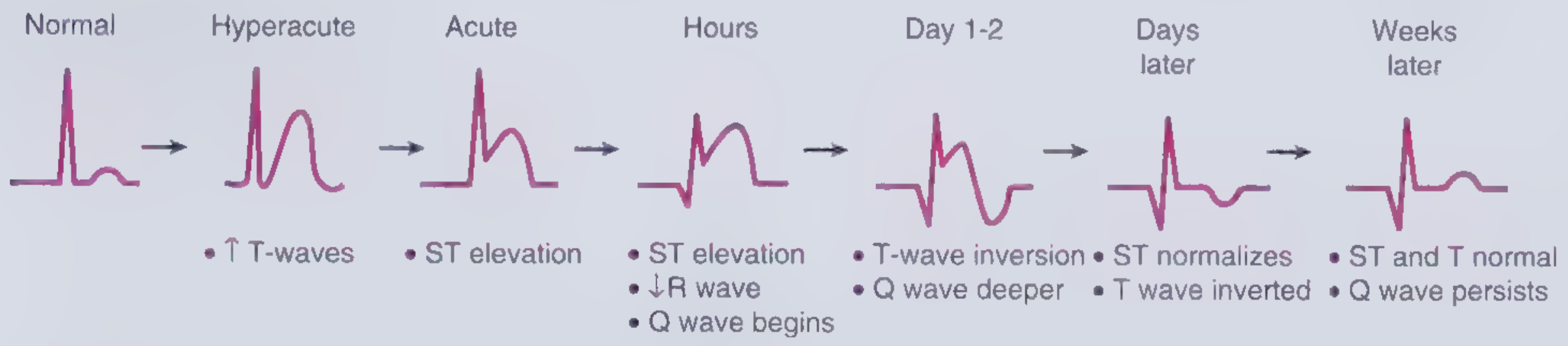
MASTER REFERENCE CHART
1. ACUTE CORONARY SYNDROMES (ACS)
| Condition | ECG Changes | Leads Affected |
|---|---|---|
| STEMI | Hyperacute tall/broad T waves (earliest) → ST elevation (convex/tombstone) → Q wave formation → T wave inversion → ST normalises → Q waves persist | Depends on territory (see localisation table below) |
| NSTEMI / UA | Horizontal or downsloping ST depression ≥0.05 mV in ≥2 contiguous leads; T wave inversion ≥0.1 mV with prominent R wave | Diffuse, or territory-specific |
| Posterior STEMI | ST depression in V1-V3 (mirror image); ST elevation in posterior leads V7-V9 | V1-V3 (reciprocal), V7-V9 (direct) |
| Right ventricular MI | ST elevation in V1, V3R, V4R | V3R, V4R |
| Wellens Syndrome (critical LAD stenosis, pain-free) | Type A: Biphasic T waves in V2-V3; Type B: Deep symmetric T wave inversion in V2-V3 | V2-V3 |
| de Winter T-wave pattern | Upsloping ST depression at J point + tall symmetric T waves; no ST elevation | V1-V6 |
| Hyperacute T waves | Tall, broad, asymmetric, peaked T waves | Territory-specific |
2. STEMI LOCALISATION BY LEAD TERRITORY
| MI Territory | Leads with ST elevation / Q waves | Artery Occluded | Reciprocal Changes |
|---|---|---|---|
| Anterior | V1-V4 | LAD | III, aVF (ST depression) |
| Septal | V1-V2 | Septal branches of LAD | - |
| Lateral | I, aVL, V5-V6 | LCx or diagonal | V1-V2 (ST depression) |
| Anterolateral | V1-V6, I, aVL | Proximal LAD | III, aVF |
| Inferior | II, III, aVF | RCA (80%) or LCx (20%) | I, aVL (ST depression) |
| Posterior | V7-V9 (elevation); V1-V3 (depression) | RCA or LCx | V1-V3 |
| Right Ventricular | V1, V3R, V4R | Proximal RCA | - |
3. PERICARDITIS
| Feature | ECG Finding |
|---|---|
| ST elevation | Diffuse, saddle-shaped (concave upwards), in multiple leads (I, II, III, aVF, V2-V6) - does NOT follow a coronary territory |
| PR depression | Classic hallmark - PR segment depression in most leads (especially II, V4-V6); PR elevation in aVR |
| T wave inversion | Occurs late, after ST normalises |
| No reciprocal ST depression | Unlike STEMI (except aVR and V1) |
| No Q waves | |
| Sinus tachycardia | Common (pain, inflammation) |
| Low voltage / electrical alternans | If pericardial effusion develops |
Key differentiator from STEMI: PR depression + diffuse concave ST elevation not localised to one territory.
4. MYOCARDITIS
| Feature | ECG Finding |
|---|---|
| ST elevation (diffuse, mimics pericarditis) | Common |
| T wave inversion | Widespread |
| Bundle branch block (new) | May occur |
| AV block (1°, 2°, 3°) | Can develop |
| Ventricular arrhythmias | PVCs, VT |
| Low QRS voltages | If myocardial oedema severe |
5. LEFT VENTRICULAR HYPERTROPHY (LVH)
| Feature | ECG Finding |
|---|---|
| Increased QRS voltage | S in V1 + R in V5 or V6 ≥35 mm (Sokolow-Lyon); R in aVL ≥11 mm |
| Left axis deviation | Common |
| ST depression + T wave inversion | "Strain pattern" in lateral leads (V5-V6, I, aVL) - downsloping ST depression with asymmetric T wave inversion |
| Left atrial enlargement | Broad, notched P wave (P mitrale) in II; biphasic P in V1 |
| Prolonged QRS | Mild widening, not frank BBB |
6. RIGHT VENTRICULAR HYPERTROPHY (RVH)
| Feature | ECG Finding |
|---|---|
| Right axis deviation (>+100°) | |
| Dominant R wave in V1 | R > S in V1 |
| Deep S waves in V5-V6 | |
| ST depression + T wave inversion | Right precordial "strain" in V1-V3 |
| Right atrial enlargement | Tall peaked P wave >2.5 mm in II (P pulmonale) |
| rSR' pattern in V1 |
7. ATRIAL FIBRILLATION (AF)
| Feature | ECG Finding |
|---|---|
| No P waves | Replaced by irregular fibrillatory baseline (f waves), 350-600/min |
| Irregularly irregular RR intervals | |
| Narrow QRS (if no aberrant conduction) | |
| Variable ventricular rate | Uncontrolled: 100-180 bpm |
| Fine or coarse f waves | Best seen in V1, II |
8. ATRIAL FLUTTER
| Feature | ECG Finding |
|---|---|
| Sawtooth flutter waves | 250-350/min, best seen in II, III, aVF |
| Regular atrial rate ~300/min | |
| Regular ventricular rate | Usually 2:1 (150 bpm), 3:1, or 4:1 block |
| No isoelectric baseline | Continuous flutter activity |
9. AV BLOCK
| Type | ECG Finding |
|---|---|
| 1st degree | PR interval > 200 ms (>5 small squares); every P conducts |
| 2nd degree Mobitz I (Wenckebach) | Progressive PR prolongation until one P wave is not conducted (dropped QRS); cyclical |
| 2nd degree Mobitz II | Fixed PR interval with sudden non-conducted P waves; more ominous |
| 3rd degree (Complete) | P waves and QRS completely dissociated; P rate > QRS rate; escape rhythm (junctional = narrow, ventricular = wide) |
10. BUNDLE BRANCH BLOCKS
Left Bundle Branch Block (LBBB)
| Feature | Finding |
|---|---|
| QRS duration | ≥120 ms |
| Broad notched R in lateral leads | I, aVL, V5-V6 ("M" pattern) |
| Deep broad S in V1 | "W" pattern in V1 |
| No septal Q waves in I, V5-V6 | |
| ST/T discordant | ST depression + T inversion opposite to main QRS deflection |
| New LBBB + chest pain | Treat as STEMI equivalent (Sgarbossa criteria apply) |
Right Bundle Branch Block (RBBB)
| Feature | Finding |
|---|---|
| QRS duration | ≥120 ms |
| rSR' in V1-V2 | "M" or "rabbit ears" pattern |
| Wide slurred S wave in I, V5-V6 | |
| ST depression + T inversion | V1-V3 (discordant) |
| Can be normal variant |
11. WOLFF-PARKINSON-WHITE (WPW) SYNDROME
| Feature | ECG Finding |
|---|---|
| Short PR interval | <120 ms |
| Delta wave | Slurred upstroke of QRS |
| Widened QRS | >120 ms |
| Secondary ST-T changes | Discordant to delta wave |
| Risk | AF with very rapid ventricular rate → VF |
12. BRUGADA SYNDROME
| Feature | ECG Finding |
|---|---|
| Type 1 (diagnostic) | Coved ST elevation ≥2 mm with T wave inversion in V1-V2 (sometimes V3); "shark fin" or "coved" pattern |
| Type 2 | Saddle-back ST elevation ≥2 mm in V1-V2 |
| Type 3 | ST elevation <1 mm |
| PR prolongation | May be present |
| RBBB-like pattern | V1-V2 |
13. PULMONARY EMBOLISM (PE)
| Feature | ECG Finding |
|---|---|
| Sinus tachycardia | Most common (>40% of cases) |
| S1Q3T3 pattern | Deep S in lead I + Q wave in III + T inversion in III (classic but only ~20% of cases) |
| Right axis deviation | New |
| New RBBB | |
| T wave inversions in V1-V4 | Right heart strain; correlates with severity |
| Right atrial enlargement | Peaked P wave in II |
| Sinus tachycardia or AF | |
| Low voltage | Occasionally |
14. HYPERKALAEMIA
| Serum K⁺ | ECG Finding |
|---|---|
| 5.5-6.5 mmol/L | Tall, peaked, narrow ("tented") T waves - earliest sign |
| 6.5-7.5 mmol/L | PR prolongation, P wave flattening/disappearance |
| 7.0-8.0 mmol/L | Widened QRS (sine wave appearance) |
| >8.0 mmol/L | VF / asystole |
15. HYPOKALAEMIA
| Feature | ECG Finding |
|---|---|
| U waves | Prominent U wave (follows T wave), best in V2-V3 |
| T wave flattening or inversion | |
| ST depression | |
| QT(U) prolongation | Risk of torsades de pointes |
| PR prolongation |
16. HYPERCALCAEMIA vs HYPOCALCAEMIA
| Condition | ECG Finding |
|---|---|
| Hypercalcaemia | Short QT interval; short ST segment; J-wave (Osborn wave in severe cases) |
| Hypocalcaemia | Prolonged QT interval (specifically QTc); prolonged ST segment; T waves normal; risk of torsades |
17. HYPOTHERMIA
| Feature | ECG Finding |
|---|---|
| Osborn wave (J wave) | Positive deflection at J point, best in V4-V6 and II; pathognomonic |
| Bradycardia | Progressive |
| Prolonged PR, QRS, QT | All intervals prolonged |
| AF/Junctional rhythm | Common |
| VF | At core temp <28°C |
18. LONG QT SYNDROME
| Feature | ECG Finding |
|---|---|
| QTc prolonged | >440 ms in males, >460 ms in females (>500 ms = high risk) |
| Abnormal T wave morphology | Notched, bifid, or late-appearing T waves |
| Risk | Torsades de Pointes → VF |
19. HYPERTROPHIC CARDIOMYOPATHY (HCM)
| Feature | ECG Finding |
|---|---|
| LVH pattern | High voltage |
| Deep narrow "dagger" Q waves | II, III, aVF, V5-V6 (septal depolarisation abnormality) - NOT infarction Q waves |
| ST depression + T wave inversion | Lateral leads |
| Left axis deviation | |
| AF | Common |
20. DILATED CARDIOMYOPATHY (DCM)
| Feature | ECG Finding |
|---|---|
| Low QRS voltage | Or paradoxically increased |
| LBBB | Common |
| Left axis deviation | |
| ST-T changes | Non-specific |
| Atrial and ventricular arrhythmias | |
| Intraventricular conduction delay |
21. DIGOXIN EFFECT vs TOXICITY
| State | ECG Finding |
|---|---|
| Therapeutic (Digoxin effect) | Scooped/reverse tick ST depression ("Salvador Dali moustache"), shortened QT, T wave inversion in lateral leads |
| Toxicity | Bradycardia, AV block (any degree), PAT with block (classic), bidirectional VT, multiple arrhythmias simultaneously |
22. ACUTE COR PULMONALE / CHRONIC PULMONARY DISEASE
| Feature | ECG Finding |
|---|---|
| Right axis deviation | |
| P pulmonale | Peaked P > 2.5 mm in II |
| RVH pattern | |
| Low voltage | In COPD (hyperinflation) |
| Multifocal atrial tachycardia (MAT) | COPD-specific: ≥3 different P wave morphologies with irregular rate |
| S1S2S3 pattern |
QUICK-REFERENCE SUMMARY TABLE
| Condition | Key ECG Signature |
|---|---|
| STEMI | ST elevation (convex), Q waves, in territory |
| NSTEMI/UA | ST depression, T inversion, no Q waves |
| Pericarditis | Diffuse saddle-shaped ST elevation + PR depression |
| PE | S1Q3T3 + sinus tachycardia + T inversion V1-V4 |
| LVH | High voltage + lateral strain (ST dep + T inv) |
| LBBB | Wide QRS, M-pattern V5-V6, W-pattern V1 |
| RBBB | rSR' V1, wide S in I/V5-V6 |
| WPW | Short PR + delta wave |
| Brugada | Coved ST elevation V1-V2 |
| Hyperkalaemia | Peaked T → wide QRS → sine wave |
| Hypokalaemia | U waves + flat T waves + ST depression |
| Hypothermia | Osborn (J) wave + bradycardia |
| Long QT | Prolonged QTc >440/460 ms |
| HCM | Deep dagger Q waves II, III, aVF, V5-V6 |
| Digoxin effect | Scooped ST depression + short QT |
| AF | No P waves + irregularly irregular |
| 3rd degree AV block | P-QRS dissociation |
Sources: Rosen's Emergency Medicine (9e), Braunwald's Heart Disease (Vol 1-2), Harrison's Principles of Internal Medicine (22e, 2025), Frameworks for Internal Medicine, Tintinalli's Emergency Medicine (9e)
How to do examination for general physical examination
Finding Sources
Finding Sources
Reading File
Reading File
Reading File
Finding Sources
Finding Sources
Finding Sources
Reading File
Finding Sources
Reading File
Reading File
I now have rich content from Harrison's, Goldman-Cecil, and Braunwald's. Let me compile the full, detailed guide.
General Physical Examination (GPE) - Step-by-Step How-To Guide
Sourced from Harrison's Principles of Internal Medicine (22e, 2025), Goldman-Cecil Medicine, and Braunwald's Heart Disease.
BEFORE YOU START
- Position: Patient lying comfortably in bed at 45°, adequately exposed but with dignity maintained
- Lighting: Good natural or bright overhead light
- Equipment ready: Thermometer, sphygmomanometer (correct cuff size), stethoscope, pen torch, tongue depressor
- Order: Always examine from the right side of the patient
- First impression: Stand at the foot of the bed and observe before touching - this is the "end-of-bed diagnosis"
STEP 1 - GENERAL APPEARANCE (End-of-Bed Assessment)
Before touching the patient, observe and note:
| What to Assess | What to Look For |
|---|---|
| Consciousness | Alert, drowsy, confused, unconscious |
| Distress | Comfortable at rest vs. in pain / breathless / restless |
| Built and nutrition | Obese, normal, thin, cachectic / emaciated |
| Apparent age vs. stated age | Looking older than age = chronic illness |
| Posture | Sitting upright (orthopnoea), leaning forward (pericarditis, COPD), curled up (peritonitis) |
| Special facies | Moon face (Cushing's), coarse features (acromegaly/hypothyroid), malar flush (mitral stenosis/SLE) |
| Obvious abnormalities | Jaundice, pallor, cyanosis, oedema, deformities, rashes |
| Breathing pattern | Laboured, rapid, Kussmaul, Cheyne-Stokes |
| Speech | Hoarse, dysarthric, normal |
| Surroundings | Oxygen mask, IV lines, medications on bedside table, wheelchair |
"The most important vital sign is what the patient looks like." - Goldman-Cecil Medicine
STEP 2 - VITAL SIGNS
Measure and record all five. Never skip any.
A. Temperature
- Method: Oral electric thermometer preferred; axillary is 0.5°C lower; rectal 0.5°C higher
- Technique: Oral - place under tongue, lips closed, for 1 minute
- Normal: 36.5-37.5°C (oral)
- Interpret:
- Fever (>37.5°C): infection, inflammation, malignancy
- Hypothermia (<35°C): exposure, sepsis, hypothyroidism, alcohol/barbiturate OD
B. Pulse
- Site: Right radial artery (thumb side of wrist, between flexor carpi radialis and radial styloid)
- Technique: Use index + middle + ring fingers gently over artery; count for full 60 seconds if irregular, 30 seconds if regular
- Assess systematically:
| Parameter | Normal | Abnormal Finding |
|---|---|---|
| Rate | 60-100 bpm | Tachy >100, Brady <60 |
| Rhythm | Regular | Irregular (regular irregular = AF, irregular irregular = AF, ectopics) |
| Volume | Normal | High (bounding - aortic regurg, CO2 retention, fever); Low (weak/thready - shock, AS) |
| Character | Normal upstroke | Slow rising (AS); Collapsing/waterhammer (AR); Bisferiens (HOCM); Pulsus alternans (severe LVF) |
| Vessel wall | Soft/springy | Hard, calcified, tortuous (arteriosclerosis - "pipe stem" artery) |
- Also feel: Both radials simultaneously (radioulnar delay); then radial vs femoral (radio-femoral delay = coarctation of aorta)
C. Blood Pressure
- Cuff selection: Bladder should encircle 80% of arm; too small = falsely high; too large = falsely low
- Technique:
- Patient seated or lying, arm at heart level, relaxed for 5 minutes
- Palpate radial artery; inflate cuff until radial pulse disappears + 30 mmHg more
- Place stethoscope over brachial artery in antecubital fossa
- Deflate at 2 mmHg/second
- Systolic = first Korotkoff sound (Phase 1)
- Diastolic = disappearance of sound (Phase 5)
- Take in BOTH arms on first visit - >10 mmHg difference is significant (subclavian stenosis, aortic dissection)
- Postural BP: Take lying and standing (after 2 minutes standing) - drop >20 mmHg systolic / >10 mmHg diastolic = orthostatic hypotension
- Normal: <120/80 mmHg
D. Respiratory Rate
- Technique: Count breaths for a full 60 seconds while appearing to check the pulse (patients alter breathing if they know you are watching)
- Count chest/abdominal movement as one breath
- Normal: 12-20 breaths/min
- Observe: Depth (shallow/deep), rhythm, use of accessory muscles, symmetry
- Tachypnoea (>20/min): pneumonia, PE, acidosis, pain, anxiety
- Bradypnoea (<12/min): opioids, sedatives, brainstem lesion
E. Oxygen Saturation (SpO2)
- Method: Pulse oximeter on finger (ensure warm, well-perfused digit; remove nail polish)
- Normal: ≥95% on room air
- <94% = significant hypoxaemia requiring investigation
- Note: unreliable in CO poisoning (falsely normal), severe anaemia, peripheral vasoconstriction
F. Weight, Height, BMI
| Measurement | Method |
|---|---|
| Weight | Patient standing on calibrated scale, minimal clothing |
| Height | Stadiometer, standing upright without footwear |
| BMI | Weight (kg) / Height² (m²) |
| Waist circumference | Measure at smallest area between lower rib and umbilicus |
| BMI Category | Range |
|---|---|
| Underweight | <18.5 |
| Normal | 18.5-24.9 |
| Overweight | 25-29.9 |
| Obese | ≥30 |
- Waist circumference >102 cm (men) / >88 cm (women) = metabolic risk
STEP 3 - SKIN (Head-to-Toe Inspection)
Examine skin systematically as you proceed through the rest of the examination.
Pallor
- Where to look: Lower palpebral conjunctiva (pull down lower eyelid), palmar creases, tongue, nail beds
- Conjunctival pallor = most reliable sign of anaemia
- Palmar crease pallor: significant when pallor visible in creases themselves
Jaundice (Icterus)
- Where to look: Sclerae first (earliest and most sensitive), then skin, then hard palate
- Best assessed in natural daylight
- Scleral icterus detectable when serum bilirubin > ~35 µmol/L (2 mg/dL)
- Note: carotenaemia causes yellow skin but spares the sclerae
Cyanosis
| Type | Where to Check | Cause |
|---|---|---|
| Central cyanosis | Tongue, lips, oral mucosa | Right-to-left shunt, severe respiratory failure, methaemoglobinaemia |
| Peripheral cyanosis | Fingertips, toes, ear lobes | Reduced peripheral flow (cold, shock, heart failure, Raynaud's) |
| Differential cyanosis | Lower limbs only, upper normal | Patent ductus arteriosus + pulmonary hypertension |
- Central cyanosis visible when deoxy-Hb > 5 g/dL
Oedema
-
Technique: Press firmly with thumb for 5-10 seconds over bony prominence (dorsum of foot, medial malleolus, shin, sacrum in bedridden)
-
Pitting oedema: Pit persists after pressure release
-
Grading:
- 1+ : Trace, slight pit, rebounds quickly
- 2+ : Pit rebounds in <15 seconds
- 3+ : Deep pit, rebounds in 15-30 seconds, limb appears swollen
- 4+ : Very deep pit, takes >30 seconds, gross swelling
-
Sacral oedema: Always check in bedridden patients (dependent area)
-
Non-pitting oedema: lymphoedema, myxoedema (hypothyroidism)
STEP 4 - HANDS
Start with the hands - they give enormous clinical information.
Nails
| Sign | Appearance | Clinical Significance |
|---|---|---|
| Clubbing | Loss of nail-fold angle, fluctuant nail bed, drum-stick appearance | Lung cancer, fibrosing alveolitis, bronchiectasis, cyanotic CHD, infective endocarditis, liver cirrhosis |
| Koilonychia | Spoon-shaped (concave) nails | Iron deficiency anaemia |
| Leuconychia | White nails | Hypoalbuminaemia (liver disease, nephrotic syndrome) |
| Splinter haemorrhages | Linear reddish-brown lines in nail bed (mid-nail position) | Infective endocarditis (peripheral emboli); trauma (near tip) |
| Beau's lines | Transverse grooves across nail | Severe systemic illness, chemotherapy (growth arrest) |
| Yellow nails | Slow-growing, yellowish | Yellow nail syndrome (lymphoedema, pleural effusion) |
| Terry's nails | White proximally, normal pink distally | Liver cirrhosis, heart failure, diabetes |
| Half-and-half nails | Proximal white, distal red/brown | Chronic renal failure |
Clubbing - How to Grade
| Grade | Description |
|---|---|
| Grade 1 | Fluctuation and softening of nail bed (ballotable nail) |
| Grade 2 | Loss of normal angle between nail and nail fold (Lovibond angle >180°); Schamroth's sign positive |
| Grade 3 | Increased curvature of nail (drum-stick appearance) |
| Grade 4 | Hypertrophic pulmonary osteoarthropathy (periosteal new bone formation, wrist/ankle pain) |
Schamroth's sign: Place dorsal surfaces of corresponding fingers of both hands together - loss of the normal diamond-shaped window = positive = clubbing
Palms and Fingers
| Sign | Location | Clinical Significance |
|---|---|---|
| Palmar erythema | Thenar and hypothenar eminences | Liver disease, pregnancy, thyrotoxicosis, rheumatoid arthritis |
| Dupuytren's contracture | Palmar fascia, ring > little finger | Liver cirrhosis, diabetes, familial, trauma |
| Xanthomata | Tendon sheaths, extensor surfaces | Hypercholesterolaemia (Type II), palm crease xanthomata = Type III |
| Janeway lesions | Palms and soles - flat, non-tender haemorrhagic | Infective endocarditis (septic emboli) |
| Osler's nodes | Fingertip pads - tender, raised nodules | Infective endocarditis (immune complex) |
| Thenar wasting | Base of thumb | Carpal tunnel syndrome, median nerve palsy |
| Hypothenar wasting | Little finger side | T1 lesion, ulnar nerve palsy |
Temperature and Moisture
- Warm, moist: Thyrotoxicosis, fever, anxiety
- Warm, dry: Normal, vasodilated
- Cold, clammy: Shock, anxiety, sympathetic activation
- Dry, rough: Hypothyroidism
Tremor
- Ask patient to extend arms with fingers spread - observe for:
- Fine tremor (thyrotoxicosis, anxiety, beta-agonists)
- Resting tremor (Parkinson's) - disappears on movement
- Asterixis (liver flap) - negative myoclonus; ask patient to cock wrists back with arms extended, count 15 seconds - flapping = hepatic encephalopathy, CO2 retention, uraemia
STEP 5 - FACE AND HEAD
Eyes
Work from outside in:
| Structure | What to Look For | Significance |
|---|---|---|
| Periorbital area | Puffiness, xanthelasma | Nephrotic syndrome, hypothyroidism; hyperlipidaemia |
| Sclerae | Icterus (yellow), injection, blue (osteogenesis imperfecta) | Jaundice; connective tissue disease |
| Conjunctivae | Pallor (pull down lower lid), chemosis | Anaemia; allergy, SVC obstruction |
| Cornea | Arcus senilis (white ring), Kayser-Fleischer rings (brown-green at periphery) | Dyslipidaemia if <50 years; Wilson's disease |
| Pupils | Size, equality, shape, reaction to light and accommodation | CN III lesion, Horner's, Argyll Robertson |
| Exophthalmos | Protrusion of globe (can you see sclera above iris?) | Graves' disease (thyrotoxicosis) |
Mouth
- Lips: Central cyanosis, angular stomatitis (B12/iron deficiency), herpes labialis
- Gums: Hypertrophy (phenytoin, ciclosporin, leukaemic infiltration), bleeding, lead line
- Teeth: Dental hygiene (endocarditis risk), notching (Hutchinson's teeth = congenital syphilis)
- Tongue:
- Glossitis (smooth, beefy red) = B12/folate/iron deficiency
- Macroglossia = hypothyroidism, amyloid, acromegaly
- Deviation on protrusion = CN XII palsy (to weak side)
- Central cyanosis (underside of tongue)
- Breath odour: Uraemic fetor (ammoniacal, "fishy") = renal failure; hepatic fetor (sweet/musty/"fetor hepaticus") = liver failure; ketotic (pear drops/acetone) = diabetic ketoacidosis; faeculent = bowel obstruction
Face - Specific Facies
| Facies | Features | Condition |
|---|---|---|
| Myxoedematous | Puffiness, coarse features, loss of lateral 1/3 eyebrow, pallor, dry skin | Hypothyroidism |
| Thyrotoxic | Exophthalmos, lid lag, stare, lid retraction, tremor | Hyperthyroidism |
| Cushingoid | Moon face, facial plethora, acne, hirsutism | Cushing's syndrome |
| Acromegalic | Prognathism, large nose, thick lips, widely spaced teeth | Acromegaly |
| Mitral facies | Malar flush (purple-red patches over cheeks) | Mitral stenosis |
| SLE butterfly rash | Malar erythema sparing nasolabial folds | Systemic lupus |
| Parkinson's facies | Mask-like, expressionless, reduced blinking | Parkinson's disease |
| Marfan's | Long, narrow face, high arched palate | Marfan syndrome |
STEP 6 - NECK
Jugular Venous Pressure (JVP)
- Position: Patient at 45° reclined, head slightly turned to left
- Which vein: Internal jugular (runs between the two heads of SCM), preferred over external jugular
- Technique:
- Look for the biphasic pulsation between the clavicle and the ear
- JVP pulsates - can be obliterated by gentle pressure at the base of the neck (carotid pulse cannot)
- JVP rises with abdominal pressure (hepatojugular reflux)
- Measure vertical height of the meniscus above the sternal angle (angle of Louis)
- Normal: <4 cm above sternal angle at 45°
- Elevated JVP: Heart failure, pericardial effusion/tamponade, constrictive pericarditis, SVC obstruction, fluid overload
- JVP waveform: a wave (atrial contraction); c wave (tricuspid closure); x descent; v wave (venous filling); y descent
Thyroid Gland
- Inspection: Ask patient to swallow a sip of water - thyroid moves upward on swallowing; note midline vs. lateral, size
- Palpation: Stand behind the patient; use both hands with fingertips on the gland, ask to swallow again
- Note: size, consistency (soft/firm/hard), surface (smooth/nodular), tenderness, tracheal position
- Percussion: For retrosternal extension - percuss over manubrium
- Auscultation: Bell of stethoscope over the gland - bruit = hyperthyroidism (increased vascularity)
Lymph Nodes
Systematic order (use gentle circular finger pad palpation):
- Submental
- Submandibular
- Pre-auricular (parotid)
- Post-auricular (mastoid)
- Occipital
- Anterior cervical chain (along anterior border of SCM)
- Posterior cervical chain (along posterior border of SCM)
- Supraclavicular - especially left side (Virchow's node / Troisier's sign = abdominal malignancy)
- Axillary (support patient's arm with your arm; examine all five groups)
- Epitrochlear
- Inguinal (horizontal and vertical chains)
For each node, document:
| Feature | Options |
|---|---|
| Size | cm |
| Consistency | Soft, firm, hard, rubbery |
| Tenderness | Tender vs. non-tender |
| Mobility | Mobile vs. fixed / matted |
| Surface | Smooth vs. irregular |
| Overlying skin | Normal vs. red, sinus |
Hard, non-tender, fixed, matted nodes = malignancy; Soft, tender = reactive (infection); Rubbery = lymphoma
STEP 7 - EYES (Fundoscopy - when indicated)
Using ophthalmoscope (dilate pupil if needed):
| Finding | Significance |
|---|---|
| Papilloedema | Raised intracranial pressure |
| AV nipping, flame haemorrhages, cotton wool spots, hard exudates | Hypertensive retinopathy |
| Microaneurysms, dot/blot haemorrhages, new vessel formation | Diabetic retinopathy |
| Pale disc | Optic atrophy |
| Roth spots (white-centred haemorrhages) | Infective endocarditis |
STEP 8 - ADDITIONAL GENERAL SIGNS
Dehydration Assessment
| Sign | Method |
|---|---|
| Skin turgor | Pinch skin on dorsum of hand or over sternum; normally springs back immediately; slow recoil = dehydration (unreliable in elderly) |
| Dry mucous membranes | Inspect mouth and tongue |
| Sunken eyes | Inspect |
| Capillary refill | Press fingernail for 5 seconds; release - normal refill <2 seconds; >2 sec = poor perfusion |
| Postural hypotension | BP lying and standing |
Mental Status (Assess Throughout)
- AVPU: Alert / Voice / Pain / Unresponsive
- GCS (formal, if altered):
- Eyes (4), Verbal (5), Motor (6) - maximum 15, minimum 3
- Orientation: Ask name, date, place
- Mood and affect: Observe during conversation
QUICK-REFERENCE: GPE SEQUENCE AT A GLANCE
STEP 1:  End-of-bed observation → general appearance, distress, built
STEP 2:  Vital signs → Temp → Pulse → BP → RR → SpO2 → Weight/Height/BMI
STEP 3:  Skin → Pallor, Jaundice, Cyanosis, Oedema
STEP 4:  Hands → Nails (clubbing, koilonychia, etc.) → Palms → Temperature → Tremor
STEP 5:  Face & Head → Eyes → Mouth/Tongue → Specific facies
STEP 6:  Neck → JVP → Thyroid → Lymph nodes (cervical/axillary/inguinal)
STEP 7:  Fundoscopy (if indicated)
STEP 8:  Additional → Hydration → Capillary refill → Mental status
        ↓
     SYSTEMIC EXAMINATION (CVS → Resp → Abdomen → Neuro → MSK)
KEY "NEVER MISS" SIGNS IN GPE
| Sign | What It Means |
|---|---|
| Virchow's node (left supraclavicular) | GI malignancy (Troisier's sign) |
| Asterixis (liver flap) | Hepatic encephalopathy / CO2 retention / uraemia |
| Clubbing | Respiratory/cardiac/GI disease |
| Splinter haemorrhages (mid-nail) | Infective endocarditis |
| Osler's nodes + Janeway lesions | Infective endocarditis |
| Xanthelasma + corneal arcus <50y | Hypercholesterolaemia |
| Kayser-Fleischer rings | Wilson's disease |
| Buffalo hump + moon face + purple striae | Cushing's syndrome |
| Pallor + koilonychia + angular stomatitis | Iron deficiency anaemia |
| Malar rash + butterfly distribution | SLE |
| Exophthalmos + lid retraction + tremor | Thyrotoxicosis |
Sources: Harrison's Principles of Internal Medicine 22e (2025), Goldman-Cecil Medicine, Braunwald's Heart Disease, Textbook of Family Medicine 9e
This is a shared conversation. Sign in to Orris to start your own chat.