
30 years male presented with heart burns and dyspepsia. A breath test and biopsy were ordered to confirm the diagnosis. What is your diagnosis? Write about the morphology and complications of this disease.
Helicobacter pylori gastritis histology biopsy
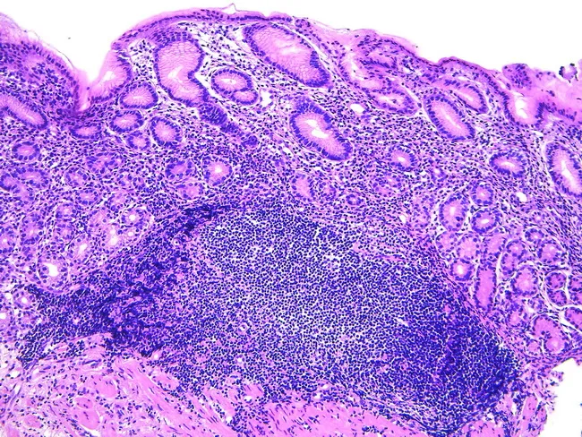
Gastric biopsy histology showing acquired MALT following Helicobacter pylori infection. Hematoxylin and eosin stained sections reveal dense lymphoid infiltration in the lamina propria with formation of lymphoid aggregates and germinal centers, partially effacing normal mucosal architecture. The overlying gastric epithelium remains intact in areas, but lymphoepithelial lesions may be present, indicating invasion of ducts or epithelium by neoplastic B-cells. The stomach normally lacks organized lymphoid tissue; the observed lymphoid proliferation represents mucosa-associated lymphoid tissue expansion, consistent with extranodal marginal zone B-cell lymphoma of mucosa-associated lymphoid tissue (MALT lymphoma). The cellular population is typically small to medium-sized lymphocytes with irregular chromatin, sometimes with plasmacytoid differentiation; scattered immunoblasts may occur. Chronic inflammation, such as Helicobacter pylori gastritis, provides the antigenic stimulus driving MALT development and potential regression after microbial eradication. Clinically, MALT lymphoma of the stomach may respond to antibiotic therapy, with lesion regression upon H. pylori eradication, or require staging and systemic therapy if refractory or advanced. Differential includes reactive lymphoid hyperplasia and transformation to diffuse large B-cell lymphoma. Diagnostic significance lies in recognizing MALT patterns to guide treatment, prognosis, and surveillance; correlate with endoscopic findings and serology for infection status.
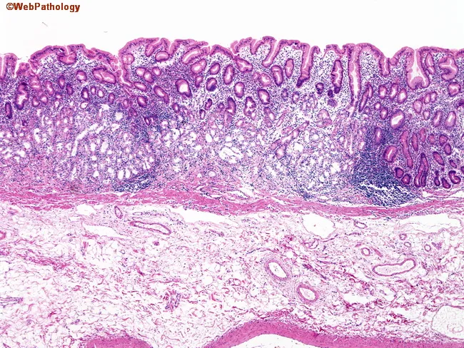
This histopathology image depicts a gastric mucosa biopsy from the fundic/corpus region examined under light microscopy after hematoxylin and eosin staining. The specimen demonstrates prominent thinning of the fundic mucosa with loss of oxyntic/parietal cells, consistent with gastric mucosal atrophy. The lamina propria is densely infiltrated by lymphocytes and plasma cells (lymphoplasmacytic infiltrate), typical of autoimmune gastritis. Glandular architecture is attenuated; residual fundic glands appear irregular with cystic dilatation and foveolar hyperplasia may be absent. The submucosa and deeper layers appear relatively uninvolved in this field. The morphological pattern, characterized by corpus-predominant atrophy and parietal cell loss, supports an autoimmune process rather than Helicobacter pylori gastritis, though correlation with endoscopy and serology is essential. Clinically, such histology implies hypochlorhydria/achlorhydria and potential pernicious anemia risk due to intrinsic factor deficiency, guiding further workup (gastrin levels, autoantibodies, B12 status). This image serves as a representative teaching example for pathology of autoimmune gastritis, highlighting key features: parietal cell depletion, mucosal atrophy, dense plasma cell rich inflammation, and the need to verify site of origin for biopsy through endoscopic correlation. Potential clinical applications include diagnostic validation, educational case discussion, and research on corpus-predominant atrophic gastritis.
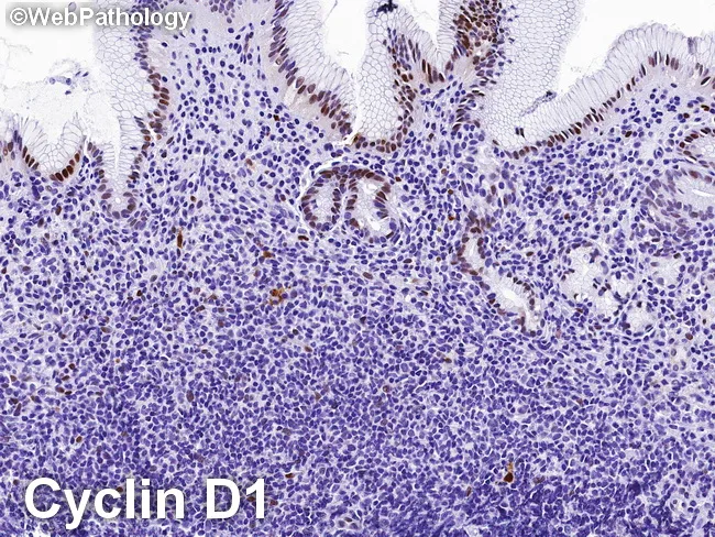
High-power gastric mucosal biopsy prepared for light microscopy with cyclin D1 immunohistochemistry demonstrates features characteristic of extranodal marginal zone B‑cell lymphoma of MALT type involving the stomach. In this specimen, a dense, monomorphic infiltrate of small to medium-sized lymphocytes expands the lamina propria and surrounds gastric glands, forming classic lymphoepithelial lesions. The neoplastic cells typically exhibit irregular, slightly indented nuclei with scant cytoplasm and inconspicuous nucleoli; plasmacytoid differentiation may be present in focal areas. Immunohistochemical staining is negative for cyclin D1, supporting a MALT phenotype and helping distinguish from mantle cell lymphoma, which is cyclin D1 positive. The neoplastic cells reliably express B‑cell markers (CD20/CD79a) and often show light-chain restriction, consistent with a monoclonal B‑cell population arising in mucosa‑associated lymphoid tissue. Background reactive lymphoid infiltrates and H. pylori–associated gastritis can accompany the neoplasm. Clinically, MALT lymphoma of the stomach is frequently associated with Helicobacter pylori infection and is potentially amenable to antibiotic eradication therapy, with response rates correlating with stage and H. pylori status. Radiologic and endoscopic correlation may reveal regional gastric wall involvement but histology remains essential for diagnosis, grading, and staging. This image underlines the diagnostic utility of cyclin D1 negativity in differentiating gastric MALT lymphoma from cyclin D1‑positive mantle cell lymphoma.
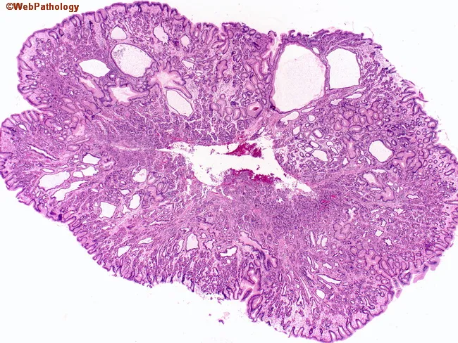
Imaging modality: Histopathology with light microscopy of a gastric mucosa biopsy. The slide shows a polypoid proliferation in the gastric body/fundus region composed of cystically dilated fundic glands embedded in fibromuscular lamina propria. The epithelium lining these glands is largely atrophic with minimal inflammatory change, and there is preservation of mucous foveolar cells around the periphery. The glands are variably sized, with round to elongated lumina, and the overall architecture is organized without destructive invasion. No dysplasia or neoplastic transformation is evident. Background mucosa lacks significant Helicobacter pylori-associated gastritis. The glands demonstrate oxyntic differentiation, characterized by parietal and chief cell-rich acini, consistent with fundic gland polyps. The lesion is localized and nonpedunculated; surface epithelium is intact. Clinically, fundic gland polyps are usually small (<2 cm) and benign; sporadic and syndromic forms exist, the latter associated with familial cancer syndromes and requiring tailored surveillance. The diagnostic significance is that these features distinguish fundic gland polyps from inflammatory or neoplastic gastric polyps, reducing immediate oncologic concerns but mandating appropriate clinical-follow-up per syndrome status. In differential diagnosis, consider gastric hyperplastic polyps, adenomas and inflammatory polyps. This image is educational for pathology residents illustrating typical fundic gland polyp histology and the benign behavior in non-syndromic cases.
Diagnosis: Helicobacter pylori Gastritis (Chronic Active Gastritis)
The Organism and Diagnostic Tests
- Urea breath test: The patient ingests labeled (¹³C or ¹⁴C) urea. H. pylori urease cleaves urea → ammonia + labeled CO₂, which is detected in exhaled breath. Highly sensitive and specific.
- Biopsy: Antral biopsies allow direct histologic identification, rapid urease test (CLO test), culture, and PCR for H. pylori DNA.
Pathogenesis
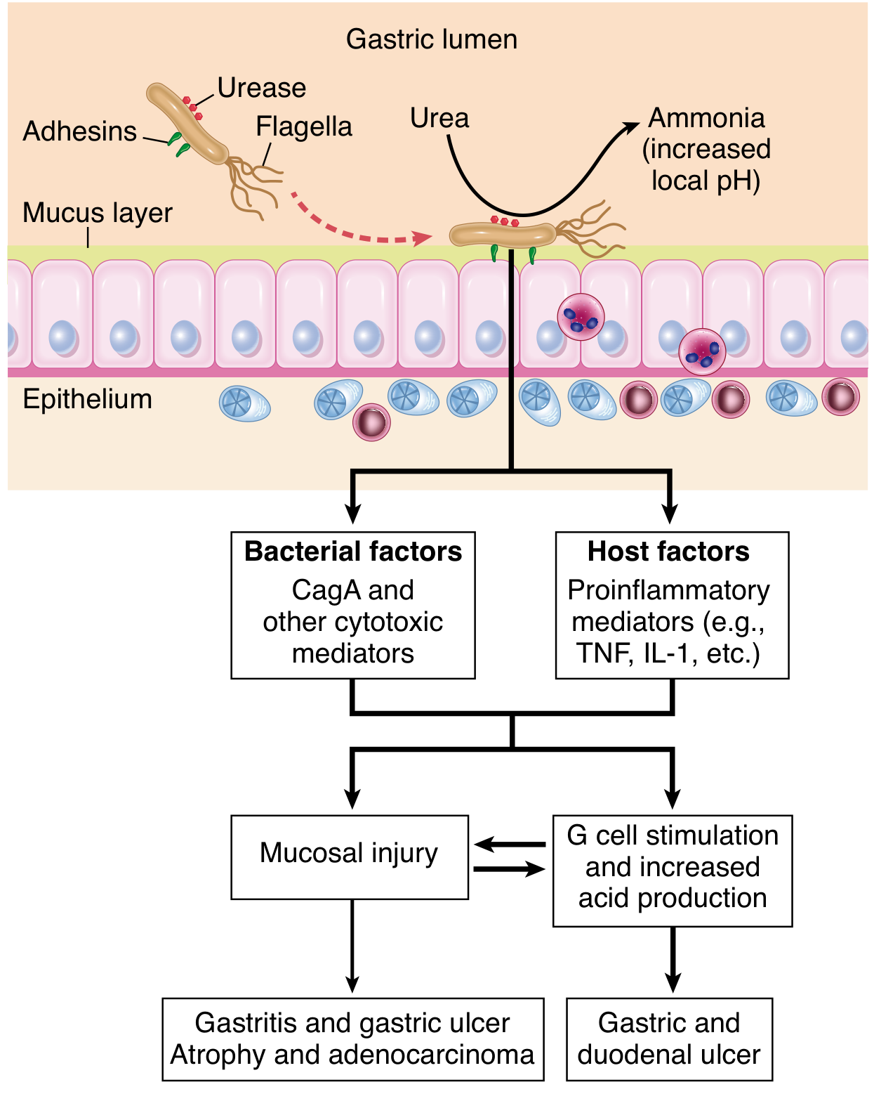
- Flagella - allow motility through viscous gastric mucus
- Urease - converts endogenous urea to ammonia, raising local pH and protecting the organism from gastric acid
- Adhesins - enhance binding to foveolar epithelial cells
- Toxins (CagA/VacA) - CagA is injected into epithelial cytoplasm, activating inflammatory signaling; stimulates release of IL-8 (a potent neutrophil chemotactic factor), initiating mucosal damage
Morphology
- H. pylori-infected antral mucosa appears erythematous with a coarse or nodular surface
- When inflammation is intense, thickened rugal folds can be seen, which may mimic infiltrative malignancy endoscopically
| Feature | Description |
|---|---|
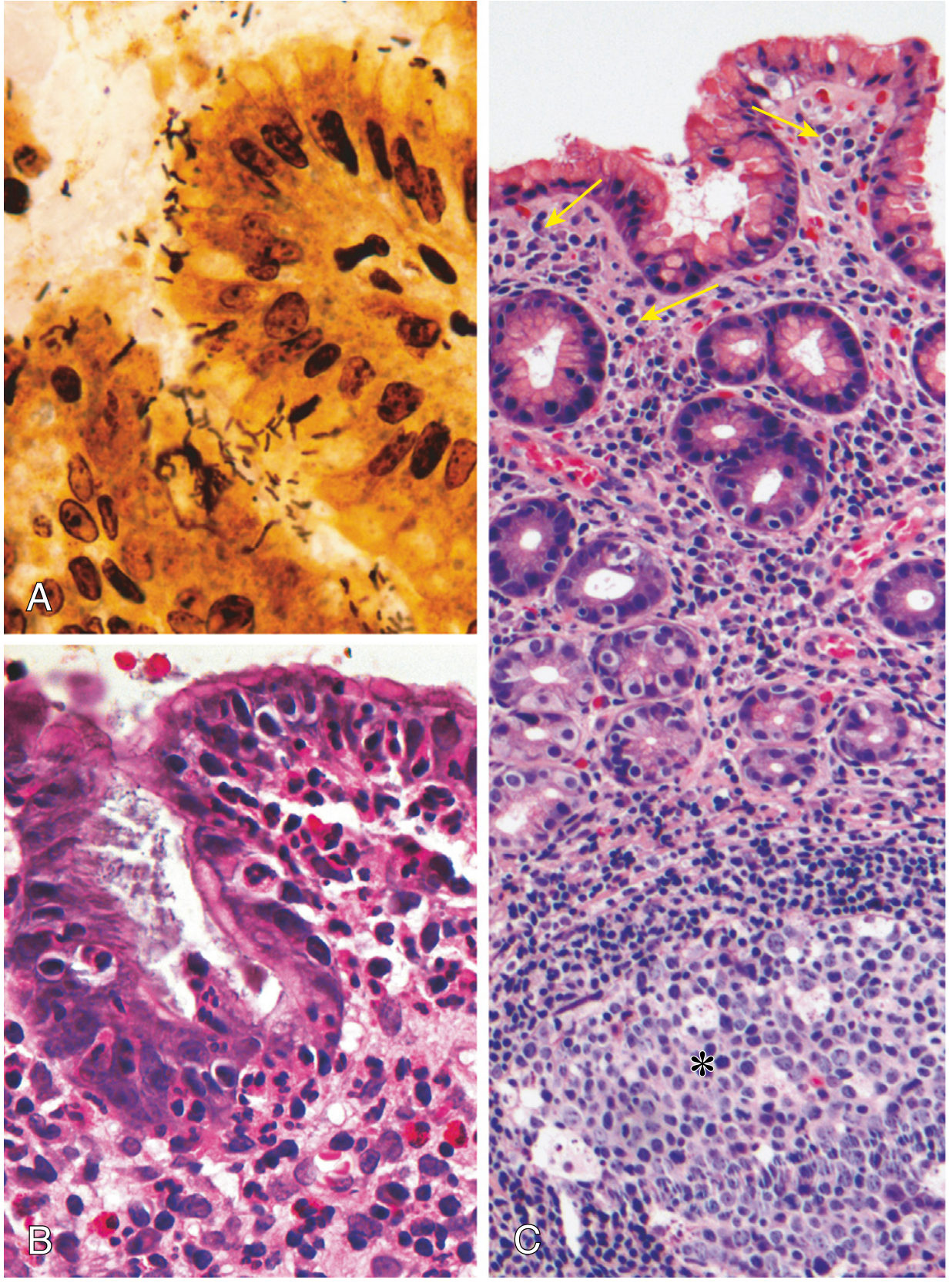
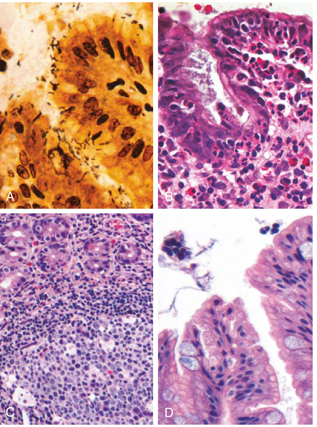
| Organism location | Concentrated within surface mucus overlying foveolar cells (surface and neck regions of glands). Visible on H&E; best seen on Giemsa, Warthin-Starry silver stain, or immunostains |
| Neutrophil infiltration | Variable numbers within lamina propria; neutrophils cross the basement membrane into the epithelium, accumulating in gland lumens to form pit abscesses |
| Plasma cells | Large numbers in the superficial lamina propria, often in clusters or sheets - this is the hallmark of chronic active inflammation |
| Lymphocytes & macrophages | Increased numbers in lamina propria |
| Lymphoid aggregates | Submucosal aggregates with germinal centers - induced MALT (mucosa-associated lymphoid tissue) |
| Intestinal metaplasia | Goblet cells and columnar absorptive cells replacing gastric epithelium - precancerous lesion |
| Hyperplastic/inflammatory polyps | Due to regenerative changes from chronic mucosal damage; elongated foveolar glands in an inflamed stroma |
Complications
| Complication | Mechanism |
|---|---|
| Peptic ulcer disease | Antral infection → gastrin hypersecretion → increased acid → gastric and duodenal ulcers. H. pylori found in ~95% of duodenal ulcers and ~70% of gastric ulcers |
| Gastric ulcer | Direct mucosal injury by CagA toxins and ammonia; disruption of the mucosal barrier |
| Chronic atrophic gastritis | Long-standing infection spreads to body/fundus → loss of parietal cells → hypochlorhydria |
| Intestinal metaplasia | Replacement of gastric epithelium by intestinal-type cells; a precancerous change |
| Gastric adenocarcinoma | Atrophy + intestinal metaplasia → dysplasia → carcinoma. H. pylori is classified as a Group 1 carcinogen by IARC. CagA+ strains carry higher risk |
| MALT lymphoma | Induced MALT lymphoid aggregates can undergo malignant transformation to extranodal marginal zone B-cell lymphoma. H. pylori eradication alone can cause regression of early MALT lymphoma |
| Hyperplastic polyps | Inflammatory/regenerative polyps from chronic mucosal damage |
Summary Cascade
H. pylori colonization (antrum)
        ↓
Urease → ammonia → mucosal injury + G-cell stimulation
        ↓
Chronic active gastritis (neutrophils + plasma cells + lymphocytes)
        ↓
     ┌──────────────────────────────────┐
     ↓                                  ↓
Increased acid secretion           Atrophy + intestinal metaplasia
     ↓                                  ↓
Peptic ulcer (gastric/duodenal)    Gastric adenocarcinoma
                                        ↓
                                   MALT lymphoma
Treatment Principle
1. A 60 years old male smoker, presented to OPD with complaints of altered bowel habits with bleeding per rectum and abdominal pain. Patient was referred to surgical oncologist and he suggested endoscopic biopsy. a) What is your probable diagnosis? b) Write in detail about etiopathogenesis and morphology of this lesion.