
Ecmo, indication and procedure in ICU
Reading File
Now let me search the medical library and web simultaneously for comprehensive ECMO content.
Finding Sources
Searching the Web
ECMO indications procedure ICU critical care 2024 guidelines
Searching PubMed
extracorporeal membrane oxygenation ECMO indications outcomes
Good. Now let me read the textbook content in parallel with extracting the StatPearls article.
Reading File
Reading File
Reading Web Page
https://www.ncbi.nlm.nih.gov/books/NBK576426
Good data. Let me now read deeper into the Fishman's pulmonary textbook chapter for VV-ECMO detail, and also fetch the ECMO circuit diagram image.
Reading File
Loading Image
Excellent - I have very rich content from the textbooks, StatPearls, and the ECMO circuit diagram. Let me now compile the comprehensive answer.
Extracorporeal Membrane Oxygenation (ECMO) in the ICU
What is ECMO?
ECMO (extracorporeal membrane oxygenation) - also called extracorporeal life support (ECLS) - is a form of prolonged cardiopulmonary bypass that temporarily takes over the function of the heart and/or lungs when they fail to sustain life despite maximal conventional treatment. It was derived from the Gibbon cardiopulmonary bypass circuit first used successfully in 1953.
The basic circuit consists of:
- An inflow cannula that drains venous blood from the patient
- A centrifugal pump to drive flow
- A membrane oxygenator (adds O₂, removes CO₂)
- A heat exchanger to regulate temperature
- An outflow cannula that returns blood to the patient
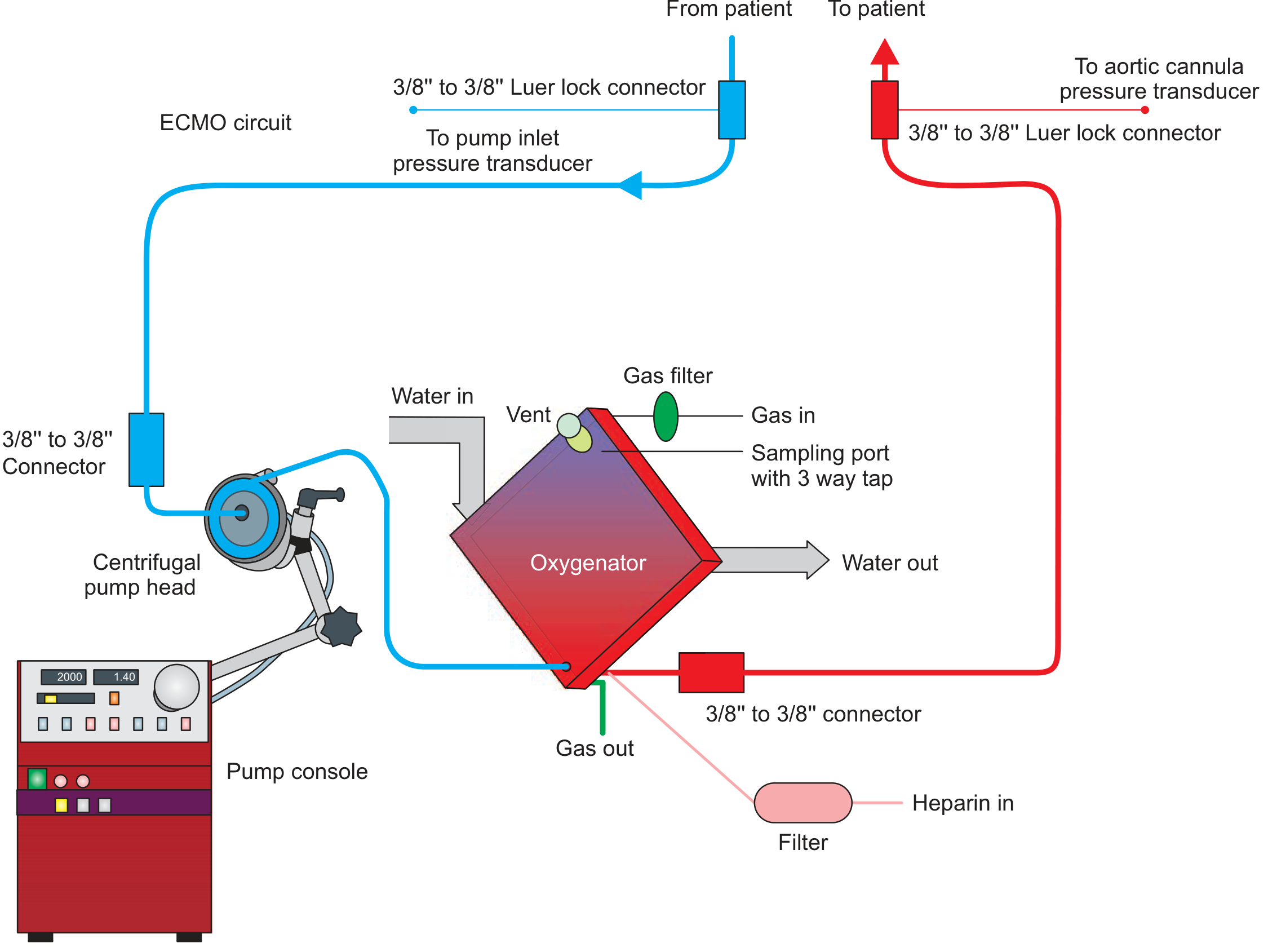
Figure: Basic ECMO circuit showing centrifugal pump head, oxygenator, pump console, gas filter, heparin infusion, and pressure transducers - Fishman's Pulmonary Diseases and Disorders
Two Main Types
1. Veno-Venous (VV) ECMO - Lung Support Only
- Blood drains from a vein and returns to a vein
- Provides gas exchange support (O₂ and CO₂) but no hemodynamic support
- Patient's own heart must still function
- Most common configuration: right femoral vein (inflow) + right internal jugular vein (outflow), with tip of inflow cannula at the IVC-atrial junction
2. Veno-Arterial (VA) ECMO - Heart + Lung Support
- Blood drains from a vein and returns to an artery
- Provides both gas exchange AND hemodynamic/circulatory support
- Common configuration: femoral vein drainage + femoral artery return (peripheral VA ECMO)
- Connected in parallel with the heart and lungs
- Can lead to LV distension and pulmonary edema - often requires additional LV unloading (IABP, Impella, inotropes)
Indications
VV-ECMO Indications (Respiratory)
| Condition | Threshold |
|---|---|
| Severe hypoxemic respiratory failure | PaO₂/FiO₂ < 80 mmHg despite optimal management, including prone positioning |
| Hypercapnic respiratory failure | pH < 7.25 despite optimal MV (RR 35, Pplat ≤ 30 cm H₂O); or PaCO₂ > 80 mmHg |
| Bridge to lung transplantation | Primary graft dysfunction post-transplant (< 7 days) |
Specific conditions:
- ARDS (pneumonia of any etiology, aspiration, inhalation injury, alveolar proteinosis, obstetric pathology)
- Airway obstruction, pulmonary contusion, bronchopleural fistula
- Status asthmaticus refractory to treatment
- Massive pulmonary hemorrhage / massive hemoptysis
- Pulmonary vasculitis
- Bridge to or support during lung transplantation
The ELSO (Extracorporeal Life Support Organization) triggers ECMO for PaO₂/FiO₂ < 100 with FiO₂ > 0.9 and/or Murray score ≥ 3, or oxygenation index > 80 despite 6 hours of optimal therapy.
VA-ECMO Indications (Cardiac)
- Cardiogenic shock refractory to inotropes and vasopressors
- Cardiac arrest - used as extracorporeal CPR (ECPR)
- Bi-ventricular failure
- Acute myocarditis (especially fulminant myocarditis - recent meta-analysis confirms LV unloading improves survival in pediatric fulminant myocarditis on ECMO, PMID 40211564)
- Post-cardiotomy shock (failure to wean from bypass)
- Bridge to VAD implantation or cardiac transplant
- Myocardial stunning with expected recovery
- Massive pulmonary embolism with hemodynamic collapse
- Refractory ventricular arrhythmias
- Drug/toxin-induced cardiac failure
ECPR (Extracorporeal CPR)
VA ECMO applied rapidly during cardiac arrest where conventional CPR fails to achieve ROSC. Time-sensitive: cannulation should begin within 10-20 minutes of failed resuscitation, and ECMO flow established within 1 hour of arrest. Requires dedicated team, clear roles, and defined logistics.
Contraindications
Absolute
- Irreversible end-organ damage (brain death, severe pre-existing neurological injury)
- Advanced malignancy or other terminal illness with no realistic recovery
- Uncontrolled bleeding that cannot be managed (relative to anticoagulation requirements)
- Severe aortic regurgitation (VA ECMO contraindicated)
Relative
- Prolonged mechanical ventilation at high pressures (> 7-10 days) - reduces lung recovery potential
- Immunosuppression (relative, context-dependent)
- Advanced age (no absolute cutoff; patient selection-dependent)
- Morbid obesity limiting cannulation
- Peripheral vascular disease limiting femoral access (consider axillary or carotid approach instead)
Procedure in the ICU
Pre-Cannulation Preparation
- Confirm diagnosis and failure of maximal conventional therapy
- Airway secured (intubation), central venous access, arterial line
- Baseline labs: ABG, CBC, coagulation profile, BMP, blood crossmatch
- Bedside echo to assess cardiac function and guide cannula placement
- Consent (or emergency waiver)
Cannulation Technique
Peripheral VV-ECMO (most common in ICU):
- Ultrasound-guided access to the right common femoral vein (inflow) with a large-bore multi-orifice cannula (21-25 Fr)
- Seldinger technique: wire → serial dilation → cannula placement
- Outflow cannula placed via right internal jugular vein (15-20 Fr), tip advanced to right atrium/SVC junction
- Position confirmed by fluoroscopy or transesophageal echocardiography (TEE)
- Alternatively, a dual-lumen single cannula (e.g., Avalon cannula) can be placed via the right IJ alone - drains from SVC and IVC, returns to right atrium directed toward tricuspid valve
Peripheral VA-ECMO:
- Inflow: femoral vein (venous drainage cannula, 21-25 Fr)
- Outflow: femoral artery (arterial return cannula, 15-17 Fr)
- A distal perfusion catheter (5-6 Fr) is inserted into the superficial femoral artery to prevent ipsilateral limb ischemia
- Position confirmed by fluoroscopy or echo
Central cannulation (in post-cardiac surgery patients already on bypass): Right atrium/SVC for inflow, aorta for outflow - higher flows possible but more invasive.
Circuit Initiation and Settings
- Start flow at 2-3 L/min, gradually increase to 4-6 L/min (targeting 60-80 mL/kg/min)
- Sweep gas (gas flow through oxygenator): primarily controls CO₂ removal
- FDO₂ of gas blender: controls oxygenation
- Once on VV-ECMO: institute "lung rest" - reduce ventilator settings (TV 3-4 mL/kg IBW, Pplat < 25 cmH₂O, FiO₂ 0.3-0.4, PEEP 8-10 cmH₂O)
- On VA-ECMO: vasopressors titrated to MAP > 60 mmHg, < 80 mmHg to avoid LV overdistension
Anticoagulation
- Unfractionated heparin infusion is standard (target aPTT 60-80 sec, or ACT 180-220 sec)
- Initiated at cannulation (heparin bolus 50-100 U/kg)
- Platelet monitoring required (target > 50,000-80,000/µL; circuit consumption causes thrombocytopenia)
- Bivalirudin used as alternative when HIT is suspected
Monitoring on ECMO
- Continuous: SpO₂, ETCO₂, circuit pressures (pre/post pump, pre/post oxygenator), flow
- Regular: ABG, ACT/aPTT, CBC, metabolic panel, lactate, CXR
- Echo: assess cardiac function, volume status, cannula position
- Watch for recirculation (VV-ECMO) - blood returns from outflow and is immediately drawn back into inflow cannula; reduces effective oxygen delivery
Weaning and Decannulation
VV-ECMO Weaning
- Gradual reduction of sweep gas flow (reduce to 1-2 L/min)
- Monitor SaO₂ and PCO₂ tolerance
- Trial off sweep gas ("sweep off" trial) with ventilator at moderate settings
- If patient maintains acceptable gas exchange for 1-4 hours: decannulate
- Ventilator settings at wean: FiO₂ ≤ 0.6, Pplat < 30, PEEP 8-10 cmH₂O
VA-ECMO Weaning
- Gradual flow reduction (500 mL/min every 6-12 hours)
- Echo at each step to assess LV/RV contractility, cardiac output
- Minimum flow ~1 L/min before trial off
- Maintain anticoagulation throughout weaning
Decannulation
- Remove cannulas with manual pressure (percutaneous) or surgical closure
- Confirm hemostasis before sedation reversal
Complications
| Category | Specific Complications |
|---|---|
| Mechanical | Oxygenator failure, pump thrombosis, tubing rupture, air embolism |
| Cannula-related | Malposition, vessel perforation, hemorrhage, arterial dissection, pseudoaneurysm |
| Vascular | Limb ischemia (femoral artery cannulation - requires distal perfusion cannula), DVT |
| Hemorrhagic | Bleeding at cannulation site, intracranial hemorrhage, retroperitoneal hemorrhage |
| Thrombotic | Circuit clotting, stroke, systemic thromboembolism |
| Cardiac (VA) | LV distension, pulmonary edema, "north-south syndrome" (differential hypoxia) in peripheral VA ECMO |
| Infectious | Circuit-related bacteremia, cannula-site infection (incidence rising with prolonged support) |
| Metabolic | Hemolysis, thrombocytopenia, hypokalemia |
North-South (Harlequin) Syndrome is specific to peripheral VA-ECMO: oxygenated blood returns retrogradely via femoral artery but mixes with blood ejected from a poorly functioning heart; the upper body (including coronaries and brain) may receive hypoxic blood from native cardiac output while the lower body receives well-oxygenated ECMO blood. Solution: ensure adequate ventilation, add VV circuit component, or switch to central cannulation.
Key Clinical Points for the ICU
- ECMO is a bridge - to recovery, transplant, or a durable device. It is not destination therapy.
- Timing matters: a 2024 National Inpatient Sample analysis showed each 12-hour delay from admission to VA ECMO initiation increased in-hospital mortality by 6%.
- Prone positioning while on VV-ECMO is feasible and safe (2023 PRONECMO trial showed no difference in 60-day ECMO-free survival but confirmed feasibility).
- Lung-protective (ultra-protective) ventilation must be maintained on VV-ECMO to minimize ventilator-induced lung injury.
- ECMO programs require dedicated, well-trained multidisciplinary teams - outcomes are highly institution-dependent.
Sources: Fishman's Pulmonary Diseases and Disorders (2-Volume Set), p. 2642 | Fischer's Mastery of Surgery 8th ed., p. 346 | StatPearls - ECMO in Adults | CCSO ECMO Consultation Guidelines 2024 | Recent evidence: PMID 38810214, 40211564
This is a shared conversation. Sign in to Orris to start your own chat.