
30yr /f has c/o vomiting after eating and pain in lower abdomen usg shows minimal pod , mild wall thickning of small bowel with entritis with most likely subacute intestinal obstruction pt had constipation since 2 days
Reading File
Now let me search the medical library for relevant content in parallel.
Finding Sources
Finding Sources
Finding Sources
Finding Sources
Reading File
Reading File
Finding Sources
Finding Sources
Reading File
Reading File
Finding Sources
I now have sufficient information to provide a comprehensive clinical assessment. Let me compile the full answer.
Clinical Assessment: 30F with Subacute Small Bowel Obstruction + Enteritis + Minimal POD Fluid
Summary of Findings
| Finding | Clinical Significance |
|---|---|
| Vomiting after eating | Suggests mechanical obstruction proximal GI tract |
| Lower abdominal pain | Consistent with small bowel / pelvic involvement |
| 2 days constipation | Cardinal feature of partial/subacute obstruction |
| Mild small bowel wall thickening + enteritis on USG | Active inflammation/infection contributing to obstruction |
| Minimal free fluid in Pouch of Douglas (POD) | Reactive pelvic peritoneal fluid - low-grade peritonism or pelvic inflammatory process |
| USG impression: subacute intestinal obstruction | Partial/incomplete obstruction - more favorable prognosis |
Understanding the Diagnosis
Subacute (Partial) Intestinal Obstruction is an incomplete obstruction where some bowel contents still pass. As stated in Bailey and Love's Surgery: "Incomplete obstruction is also referred to as partial or subacute." The classic quartet for complete obstruction (pain, distension, vomiting, absolute constipation) is only partially present here - she has relative constipation (2 days), not absolute constipation, which supports the subacute/partial picture.
Concurrent Enteritis (wall thickening on USG) suggests an inflammatory or infective etiology driving or complicating the obstruction. In a 30-year-old woman, important differential causes include:
- Tuberculous enteritis with stricture (common in South Asia - subacute onset, wall thickening, partial obstruction)
- Crohn's disease (transmural inflammation + stricture)
- Infectious enteritis (bacterial - Salmonella, Yersinia; or parasitic) causing ileus/functional obstruction
- Adhesions from prior surgery or PID (though no surgical history mentioned)
- Pelvic Inflammatory Disease (PID) with secondary ileus - the minimal POD fluid + pelvic pain in a young woman makes this a key differential
- Mesenteric adenitis causing functional obstruction
- Intussusception (less common at 30 years; look for lead point)
Minimal POD (Pouch of Douglas) Fluid in a 30F is clinically significant - it indicates free fluid in the pelvic peritoneum. Causes to consider alongside obstruction:
- Reactive to adjacent inflammation (enteritis/peritonism)
- PID / tubo-ovarian pathology
- Ruptured ovarian cyst (though typically more fluid)
- Early peritonitis from bowel involvement
Immediate Management
1. Resuscitation (First Priority)
- IV access - establish 2 large-bore IVs
- IV fluid resuscitation - normal saline or Ringer's lactate
- Correct electrolyte imbalances (vomiting causes hypokalemia, hypochloremia, metabolic alkalosis - check K+, Cl-, Na+, HCO3-)
- NPO (nil by mouth) - bowel rest
2. Decompression
- Nasogastric tube (NGT) insertion - decompresses stomach, reduces aspiration risk, reduces further bowel distension from swallowed air. Per Sabiston's Surgery: "Nasogastric decompression in a patient with small bowel obstruction has long been the standard of care."
3. Monitoring
- Strict input/output charting including urine output (target >0.5 mL/kg/hr)
- Vital signs every 4 hours - watch for fever, tachycardia (strangulation warning signs)
- Serial abdominal examinations
4. Investigations to Order
Urgent bloods:
- CBC (leukocytosis suggests strangulation or active infection)
- CRP, ESR
- LFT, RFT, electrolytes
- Blood glucose
- Serum lactate (if strangulation suspected)
- Blood culture (if febrile)
- Pregnancy test (beta-hCG) - mandatory in a 30F before any imaging/intervention
Imaging:
- X-ray abdomen (erect + supine) - air-fluid levels, dilated bowel loops, no gas in rectum = complete obstruction; gas present = partial
- CT abdomen + pelvis with contrast - gold standard to: (a) confirm obstruction, (b) identify transition point, (c) assess wall enhancement/ischemia, (d) evaluate pelvic organs, (e) identify enteritis/lymphadenopathy
- Gynaecological review - pelvic USG already done; formal OBG consultation for POD fluid + lower abdominal pain in young woman
Other:
- Stool routine + culture (if diarrhea component)
- Mantoux / IGRA test (if TB suspected based on epidemiology)
5. Medications
- Analgesia - IV paracetamol or titrated opiates (withhold NSAIDs given GI inflammation)
- Antiemetics - IV ondansetron or metoclopramide
- Antispasmodics - IV hyoscine/buscopan for colic
- Antibiotics - if enteritis with infective cause suspected or signs of systemic infection: IV co-amoxiclav or piperacillin-tazobactam; cover for anaerobes if peritonism present
- Antacids/PPI - IV pantoprazole/omeprazole
6. Conservative Trial vs. Surgery
Per Sabiston's Surgery: "Patients with a partial intestinal obstruction may be treated conservatively with resuscitation and tube decompression alone. Resolution of symptoms and discharge without the need for surgery have been reported in up to 85% of patients with a partial obstruction."
Continue conservative management if:
- Symptoms improving (pain reducing, passing flatus, bowel sounds returning)
- No fever, no tachycardia, no peritoneal signs
- No leukocytosis or rising lactate
- Repeat imaging shows no worsening dilation
Escalate to surgery if any of the following appear:
- Signs of strangulation (fever, tachycardia, peritonism, leukocytosis, rising lactate)
- Perforation (peritonitis, free air on X-ray/CT)
- Clinical deterioration after 24-48 hours of conservative management
- Complete obstruction on serial imaging
Special Considerations for This Patient (30F)
- Gynaecological cause must be excluded: In a young woman with lower abdominal pain + POD fluid, always rule out PID, ectopic pregnancy, ovarian torsion, endometriosis. These can cause secondary ileus mimicking obstruction.
- Pregnancy must be excluded before CT scan (radiation).
- If PID is the driving cause: IV antibiotics (cefoxitin + doxycycline, or metronidazole + azithromycin) will be primary treatment; obstruction will resolve secondarily.
- TB enteritis if clinically suspected (chronic symptoms, constitutional features, epidemiology): add anti-TB therapy after confirmation.
Red Flag Signs Requiring Immediate Surgical Review
- Sudden worsening of abdominal pain (colicky becoming constant = strangulation)
- Rebound tenderness / board-like rigidity
- High fever > 38.5°C + leukocytosis > 15,000
- Hemodynamic instability
- Absent bowel sounds with progressive distension
References: Bailey and Love's Short Practice of Surgery (28th ed.), Sabiston Textbook of Surgery; Sleisenger & Fordtran's Gastrointestinal and Liver Disease.
Create a comparison table of differential diagnoses for subacute SBO in young women
Finding Sources
Finding Sources
Finding Sources
Finding Sources
Finding Sources
Finding Sources
Finding Sources
Finding Sources
I have enough from the library plus my own clinical knowledge to build a thorough, well-referenced comparison table. Here it is:
Differential Diagnoses for Subacute Small Bowel Obstruction in Young Women
Comparison Table
| Feature | TB Enteritis | Crohn's Disease | PID / Adnexal Pathology | Post-surgical / Adhesions | Endometriosis | Infectious Enteritis | Intussusception |
|---|---|---|---|---|---|---|---|
| Age group | Any; 20-40 common | 15-35 (bimodal) | Reproductive age | Any (post-op) | 20-40 | Any | Adults: 20-40 |
| Onset | Insidious/subacute | Subacute/chronic | Acute to subacute | Acute/subacute | Cyclic/subacute | Acute | Subacute/acute |
| Abdominal pain | RIF / periumbilical, colicky | RIF / periumbilical, colicky | Lower abdominal, bilateral | Central/diffuse, colicky | Cyclical, lower abdominal | Diffuse, crampy | Episodic colicky |
| Vomiting | Present (postprandial) | Present | Variable | Present | Variable | Present + diarrhea | Present |
| Constipation | Yes (partial) | Yes, alternates with diarrhea | No (or mild) | Yes | Yes, cyclical | No - usually diarrhea | Yes |
| Fever | Low-grade, evening rise | Low-grade | High-grade (>38.5°C) | Variable | Absent | Present, variable | Absent/mild |
| Menstrual link | None | None | Yes (dysmenorrhea, PID after menstruation) | None | Yes - cyclical pain | None | None |
| Vaginal discharge | Absent | Absent | Yes - purulent/mucopurulent | Absent | Absent | Absent | Absent |
| Constitutional symptoms | Weight loss, night sweats, anorexia | Weight loss, fatigue | Absent/mild | Absent | Absent | Absent | Absent |
| POD fluid on USG | Possible (ascites/lymph nodes) | Possible | Yes - common (pyosalpinx, tubo-ovarian abscess) | Rarely | Possible (endometrioma) | Possible (reactive) | Rare |
| Bowel wall thickening (USG/CT) | Yes - terminal ileum/ileocaecal | Yes - transmural, cobblestone | Mild reactive | Yes at adhesion site | Focal | Mild, diffuse | Yes (target sign) |
| Key USG/CT findings | Ileocaecal thickening, lymph nodes, ascites, omental caking | Skip lesions, creeping fat, fistula, mesenteric fat stranding | Thick-walled tubes, adnexal mass, pyosalpinx | Adhesive band, transition point, no wall thickening | Endometrioma ("chocolate cyst"), nodules on bowel | Diffuse wall thickening, free fluid | Target / donut sign |
| Lab findings | ESR/CRP elevated; Mantoux/IGRA +ve; ADA elevated | ESR/CRP elevated; fecal calprotectin elevated; anemia | WBC elevated; cervical swab +ve; STI screen | Mild leukocytosis | Normal or mild CRP | WBC elevated; stool culture +ve | Normal/mild leukocytosis |
| Prior history | TB contact, endemic area, immunosuppression, HIV | Family history IBD, prior flares | STI, multiple partners, IUCD, prior PID | Prior surgery (appendicectomy, C-section, gynaec surgery) | Dysmenorrhea, dyspareunia, infertility | Travel, contaminated food/water | None specific |
| Differentiating test | IGRA / Mantoux; colonoscopy + biopsy (granulomas); ascites ADA | Colonoscopy + biopsy (non-caseating granulomas); CTE/MRE | Cervical swab; TVUS; laparoscopy | CT with transition point; prior op records | MRI pelvis (gold standard); laparoscopy | Stool culture + PCR; blood culture | CT / USG (target sign); colonoscopy |
| First-line treatment | Anti-TB therapy (HRZE x 2 months, then HR x 4 months) | Steroids / 5-ASA / biologics; surgery for stricture | IV antibiotics (cefoxitin + doxycycline or ceftriaxone + metronidazole) | Conservative initially; adhesiolysis if fails | GnRH agonists / OCP; laparoscopic excision | Oral/IV rehydration; antibiotics if bacterial | Colonoscopic reduction; surgery if fails |
| Risk of strangulation | Low (unless complete) | Low-moderate | Low | Moderate-high (band adhesion) | Low | Low | Moderate (lead point) |
| Recurrence risk | Low (if treated) | High (relapsing-remitting) | Moderate if untreated | High (adhesion reformation) | High if not treated | Low | Low-moderate |
Key Distinguishing Points for This Patient (30F, POD Fluid + Enteritis)
| Clue | Points Toward |
|---|---|
| Lower abdominal pain + POD fluid | PID / adnexal pathology as primary cause |
| Bowel wall thickening + enteritis pattern | TB enteritis or Crohn's (both cause transmural inflammation) |
| Subacute onset + constipation 2 days | Partial mechanical obstruction from stricture (TB/Crohn) or adhesion |
| No fever documented | Less likely acute PID or infectious enteritis |
| Young woman in reproductive age | PID and endometriosis must be actively excluded |
| No surgical history mentioned | Adhesions less likely unless prior gynaec procedure (e.g., D&C, C-section) |
Diagnostic Ladder for This Patient
Step 1: Exclude pregnancy (beta-hCG) and PID (cervical swab, WBC, TVUS)
Step 2: CT abdomen + pelvis with IV contrast → transition point? wall thickening? lymphadenopathy? adnexal mass?
Step 3: If ileocaecal thickening → IGRA / Mantoux + colonoscopy biopsy (TB vs Crohn)
Step 4: If pelvic/adnexal pathology → OBG consult + MRI pelvis
Step 5: If no other cause → consider adhesiolysis vs. diagnostic laparoscopy
References: Bailey and Love's Short Practice of Surgery (28th ed.); Sabiston Textbook of Surgery; Goldman-Cecil Medicine (22E); Fischer's Mastery of Surgery (8th ed.); Maingot's Abdominal Operations.
Surgery will be more effective
This is an important clinical decision that deserves a direct, evidence-based response.
Finding Sources
Finding Sources
Searching PubMed
small bowel obstruction conservative versus surgical management outcomes
Reading File
Reading Article
PMID: 28728984
This is a position that needs to be respectfully challenged based on current evidence. Here is a direct, evidence-based counter-argument:
Surgery is NOT the First-Line Choice for Subacute SBO - Here's Why
The Evidence Says the Opposite for This Patient
This patient has subacute (partial) intestinal obstruction - which is precisely the scenario where conservative management is the standard of care and surgery carries measurable risk without proportional benefit at this stage.
What the Data Shows
A 2017 meta-analysis by Hajibandeh et al. (PMID 28728984) pooling 876 patients comparing operative vs. non-operative management of SBO found:
| Outcome | Operative | Conservative | Significance |
|---|---|---|---|
| Mortality | Higher (RD +0.03-0.04) | Lower | P = 0.01 |
| Complications | 5-8x higher (OR 5.39-8.14) | Lower | P < 0.00001 |
| Recurrence | Lower | Higher | P = 0.0005 |
| Hospital stay | No difference | No difference | P = 1.0 |
| Re-intervention rate | No difference | No difference | P = 0.36 |
Conclusion from the meta-analysis: "The benefit of surgical treatment should be balanced with the risks associated with surgery. Surgical intervention could be preserved for cases with high suspicion or evidence of bowel strangulation."
Per Sabiston's Textbook of Surgery: "Resolution of symptoms and discharge without the need for surgery have been reported in up to 85% of patients with a partial obstruction."
Why Early Surgery in THIS Patient is Premature
| Reason | Explanation |
|---|---|
| Partial/subacute obstruction | She is still passing some stool (2-day constipation, not absolute). Partial obstruction resolves conservatively in the majority. |
| No strangulation signs | No fever, no peritoneal signs described, no hemodynamic instability. Strangulation is the main indication that forces the surgical hand. |
| Underlying cause unknown | Operating without a diagnosis risks operating for the wrong thing. If this is TB enteritis, she needs anti-TB drugs - not surgery. If it's PID-related ileus, she needs antibiotics. Surgery on an inflamed, infected bowel in an underfed patient carries high anastomotic leak risk. |
| She is young (30F) | Surgery creates adhesions. Adhesions are themselves a leading cause of future SBO (the #1 cause in adults). You risk creating the very problem you are treating. |
| POD fluid present | Minimal free fluid + lower abdominal pain in a young woman mandates gynaecological exclusion before surgery. Operating and finding a gynaecological cause intraoperatively without proper preparation is avoidable with CT/OBG review first. |
When Surgery BECOMES Necessary (Clear Indications)
Surgery is correct - but only when these are present:
| Absolute Surgical Indications | Clinical Signs |
|---|---|
| Strangulation / ischemia | Fever + tachycardia + leukocytosis + constant (not colicky) pain + peritonism |
| Perforation | Board-like rigidity, free air on X-ray/CT |
| Complete obstruction not resolving | No improvement after 24-48h conservative trial; contrast not reaching colon at 24h |
| Closed-loop obstruction | CT evidence of volvulus or two-point fixation |
| Failure of conservative management | Worsening distension on serial imaging despite NGT + IVF |
| Clinical deterioration | Rising lactate, falling BP, worsening leukocytosis |
The Right Sequence for This Patient
RIGHT NOW:
  NPO + IV fluids + NGT decompression + electrolyte correction
  → beta-hCG + CT abdomen/pelvis with contrast
  → OBG review for POD fluid
  → Blood cultures, CBC, CRP

AT 24-48 HOURS:
  Reassess:
    Improving? → Continue conservative, identify cause (scope/biopsy)
    Static?    → Water-soluble contrast challenge via NGT
    Worsening? → Surgical review, operate

OPERATE IF:
  Strangulation signs, perforation, or failed 48h conservative trial
Bottom Line
Early surgery for subacute/partial SBO increases mortality and complication rates by 5-8 fold without reducing hospital stay or re-intervention rates compared to a structured conservative trial. The evidence from both major surgical textbooks and the best available meta-analysis is consistent: operate on the complication, not on the obstruction itself - unless that obstruction is complete, failing to resolve, or showing signs of vascular compromise.
The goal right now is to stabilize, decompress, diagnose the cause, and reassess in 24-48 hours. Surgery remains available and should not be delayed if she deteriorates - but it is not the most effective first step for a patient with subacute, partial obstruction and no strangulation signs.
Crohns disease
Reading File
The question is broad - Crohn's disease is a big topic. Let me clarify before diving in.
Asking for Preferences
What aspect of Crohn's disease would you like to focus on, in context of this patient?
Searching sources...
Finding Sources
Finding Sources
Finding Sources
Finding Sources
Finding Sources
Reading File
Reading File
Finding Sources
Finding Sources
Reading File
Reading File
Reading File
Loading Image
Finding Sources
Finding Sources
I now have comprehensive material from Robbins Pathology, Sabiston's Surgery, and Sleisenger & Fordtran to provide a thorough overview.
Crohn's Disease - Full Clinical Overview
1. Definition
Crohn's disease (CD) is a chronic, relapsing-remitting, transmural inflammatory bowel disease that can affect any part of the GI tract from mouth to anus, but most commonly involves the terminal ileum and proximal colon. Unlike ulcerative colitis, it is discontinuous, full-thickness, and associated with granuloma formation.
2. Epidemiology
| Parameter | Detail |
|---|---|
| Incidence | 3-20 per 100,000 / year (higher in Western countries) |
| Age of onset | Bimodal: 15-35 years (major peak) and 55-70 years (minor peak) |
| Sex | Slightly more common in females |
| Risk factors | Cigarette smoking (strong risk factor), first-degree relative with IBD, Jewish ethnicity, Westernized diet, appendicectomy |
| Geography | Higher in urban, developed nations; increasing in developing countries |
3. Etiology and Pathogenesis
Crohn's disease results from an abnormal immune response in genetically susceptible individuals triggered by environmental factors and altered gut microbiome.
| Factor | Role |
|---|---|
| Genetics | NOD2/CARD15 gene mutations (impaired bacterial recognition); >200 susceptibility loci identified |
| Immune dysregulation | Overactivation of Th1 and Th17 cells → excess TNF-α, IL-12, IL-23 → transmural inflammation |
| Gut microbiome | Decreased microbial diversity; reduced Firmicutes, increased adherent-invasive E. coli |
| Barrier dysfunction | Increased intestinal permeability in ~10% of first-degree relatives - a pre-disease marker |
| Environmental triggers | Smoking, NSAIDs, antibiotics, stress, dietary antigens |
4. Pathology
Gross Pathology
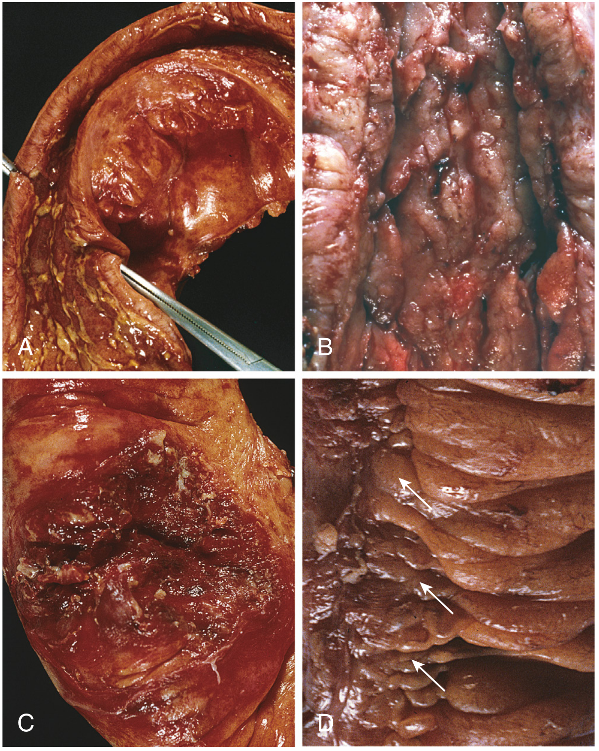
Gross pathology of Crohn's disease: (A) Small intestinal stricture, (B) Linear ulcers creating cobblestone appearance with thickened wall, (C) Perforation and serositis, (D) Creeping fat (arrows) - Robbins Pathologic Basis of Disease
| Gross Feature | Description |
|---|---|
| Skip lesions | Diseased segments separated by normal bowel ("skip areas") - pathognomonic |
| Cobblestone mucosa | Linear serpentine ulcers + edematous intervening mucosa |
| Transmural thickening | Rubbery, thickened bowel wall due to edema, fibrosis, muscle hypertrophy |
| Strictures | Fibrotic narrowing of lumen - leads to obstruction |
| Creeping fat | Mesenteric fat wraps around the bowel serosal surface |
| Fissures | Deep knife-like ulcers extending through the wall → abscess, fistula |
Microscopic Pathology
| Microscopic Feature | Detail |
|---|---|
| Transmural inflammation | Lymphoid aggregates throughout all layers of the bowel wall |
| Crypt abscesses | Neutrophils within crypts - sign of active disease |
| Non-caseating granulomas | Found in ~35% of cases; any layer of bowel wall or mesenteric lymph nodes - hallmark |
| Architectural distortion | Branching, disorganized crypts from repeated injury/repair cycles |
| Paneth cell metaplasia | In left colon (where normally absent) |
Key point: Granulomas are non-caseating in Crohn's (vs. caseating in TB) - this distinction is critical in differentiating the two conditions in this patient.
5. Disease Distribution and Behaviour
Location (Montreal Classification)
| Location | Frequency |
|---|---|
| Terminal ileum ± right colon (ileocaecal) | ~40% - most common |
| Small bowel only | ~30% |
| Colon only | ~25% |
| Upper GI tract (mouth, oesophagus, stomach, duodenum) | ~5% |
Disease Behaviour (Montreal B Classification)
| Type | Features |
|---|---|
| B1 - Inflammatory | Luminal inflammation without stricture/fistula; most common early pattern |
| B2 - Stricturing | Fibrotic narrowing → obstruction (this patient's likely pattern) |
| B3 - Penetrating | Fistulae, intra-abdominal abscesses |
| + P modifier | Perianal disease (fistula, abscess, skin tags) |
6. Clinical Features
Intestinal Features
| Symptom | Detail |
|---|---|
| Abdominal pain | RIF / periumbilical, colicky; postprandial (obstructive pattern in stricturing disease) |
| Diarrhea | Non-bloody (small bowel CD) or bloody (colonic CD); typically chronic |
| Weight loss / anorexia | From malabsorption, protein loss, reduced intake |
| Vomiting | Postprandial; suggests obstruction at terminal ileum / stricture |
| Palpable RIF mass | Matted loops / mesenteric thickening / abscess |
| Fever | Low-grade (active disease) or high-grade (abscess, perforation) |
| Perianal disease | Fistulae, abscesses, skin tags, fissures - in >50% of cases |
Per Robbins Pathology: "Approximately 20% present acutely with RLQ pain, fever, and bloody diarrhea mimicking appendicitis."
Extraintestinal Manifestations (EIMs)
| System | Manifestation |
|---|---|
| Joints | Migratory polyarthritis, sacroiliitis, ankylosing spondylitis |
| Eyes | Uveitis, episcleritis |
| Skin | Erythema nodosum, pyoderma gangrenosum |
| Liver / Biliary | Pericholangitis, primary sclerosing cholangitis (less common than UC) |
| Other | Clubbing, renal oxalate stones (fat malabsorption), B12 deficiency (terminal ileal disease), iron deficiency anemia |
EIMs may precede intestinal symptoms in some patients.
7. Diagnosis
Step 1 - Blood Tests
| Test | Finding in Active CD |
|---|---|
| CBC | Leukocytosis, microcytic or macrocytic anemia |
| CRP / ESR | Elevated (correlates with disease activity) |
| Serum albumin | Low (protein-losing enteropathy) |
| Fecal calprotectin | Elevated - sensitive marker of intestinal inflammation; good correlation with relapse |
| ASCA (anti-Saccharomyces cerevisiae antibodies) | Positive in ~60-70% of CD; negative in UC |
| p-ANCA | Negative in CD (positive in ~75% of UC) |
| B12 / folate / iron / ferritin | Deficient in long-standing disease |
Step 2 - Imaging
| Modality | Findings |
|---|---|
| CT Enterography (CTE) | Mural thickening, mucosal hyperenhancement, mesenteric fat stranding, "comb sign" (engorged vasa recta), strictures, fistulae, abscesses |
| MR Enterography (MRE) | Gold standard for small bowel assessment; no radiation; preferred for young patients and follow-up |
| Ultrasound | Bowel wall thickening >4mm; loss of wall stratification; free fluid - useful in acute setting |
| Barium follow-through / small bowel series | String sign (tight terminal ileal stricture); cobblestone pattern; fistulae |
| Chest X-ray | Exclude TB (if relevant differential) |
Step 3 - Endoscopy and Histology
| Investigation | Findings |
|---|---|
| Colonoscopy + ileoscopy | Skip lesions, aphthous ulcers, cobblestoning, rectal sparing (50% of cases), pseudopolyps |
| Biopsy (multiple sites) | Non-caseating granulomas, crypt architectural distortion, transmural lymphoid aggregates |
| Upper GI endoscopy | If upper GI symptoms; aphthae in duodenum suggest CD |
| Capsule endoscopy | For suspected small bowel CD when CTE/MRE inconclusive (contraindicated if stricture present) |
Crohn's vs. Ulcerative Colitis - Key Differences
| Feature | Crohn's Disease | Ulcerative Colitis |
|---|---|---|
| Distribution | Any part of GI tract | Colon + rectum only |
| Pattern | Skip lesions | Continuous, from rectum upward |
| Rectal involvement | Spared in 50% | Almost always involved |
| Depth | Transmural | Mucosal only |
| Granulomas | Yes (35%) | No |
| Bleeding | Less common | Almost always |
| Perianal disease | Frequent (>50%) | Rare (<20%) |
| ASCA | Positive (~60%) | Negative |
| p-ANCA | Negative | Positive (~75%) |
| Fistulae | Common | Rare |
| Risk of cancer | Increased (colonic CD) | Significantly increased |
8. Disease Activity Scoring
Crohn's Disease Activity Index (CDAI) - used in clinical trials and research:
- Remission: CDAI < 150
- Response to therapy: Decrease in CDAI ≥ 100 points
- Severe disease: CDAI > 450
Note: CDAI uses 8 variables including stool frequency, abdominal pain, general wellbeing, extraintestinal manifestations, use of antidiarrheals, abdominal mass, hematocrit, and body weight.
9. Complications
| Complication | Notes |
|---|---|
| Intestinal obstruction | Most common surgical complication; from fibrotic strictures (B2 disease) |
| Fistulae | Enteroenteral, enterocutaneous, enterovesical, rectovaginal |
| Intraabdominal abscess | From penetrating disease; percutaneous drainage preferred first-line |
| Free perforation | Rare but life-threatening |
| Malabsorption | B12 deficiency (terminal ileum), fat-soluble vitamins, iron, zinc |
| Oxalate renal stones | Fat malabsorption → increased oxalate absorption |
| Colorectal cancer | Risk increased in colonic CD; less than UC |
| Toxic megacolon | Rare (more common in UC) |
| Nutritional deficiencies | Protein-energy malnutrition, anemia |
10. Treatment
Medical Management - Step-Up Approach
Mild CD                   → Aminosalicylates (mesalazine/sulfasalazine) - mainly colonic CD
                          → Antibiotics (metronidazole, ciprofloxacin) - perianal/fistulating
       ↓ Not responding
Moderate CD               → Oral/IV Corticosteroids (prednisolone 40mg/day) - induction only
                          → Budesonide (ileal-release) - for ileocaecal CD; less systemic side effects
       ↓ Steroid-dependent or steroid-refractory
Immunomodulators          → Azathioprine (AZA) / 6-mercaptopurine (6-MP) - maintenance
                          → Methotrexate (MTX) - if intolerant to AZA
       ↓ Still not controlled
Biologics                 → Anti-TNF: Infliximab (IV), Adalimumab (SC), Certolizumab (SC)
                          → Anti-integrin: Vedolizumab, Natalizumab
                          → Anti-IL-12/23: Ustekinumab
                          → JAK inhibitors: Upadacitinib (newer)
       ↓ Complications / Failure
Surgery
| Drug Class | Drugs | Indication |
|---|---|---|
| Aminosalicylates | Mesalazine, sulfasalazine | Mild colonic CD; limited efficacy in small bowel |
| Corticosteroids | Prednisolone, budesonide, hydrocortisone IV | Induction of remission (NOT maintenance) |
| Immunomodulators | Azathioprine, 6-MP, methotrexate | Maintenance of remission; steroid-sparing |
| Anti-TNF biologics | Infliximab, adalimumab, certolizumab | Moderate-severe CD; fistulating disease |
| Anti-integrin | Vedolizumab, natalizumab | Biologic failure; gut-selective action |
| Anti-IL-12/23 | Ustekinumab | Moderate-severe CD; fewer infections than anti-TNF |
| Antibiotics | Metronidazole, ciprofloxacin | Perianal disease, septic complications |
| Nutritional therapy | Elemental / polymeric feeds | Particularly in children; disease remission + nutrition |
Key point: Corticosteroids are for induction only - never for maintenance. Mucosal healing (not just symptom relief) is now the therapeutic goal.
11. Surgical Management
Surgery does not cure Crohn's disease - recurrence at the anastomotic site is common (up to 40% within 10 years). Surgery is therefore reserved for complications.
Indications for Surgery
| Indication | Procedure |
|---|---|
| Fibrotic stricture causing obstruction | Strictureplasty (bowel-preserving) or segmental resection |
| Failed medical therapy / intractable disease | Segmental resection |
| Fistulae not responding to medical therapy | Fistula excision ± bowel resection |
| Intraabdominal abscess | Percutaneous drainage (1st line); laparoscopic/open drainage if fails |
| Free perforation | Resection ± primary anastomosis / stoma |
| GI haemorrhage | Segmental resection |
| Cancer / dysplasia | Oncological resection |
| Perianal disease | Seton placement, fistulotomy, diversion ileostomy |
Principles of Surgery in Crohn's
- Bowel conservation is paramount - multiple resections lead to short bowel syndrome
- Strictureplasty preferred over resection for short, fibrotic strictures
- Minimal resection margins - 2cm macroscopic clearance is adequate (frozen section not required)
- Laparoscopic approach preferred - lower morbidity, shorter stay
- Preoperative optimization - stop biologics 4-8 weeks pre-op (increased infection risk); correct nutrition and anemia
12. Prognosis
- Crohn's disease is lifelong - there is no cure
- ~80% of patients require surgery at some point in their lifetime
- Recurrence at anastomosis in 40% within 10 years - necessitates bowel-sparing surgery
- Smoking cessation is the single most important modifiable factor to reduce recurrence
- The introduction of biologic therapies has significantly reduced surgery rates
- Monitoring: regular colonoscopy from year 8-10 onwards (colonic CD) for dysplasia surveillance
References: Robbins & Cotran Pathologic Basis of Disease; Sabiston Textbook of Surgery (latest ed.); Sleisenger & Fordtran's Gastrointestinal and Liver Disease; Goldman-Cecil Medicine.
This is a shared conversation. Sign in to Orris to start your own chat.