
Explain me about hydrocele from standard textbook like a donkey
hydrocele diagnosis management Merck Manual professional
hydrocele treatment
Hydrocele, explained simply
What does it look and feel like?
- A painless swelling on one side of the scrotum
- It feels smooth, soft, and fluid-filled
- The swelling may become quite large gradually
- The testis may be difficult to feel because fluid surrounds it
- When a light is held behind it in a dark room, it often glows. This is called transillumination.
Why does it happen?
In babies and children
In adults
- Infection of the testis or epididymis
- Injury
- Testicular torsion
- Testicular tumor
- Previous surgery or radiotherapy
Main types
| Type | Simple meaning |
|---|---|
| Congenital / communicating | A channel remains open between abdomen and scrotum, allowing fluid to move down. It is associated with indirect inguinal hernia. |
| Primary / vaginal / non-communicating | Fluid is trapped only around the testis. This is common in adults. |
| Infantile hydrocele | The channel is partly closed, but the sac extends from the groin toward the scrotum. |
| Hydrocele of the cord | Fluid is trapped along the spermatic cord, separate from the testis. |
| Secondary hydrocele | Occurs because of infection, trauma, tumor, or another testicular/scrotal disease. |
How is it diagnosed?
-
History and examination
- Usually painless and gradually increasing in size
- It commonly transilluminates
-
Scrotal ultrasound
- Confirms fluid around the testis
- Checks that the testis itself is normal
- Helps rule out tumor, torsion, infection, hernia, or a blood collection
Treatment
Babies
- Observe initially if it is non-communicating and the child is well.
- Many resolve spontaneously by about 12 months of age.
- Surgery is considered if it:
- Persists beyond about 12 months
- Becomes bigger
- Is communicating
- Is associated with suspected inguinal hernia
Adults
- Lord's plication: The sac is opened and folded or plicated. Often used for a relatively thin-walled sac.
- Jaboulay procedure: The sac is opened and everted behind the testis. Often used for a large or thick sac.
Hydrocele versus hernia
- Hydrocele: fluid only, usually painless, usually transilluminates.
- Inguinal hernia: bowel or fat comes down into the scrotum, may be reducible, often does not transilluminate, and may become painful or obstructed.
Go to urgent care immediately if there is
- Sudden severe testicular or scrotal pain
- Redness, fever, vomiting, or marked tenderness
- A hard testicular lump
- A swelling that cannot be pushed back with abdominal pain or vomiting
Tell me about ulcer
- Peptic ulcer - stomach or duodenal ulcer
- Mouth ulcer
- Skin or leg ulcer
- Diabetic foot ulcer
- Pressure ulcer (bed sore)
Foot ulcer
NICE diabetic foot problems prevention and management foot ulcer urgent referral
diabetic foot ulcer treatment
diabetic foot ulcer plantar neuropathic ulcer clinical photograph
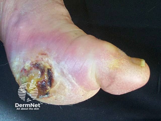
Clinical photography of a diabetic foot ulcer on the plantar surface of the foot, captured in a close, color-calibrated view to document wound morphology. The lesion is located on the plantar heel/midfoot region with exposure of subcutaneous tissue and necrotic material. The ulcer bed shows dark brown to black necrosis (eschar) overlain by yellow-brown slough, with surrounding erythema and edema indicative of local inflammation. Margins are irregular and partly undermined, and there is adjacent maceration from interdigital moisture. The overlying skin displays callused areas and fissuring, consistent with chronic pressure and neuropathic injury in long-standing diabetes. No visible sutures or foreign bodies. The image demonstrates a non-healing diabetic foot ulcer, potentially complicated by infection risk (purulent exudate limited in this view) and possible underlying peripheral vascular disease. This finding aligns with neuropathic (neuroischemic) pathways in diabetic patients and highlights impaired wound healing, reduced sensation, and repetitive trauma. Clinically, such ulcers require off-loading, meticulous debridement, wound cleansing, appropriate dressings, infection surveillance, vascular assessment, and glycemic optimization. The photograph is suitable for education, wound-tracking, and research on DFU morphology, treatment response, and dermatologic assessment of plantar ulcers. Useful search terms include diabetic foot ulcer, plantar ulcer, neuropathic ulcer, necrotic tissue, eschar, slough, infection risk, and wound care.
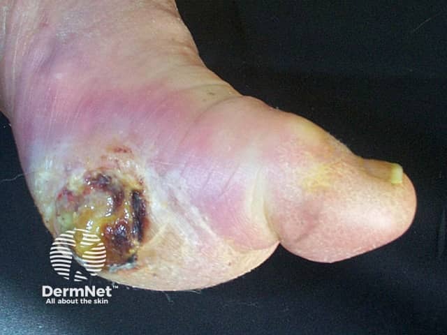
Clinical photograph of a diabetic foot ulcer on the plantar aspect of the foot. Modality and technique: digital clinical photography, macro close‑up, high‑resolution image captured under studio lighting to document wound characteristics. Anatomical location: plantar surface of the midfoot near the hindfoot weight‑bearing area; tissue involved: skin and subcutaneous tissue with an open crater. Visual features: central necrotic base with brown‑black eschar, surrounding yellow slough, irregular wound edges, and periwound edema with mild erythema. Surrounding callus and hyperkeratosis are present on adjacent plantar skin. No visible tendon or bone is apparent in this view. Assessment terms: diabetic neuropathic (neuroischemic) ulcer, chronic non‑healing wound, likely colonized/infected given tissue breakdown. Pathology at the surface includes necrotic tissue, slough, inflammatory exudate, and bacterial colonization risk. Clinical significance: indicates chronic diabetic wound with high risk of infection progression and osteomyelitis if depth is underestimated; requires debridement, offloading, moist wound care, infection control, vascular assessment, and glycemic optimization. Potential clinical uses: baseline assessment, monitoring healing, guiding offloading strategy, educating patient, and planning multidisciplinary management (podiatry, wound care, endocrinology). Synonyms: diabetic foot ulcer, plantar neuropathic ulcer, diabetic plantar wound. Clinical note: assess peripheral pulses, document wound dimensions, and obtain imaging (X-ray/MRI) if osteomyelitis is suspected to guide treatment decisions.
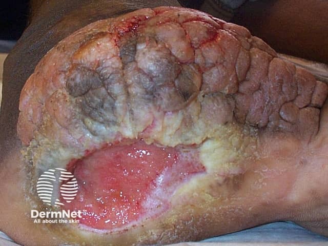
Imaging modality: Clinical photography. Perspective: plantar surface close-up of a chronic neuropathic foot ulcer. Site: plantar midfoot/ball region near the weight-bearing plantar surface of the foot with surrounding callus. Visual features: large, shallow ulcer with a reddened, raw base; yellow-gray necrotic slough rimmed by hyperemic tissue. Surrounding skin shows thickened keratin (callus), fissuring, and mild edema; moisture varies with exudate. The ulcer bed appears to granulate in parts but remains largely necrotic and at risk of secondary infection. No visible intact epidermis over the lesion. Peri-wound area demonstrates crusting and maceration from drainage. The clinical photograph documents a non-healing, neuropathy-associated lesion often seen in diabetes mellitus or peripheral neuropathy; pain is typically reduced, delaying care. In this context, the image highlights pressure-related risk, repetitive friction, and reduced protective sensation contributing to ulcer development. Diagnostic significance: supports diagnosis of a neuropathic (diabetic) plantar foot ulcer; differentiates from ischemic, venous, or pressure ulcers; emphasizes need for off-loading, debridement, moisture balance, infection surveillance, and vascular assessment. Potential uses: medical education, wound-care planning, telemedicine triage, research on wound healing, assessment of treatment response, and algorithm development for neuropathic ulcers. Documentation supports multidisciplinary care, including off-loading devices, infection control, and regular wound monitoring and prognosis.
Foot ulcer
Why does it occur?
-
Neuropathy - loss of protective sensation
- Small injuries are not felt.
- Motor neuropathy can cause claw toes, prominent metatarsal heads, and abnormal pressure points.
- Autonomic neuropathy causes dry, cracked skin, allowing bacteria to enter.
-
Pressure and friction
- Tight shoes, walking barefoot, callus, deformity, or a foreign body in the shoe.
- Neuropathic ulcers are typically on weight-bearing areas: beneath the big toe, metatarsal heads, heel, or midfoot.
-
Poor arterial blood supply - ischemia
- Peripheral arterial disease reduces oxygen and nutrient delivery, making ulcers painful and slow to heal.
- Classically, ischemic ulcers occur around the toes, heel, or foot margins.
-
Infection
- A superficial ulcer can progress to cellulitis, abscess, gangrene, or bone infection called osteomyelitis.
Types of foot ulcer
| Type | Typical site and features |
|---|---|
| Neuropathic / diabetic ulcer | Sole of foot, under metatarsal heads or heel; often surrounded by callus; may be surprisingly painless. Foot is usually warm with palpable pulses if blood supply is adequate. |
| Ischemic / arterial ulcer | Toes, heel, lateral border, pressure points; painful, pale or black wound; foot may be cold with weak or absent pulses. |
| Neuroischemic ulcer | Diabetes with both neuropathy and poor circulation. Higher risk of poor healing and amputation. |
| Pressure ulcer | Heel is common in people confined to bed or unable to move. |
| Venous ulcer | Usually around the lower leg and ankle, rather than the foot itself. |
Symptoms and signs
- A crack, blister, cut, crater, or black area of skin
- Drainage, pus, bad smell, or wet dressing
- Callus around a wound
- Redness, warmth, swelling, or tenderness
- Black tissue, called necrosis or gangrene
- Fever or feeling unwell if infection is severe
Assessment by a clinician
- Measuring and documenting the site, length, width, depth, wound base, discharge, surrounding redness, and odor
- Checking sensation with a monofilament or similar test
- Checking circulation: skin temperature, capillary refill, foot pulses, ankle-brachial pressure index or toe pressures as appropriate
- Looking for callus, deformity, footwear problems, and Charcot foot
- Checking for infection
- X-ray if the ulcer is deep, longstanding, or bone infection is suspected
- MRI may be needed when osteomyelitis remains uncertain
Treatment principles
1. Off-loading the pressure
- Avoid walking on the ulcerated area as directed by the treating team.
- A total-contact cast, removable walker, special footwear, insoles, or felt padding may be used.
- Do not use ordinary footwear that rubs or applies pressure over the wound.
2. Wound cleaning and debridement
- A trained clinician removes dead tissue, thick callus, and debris when appropriate.
- The wound is cleaned and dressed to control moisture and protect it.
- The type of dressing depends on the amount of drainage and the wound bed.
3. Treat infection when present
4. Improve blood supply
5. Control diabetes and general health
- Keep blood glucose as well controlled as safely possible.
- Optimize nutrition.
- Stop smoking.
- Treat anemia, kidney disease, edema, and other factors that delay healing.
When is it an emergency?
- Fever, chills, confusion, or feeling very unwell
- Spreading redness, swelling, severe pain, pus, or foul odor
- A black, blue, cold, or rapidly worsening toe/foot
- Red streaking up the foot or leg
- A deep ulcer where tendon or bone may be visible
- New ulcer in a person with diabetes, especially with poor circulation
- An ulcer with sudden swelling, deformity, or warmth of the foot
Prevention, especially in diabetes
- Inspect both feet every day, including soles and between toes.
- Never walk barefoot, including indoors.
- Check inside shoes before wearing them.
- Wear well-fitting shoes and clean socks.
- Do not cut corns or calluses yourself with blades or chemicals.
- Keep skin moisturized, but do not apply cream between toes.
- Arrange regular podiatry review if you have neuropathy, prior ulcer, deformity, or poor circulation.
- Seek advice promptly for a blister, cut, redness, swelling, or discharge.
Traumatic foot ulcer