
boerhaave syndrome
"Boerhaave syndrome"
Boerhaave syndrome diagnosis management guidelines
Boerhaave syndrome esophageal rupture pneumomediastinum CT chest
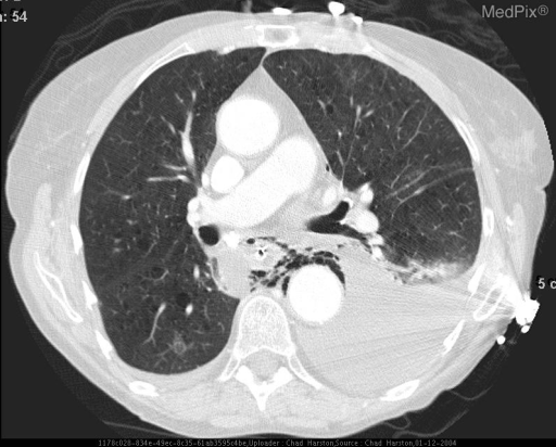
**Imaging Modality:** Axial Contrast-Enhanced Computed Tomography (CT) of the chest.
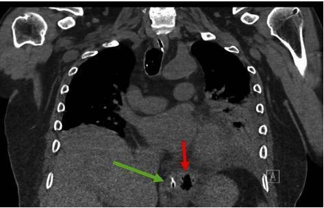
This non-contrasted coronal CT scan of the chest and upper abdomen demonstrates diagnostic features of Boerhaave's Syndrome (spontaneous esophageal perforation). A prominent red arrow points to an extraluminal pocket of gas (pneumomediastinum) located in the lower mediastinum, positioned left-lateral to the distal esophagus. Adjacent to this air collection, a green arrow identifies a nasogastric (NG) tube situated within the esophageal lumen, providing a landmark for the esophageal anatomy. The mediastinal air is seen near the diaphragmatic hiatus, a common site for pressure-induced rupture. The surrounding lung fields show partial opacification and are somewhat obscured by mediastinal structures, though no large pleural effusions are immediately apparent in this view. This imaging is critical for differentiating esophageal rupture from other causes of acute chest pain, such as aortic dissection or myocardial infarction, particularly when clinical history involves forceful vomiting or retching.
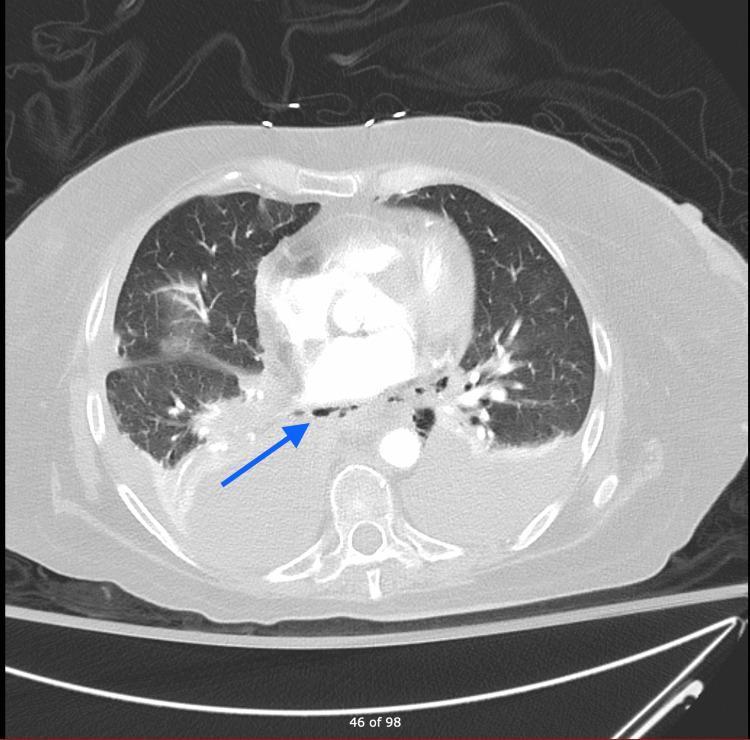
An axial view from a CT angiography (CTA) of the chest demonstrating pneumomediastinum and associated thoracic pathology. A blue arrow highlights extraluminal air pockets (pneumomediastinum) located in the posterior mediastinum, adjacent to the distal esophagus. The right lung displays increased opacity and hazy consolidation, particularly in the perihilar region, consistent with atelectasis or underlying infection. Bilateral pleural abnormalities are noted, with a visible right-sided pleural effusion and possible thickening of the pleural margins. The central vascular structures, including the aorta and heart, are visible with contrast enhancement. This clinical imaging finding is highly indicative of esophageal perforation (Boerhaave syndrome or traumatic injury) necessitating urgent evaluation. The image serves as an educational resource for identifying mediastinal air collections and secondary pulmonary manifestations of esophageal rupture.
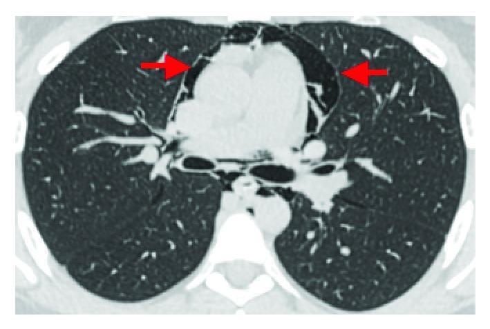
This diagnostic image is an axial contrast-enhanced CT scan of the chest presented in a lung window. The primary pathology demonstrated is pneumomediastinum, indicated by prominent collections of extraluminal air (hypodense areas) within the mediastinal space, specifically surrounding the heart and great vessels (indicated by red arrows). The air tracks along the pericardiac spaces and fascial planes. Additionally, there are signs of subcutaneous emphysema within the chest wall tissues. The lung parenchyma bilaterally appears largely unremarkable with normal bronchovascular markings and no evidence of significant consolidation, pulmonary edema, or pneumothorax. This image serves as a clinical example of Hamman's syndrome or spontaneous pneumomediastinum, often seen following intense physical exertion such as labor. The absence of contrast extravasation helps rule out esophageal rupture (Boerhaave syndrome) in this clinical context.
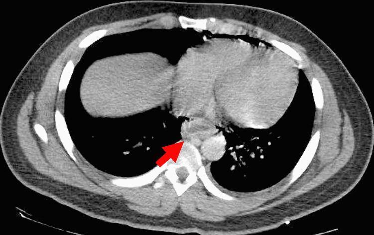
This diagnostic image is a transverse (axial) view computed tomography (CT) scan of the human chest, presented in a soft tissue/mediastinal window. Key anatomical landmarks include the lungs (appearing as dark, air-filled lateral spaces), the central mediastinum containing the heart and great vessels, and the posterior thoracic spine. A red arrow highlights a significant pathological finding in the posterior mediastinum: diffuse circumferential thickening of the esophageal wall with associated soft tissue stranding and irregularity. There is evidence of pneumomediastinum, characterized by pockets of low-density gas tracking adjacent to the esophageal margins and behind the heart. These radiological signs are highly indicative of an esophageal perforation, such as that seen in Boerhaave syndrome (effort rupture of the esophagus). The image demonstrates the critical role of CT imaging in identifying emergency gastrointestinal tract disruptions and evaluating secondary complications like mediastinitis or pneumomediastinum within a clinical setting.
Boerhaave Syndrome
History
Pathophysiology
- Barogenic rupture: a bubble of esophageal mucosa dissects through the longitudinal muscle fibers at the point where the "clasp" and oblique fibers meet - Sleisenger and Fordtran's Gastrointestinal and Liver Disease.
- ~90% of ruptures occur on the left posterolateral wall of the distal esophagus, just above the gastroesophageal junction, because this is the weakest point mechanically (cadaveric studies show 270-340 mmHg needed to rupture there). Mid-thoracic and cervical ruptures are less common.
- Defects average 2-6 cm (up to 10 cm).
- Predisposing mucosal conditions include reflux/Barrett esophagitis, infectious or eosinophilic esophagitis.
- Precipitants besides vomiting: weightlifting, forceful coughing/sneezing, straining, childbirth, seizures, blast injury.
Epidemiology
Clinical presentation
- Classic Mackler triad: vomiting, sudden severe substernal/epigastric chest pain, and subcutaneous (cervical) emphysema - present together in only a minority of patients.
- Pain may radiate to the back; patients often appear acutely ill - tachycardic, febrile, dyspneic, diaphoretic, and may develop rapid shock/sepsis.
- Hamman's crunch/sign: a crepitant, mediastinal "crunching" sound synchronous with the heartbeat, heard over the precordium - suggests pneumomediastinum but is uncommon.
- Because presentation mimics acute coronary syndrome, aortic dissection, perforated peptic ulcer, or pancreatitis, diagnosis is frequently delayed - this delay is the single biggest driver of morbidity/mortality.
Diagnosis
- Chest X-ray: pneumomediastinum, subcutaneous emphysema, pleural effusion (left-sided more common than right), pneumothorax, or pneumoperitoneum - but a normal film does not exclude the diagnosis.
- Pleural fluid may show elevated salivary amylase, which can be mistaken for pancreatitis.
- CT chest with oral water-soluble contrast is the modern test of choice - shows extraluminal air/fluid, mediastinal stranding, and sometimes the leak itself.
- Contrast esophagram (Gastrografin, then dilute barium if needed) can localize and confirm the perforation.
Management
- Resuscitation: IV fluids, broad-spectrum antibiotics (e.g., imipenem/cilastatin or a beta-lactam/beta-lactamase inhibitor covering oral/GI flora), NPO, nasogastric decompression, drainage of the mediastinum/pleural space (chest tube or thoracotomy).
- Surgery is the standard of care when diagnosed within ~24 hours - primary repair of the tear with buttressing (e.g., pleural or omental flap) plus wide drainage gives the best survival.
- Endoscopic options (covered self-expanding metal stents, clips, or endoluminal vacuum therapy) are increasingly used for early, well-contained perforations with minimal sepsis, but should be used cautiously and are not first-line if there is frank sepsis or a large, uncontained leak.
- Nonoperative/conservative management (strict NPO 7+ days, parenteral nutrition, prolonged IV antibiotics 7-14 days, drainage of collections) is reserved for small, contained leaks in stable patients, with a low threshold to convert to surgery if there's clinical deterioration, extension of the injury, or persistent fever.
- Delayed presentations (>24-48 hours) with established mediastinitis carry the highest mortality and often require more extensive debridement, sometimes esophagectomy with diversion/reconstruction.