
Circle of willi
Reading File
The Circle of Willis is the arterial anastomotic ring at the base of the brain, formed by the joining of the internal carotid and vertebrobasilar systems. It provides collateral circulation so that if one vessel is occluded, blood can still reach most brain regions.
Searching sources...
Finding Sources
Searching the Web
Circle of Willis anatomy components collateral circulation
Reading File
Reading File
Loading Image
Loading Image
Finding Sources
Reading File
Reading File
Here is a detailed breakdown of the Circle of Willis, drawn from multiple authoritative neurology and medical physiology textbooks.
Circle of Willis (Circulus Arteriosus Cerebri)
Anatomy Diagrams
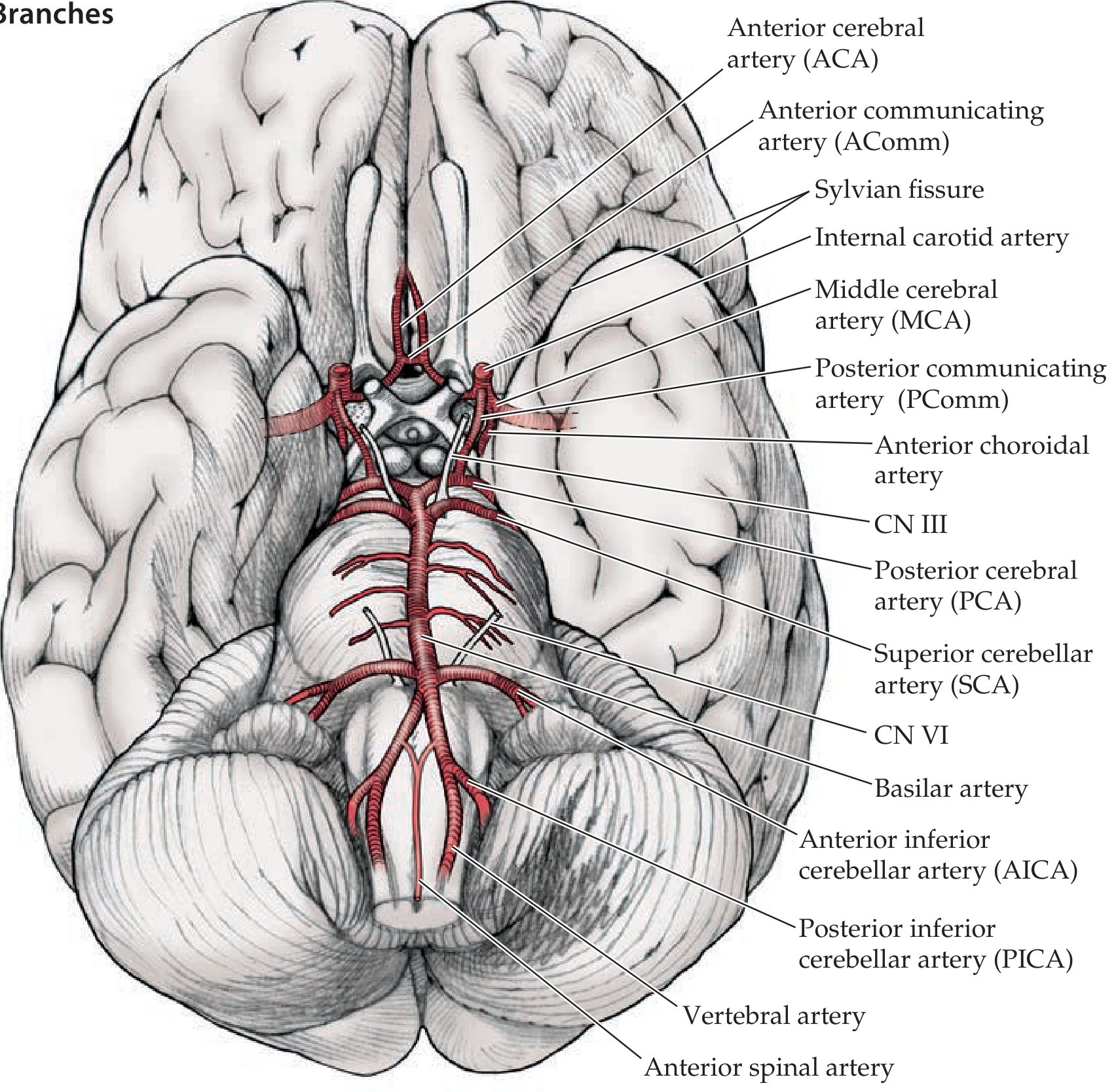
Inferior (basilar) view of brain showing the Circle of Willis and main branches:
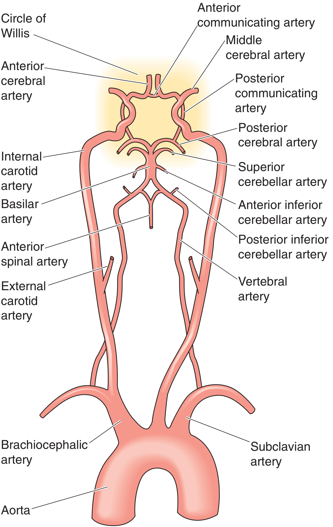
Schematic diagram showing the full arterial supply from aorta to circle:
Definition
The Circle of Willis is an anastomotic ring of arteries located in the subarachnoid space at the base of the brain. It connects the two major arterial systems supplying the brain - the internal carotid system (anterior circulation) and the vertebrobasilar system (posterior circulation).
- Neuroanatomy through Clinical Cases, 3rd Ed.
Components
The ring is formed by the following vessels:
| Artery | Origin | Role |
|---|---|---|
| Anterior cerebral artery (ACA) x2 | Terminal branch of ICA | Supplies medial frontal & parietal lobes |
| Anterior communicating artery (AComm) | Connects left and right ACAs | Links right and left anterior circulations |
| Internal carotid artery (ICA) x2 | Common carotid artery | Main inflow to anterior circulation |
| Posterior communicating artery (PComm) x2 | Branch of ICA | Links anterior (ICA) to posterior (PCA) circulation |
| Posterior cerebral artery (PCA) x2 | Top of basilar artery | Supplies occipital lobe, thalamus, midbrain |
The basilar artery (formed by the two vertebral arteries joining) feeds the PCAs but is not itself part of the ring.
Anterior vs. Posterior Circulation
- Anterior circulation: ICA → ACA + MCA (middle cerebral artery). The ACAs are joined anteriorly by the AComm artery.
- Posterior circulation: Vertebral arteries → basilar artery → PCAs.
- Link between them: PComm arteries connect the ICAs to the PCAs, joining the two circulations.
- Neuroanatomy through Clinical Cases, 3rd Ed.
Function
The primary role of the Circle of Willis is collateral circulation - if one feeding vessel is occluded, blood can be rerouted through the ring to maintain flow to the affected territory.
"In persons with an intact circle of Willis, the entire intracranial circulation can be supplied by a single patent internal carotid artery or vertebral artery."
- Goldman-Cecil Medicine
Three tiers of brain collateral circulation exist:
- Circle of Willis (most important - connects carotid and vertebrobasilar systems)
- Extracranial-intracranial anastomoses (e.g., external carotid to ophthalmic artery)
- Leptomeningeal anastomoses between terminal branches of cerebral arteries
- Localization in Clinical Neurology, 8e
Anatomical Variants (Very Common)
A complete, full-caliber circle of Willis is present in only about 25-34% of individuals - meaning most people have an incomplete ring. Common variants:
- Hypoplastic/absent A1 segment of ACA (between ICA and AComm): both ACAs then fill from one ICA.
- "Fetal" PCA: the P1 segment (between basilar and PComm) is absent/hypoplastic, so the PCA is supplied by the ICA rather than the basilar artery. This is a very common variant.
- Rare primitive anastomoses that may persist: persistent trigeminal artery (most common), persistent hypoglossal artery, persistent acoustic (otic) artery, and persistent proatlantal artery.
- Goldman-Cecil Medicine; Localization in Clinical Neurology, 8e
Key Branches Arising From/Near the Circle
| Artery | Territory |
|---|---|
| Middle cerebral artery (MCA) | Lateral frontal, parietal, temporal lobes (largest territory) |
| Anterior choroidal artery | Optic tract, hippocampus, amygdala, posterior limb of internal capsule |
| Recurrent artery of Heubner | Anterior limb of internal capsule, caudate, putamen (can be damaged during AComm aneurysm clipping) |
| Superior cerebellar artery (SCA) | Superior cerebellum |
| AICA | Anterior inferior cerebellum |
| PICA | Posterior inferior cerebellum |
Clinical Relevance
- Aneurysms most commonly form at branching points of the circle, particularly at the AComm (most common site for berry aneurysms) and the PComm junction. A PComm aneurysm can compress CN III, causing a dilated, unreactive pupil with ipsilateral ptosis and "down-and-out" eye.
- Watershed infarcts occur at the junction between territories of adjacent cerebral arteries when perfusion pressure drops globally.
- Fetal PCA is clinically important in posterior circulation strokes because it means the posterior cortex territory may be at risk from carotid (anterior) disease, not just vertebrobasilar disease.
Sources: Neuroanatomy through Clinical Cases 3rd Ed. | Goldman-Cecil Medicine | Localization in Clinical Neurology 8e | Medical Physiology (Boron & Boulpaep)
This is a shared conversation. Sign in to Orris to start your own chat.