
Difference in extra medullary and intramedullary myelopathies
intramedullary extramedullary spinal cord myelopathy diagram
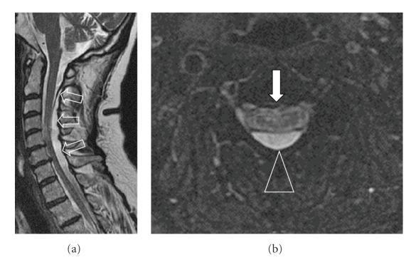
This diagnostic image consists of two MRI views of the cervical spine illustrating a dorsal intradural-extramedullary cystic lesion. (a) A sagittal T2-weighted MRI demonstrates a large, hyperintense cystic cavity extending from the cervicocranial junction to the level of the T4 vertebral body. Open arrows highlight the dorsal location of the cyst, which causes significant anterior displacement of the cervical spinal cord (myelon). Intramedullary hyperintensities, consistent with myelopathy, are visible at the C4/5 and C6/7 levels. (b) An axial T2*-weighted image at the cervical level shows the relationship between the hyperintense mass and the neural structures. A triangle marker points to a distinct thick membrane separating the cystic lesion from the spinal cord, while a filled arrow indicates intrinsic myelopathy signal changes within the cord parenchyma. This case illustrates the radiological features of a spinal arachnoid cyst and its secondary effects on the spinal cord, useful for neurology and neurosurgery education.
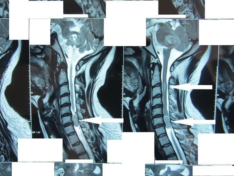
This diagnostic image consists of T2-weighted sagittal MRI views of the cervical spine, demonstrating multiple space-occupying lesions. The most prominent finding is a well-circumscribed, hyperintense extramedullary mass located at the C6-C7 level (indicated by white arrows). This lesion causes significant posterior-to-anterior displacement and compression of the spinal cord, nearly obliterating the subarachnoid space at this segment. A second, more subtle area of hyperintensity is visible within the cervical cord at a higher level, suggesting an intramedullary component or additional lesion. The vertebral bodies and disc spaces appear relatively preserved, focusing the pathology on the spinal canal. These findings are clinically significant for evaluating neurofibromatosis or multiple schwannomas, as they illustrate the relationship between extramedullary tumors and spinal cord compression (myelopathy). The images are optimized for neurosurgical planning and neuroradiology education.
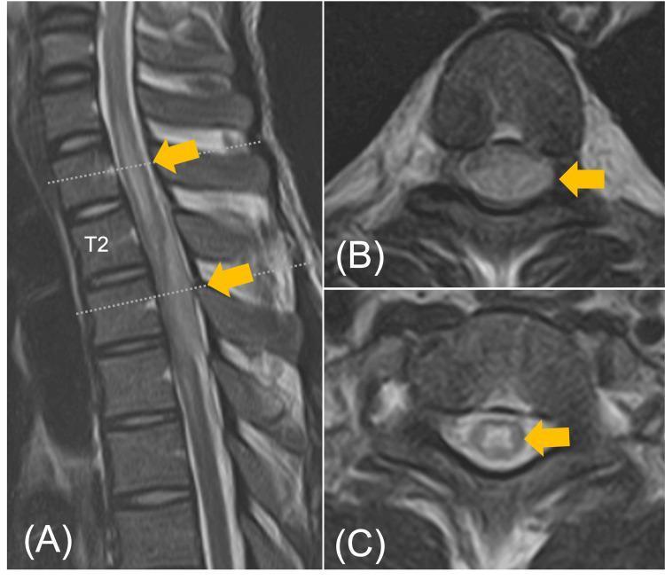
This diagnostic imaging panel displays T2-weighted magnetic resonance imaging (MRI) scans of the thoracic spine, highlighting a significant intradural extramedullary lesion. (A) Sagittal view: A hyperintense, spindle-shaped lesion is visible in the posterior aspect of the spinal canal, spanning the T2 to T4 vertebral levels. The mass causes notable ventral displacement and compression of the spinal cord. (B) Axial view at the T3 level: The lesion (indicated by a yellow arrow) occupies the dorsal aspect of the intradural space, resulting in severe compression and flattening of the cord. (C) Axial view at the T1 level: Increased intramedullary signal intensity is observed within the spinal cord, suggestive of perilesional edema or compressive myelopathy. The images demonstrate the visual findings of a thoracic spinal tuberculoma, emphasizing the mass effect on neural structures and secondary cord changes. Educational focus includes localizing intradural extramedullary pathologies and recognizing radiological signs of spinal cord compression.
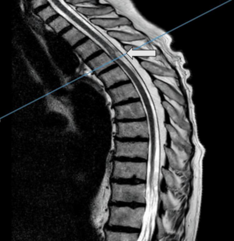
This diagnostic image is a T2-weighted sagittal MRI of the thoracic spine, highlighting the anatomical region between the upper thoracic and lumbar vertebrae. A white arrow points to an intraspinal, intradural mass located at the T4 vertebral level. The lesion exhibits altered signal intensity compared to the surrounding spinal cord tissue, characterized by a slightly hyperintense appearance. Morphologically, the mass is elongated and irregular, causing focal compression or displacement of the cord. The superior aspect of the spinal cord adjacent to the lesion shows signs of myelopathic changes, evidenced by intramedullary signal hyperintensity. The surrounding vertebral bodies and intervertebral discs maintain normal vertical alignment and signal characteristics for this imaging modality. Clinically, this imaging illustrates a rare spinal pathology, such as a spinal arachnoid web, emphasizing the diagnostic importance of MRI in evaluating myelopathy and intradural extramedullary lesions in the thoracic spine for neurosurgical planning.
Extramedullary vs. Intramedullary Myelopathies
Definition & Anatomic Classification
| Feature | Extramedullary | Intramedullary |
|---|---|---|
| Location | Outside the spinal cord, within the spinal canal | Within the spinal cord parenchyma itself |
| Subtypes | Extradural (outside dura) OR Intradural-extramedullary (inside dura, outside cord) | Intradural-intramedullary only |
| Examples | Vertebral metastases, meningiomas, schwannomas, disc herniation, epidural abscess | Ependymoma, astrocytoma, intramedullary metastases, syrinx, MS plaque, AVM |
Mechanism of Cord Injury
- Extramedullary: Cord damage occurs by compression from outside - the lesion indents the thecal sac, then displaces and compresses the cord. Damage starts from the periphery of the cord (where long tracts are arranged somatotopically with sacral fibers most peripheral).
- Intramedullary: Lesion arises within the cord parenchyma, causing direct invasion or destruction of central gray matter and adjacent tracts outward.
Clinical Features Compared
| Feature | Extramedullary | Intramedullary |
|---|---|---|
| Pain | Radicular pain is common and often early - knife-like, radiates distally, worse with coughing/sneezing/straining; back pain worse lying down | Pain may be present but often less prominent; can be bilateral, diffuse, burning |
| Sensory symptoms onset | Often contralateral to motor deficit initially; sensory level may ascend over time | Dissociated sensory loss (pain/temperature lost, touch/vibration preserved) over several segments - classic for central cord involvement |
| Sacral sensation | Usually involved early - peripheral fibers compressed first | Sacral sparing - central lesion spares the outermost sacral fibers initially |
| Motor deficit | Asymmetric spastic weakness; one leg weakens before the other | Mixed sensorimotor tract syndrome; central cord pattern (arms > legs in cervical lesions) |
| Brown-Séquard syndrome | More likely with intradural-extramedullary lesions (lateral compression) | Rarely produces a true Brown-Séquard pattern |
| Sphincter involvement | Late - bladder/bowel dysfunction comes after motor involvement | Early - sphincter disturbances appear early in the course |
| Reflexes | Hyperreflexia + Babinski below the level | Hyperreflexia below; LMN signs (areflexia, amyotrophy) at the level of the lesion |
| Fasciculations/amyotrophy | Uncommon, only if nerve roots compressed | More common - central gray matter involvement causes LMN signs at the segmental level |
| Sensory level | A relatively sharp, well-defined sensory level on the trunk | Less sharp; dissociated type (pain/temp vs. touch/vibration split) more characteristic |
| Cord expansion on MRI | Cord is compressed/displaced but not intrinsically expanded | Cord is often expanded/widened; gadolinium enhancement may occur |
Classic Syndromes
- Radicular-spinal cord syndrome - root pain + progressive myelopathy (most common presentation of intradural-extramedullary tumors)
- Transverse/sensorimotor tract syndrome - with a clear sensory level, spastic weakness, and late sphincter involvement
- Brown-Séquard hemicord syndrome (ipsilateral motor + contralateral pain/temp loss)
- Syringomyelic/central cord syndrome - segmental dissociated sensory loss (cape-like or suspended distribution), amyotrophy, early incontinence, late corticospinal weakness; sacral sensation spared early
- Mixed sensorimotor tract syndrome - ependymomas and astrocytomas commonly cause this
- Rarely, greatly elevated CSF protein + hydrocephalus (thoracolumbar intramedullary tumors)
Distinguishing Clues at the Bedside
| Clue Pointing to Extramedullary | Clue Pointing to Intramedullary |
|---|---|
| Early, prominent radicular pain | Early sphincter/bladder dysfunction |
| Pain worse lying down, improved sitting | Dissociated sensory loss (pain/temp lost, touch/vibration preserved) |
| Bilateral root pain | Segmental amyotrophy at the level |
| Motor symptoms precede sensory symptoms | Sacral sparing of sensation |
| Marked percussion tenderness over spine | Suspended (cape-like) sensory loss |
| Late sphincter disturbance | Cord widening on MRI |
Note from Goldman-Cecil Medicine: "The classic signs of intramedullary spinal cord lesions, such as dissociated sensory loss, sacral sparing, and early sphincter problems, are not sufficiently reliable alone to distinguish intramedullary from extramedullary lesions. MRI with gadolinium is the definitive diagnostic study."
Imaging
- Extramedullary: MRI shows cord displacement/compression; may show vertebral destruction (extradural) or intradural mass; cord signal change (T2 hyperintensity) if compression is prolonged
- Intramedullary: MRI shows cord expansion, intrinsic T2 hyperintensity, often gadolinium enhancement; an associated syrinx may be seen extending above and below
Common Causes by Category
| Category | Extramedullary | Intramedullary |
|---|---|---|
| Tumors | Meningioma, schwannoma (intradural-EM); vertebral metastases, lymphoma (extradural) | Ependymoma (#1 in adults), astrocytoma (#2), metastases (rare - bronchogenic ca. most common) |
| Infectious | Epidural abscess, Pott's disease (TB) | Abscess, transverse myelitis |
| Vascular | Epidural hematoma | Hematomyelia, AVM, cord infarct |
| Degenerative | Cervical spondylosis, disc herniation | - |
| Inflammatory/Demyelinating | - | Multiple sclerosis plaque, NMO |